Abstract
Coronavirus disease 2019 (COVID-19) emerged at the end of 2019, resulting in a global pandemic. As of August 4, 2020, 18.3 million confirmed cases and 695 874 confirmed deaths have been reported in the world, of which the United States experienced 4 750 578 cases and 156 594 deaths. COVID-19 is caused by a single-stranded RNA virus named severe acute respiratory syndrome coronavirus 2 (SARS-CoV-2). COVID-19 is a member of the family of coronaviruses that includes zoonotic entities of SARS-CoV-2 and Middle East respiratory syndrome coronavirus (MERS-CoV). SARS-CoV-2 and MERS-CoV emerged in 2003 and 2012, respectively, and both lead to life-threatening pneumonia. Human coronaviruses designated HCoV-NL63, HCoV-229E, and HCoV-OC43 are also part of the coronavirus family and typically result in upper respiratory infections. COVID-19 primarily targets the lung, with patients presenting with pneumonia that can result in acute respiratory distress syndrome (ARDS). However, COVID-19 can result in multiorgan systemic disease, affecting the brain, gastrointestinal system, heart, and kidneys, either directly or indirectly through the host’s inflammatory response and a hypercoagulable state.
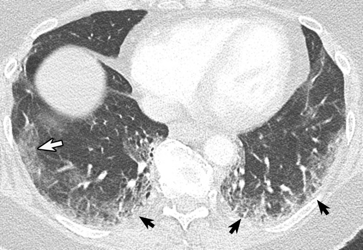
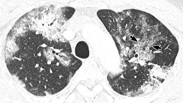
Chest radiography, CT, and point-of-care lung US have important roles in the care of patients with COVID-19. A diagnosis of COVID-19 is confirmed by reverse transcriptase–polymerase chain reaction (RT-PCR). In patients with a positive RT-PCR test result with moderate or severe clinical features of COVID-19, chest imaging can be used to evaluate the baseline severity of any lung disease (Figs 1, 2). Imaging can be used to evaluate for alternative diagnoses in patients with a negative RT-PCR test result despite a persisting clinical suspicion for COVID-19. Multiple studies have described the chest CT findings of COVID-19. Reporting systems have been published that summarize the typical imaging findings and categorize those findings according to the level of suspicion. These systems can be applied to patients under investigation and to those with incidentally depicted findings. A standard approach for interpreting imaging findings within one’s institution can enhance communication with health care providers and can be developed with knowledge of these reporting systems. Subsequent patient treatment is determined depending on the context of disease prevalence in the community and clinical features. In a low-prevalence scenario, a higher rate of false-positive examinations may ensue. In a high-prevalence scenario, a larger number of false-negative examinations, meaning more frequently atypical appearances for COVID-19, will probably be encountered. Chest radiography is shown to be less sensitive and specific than CT. Chest radiography is more readily available than CT, is easily performed, and can minimize in-hospital transmission. COVID-19 imaging findings at chest radiography correlate with chest CT findings. Point-of-care lung US performed by health care providers may aid in monitoring treatment response during hospitalization.
Figure 1.
Peripheral ground-glass opacity (GGO) manifesting in a patient 5 days after symptoms of dyspnea, nausea, and body aches. Axial CT image shows faint rounded GGO (white arrow) in the right lower lobe laterally, with mild peripheral GGO in both lower lobes (black arrows).
Figure 2.
Crazy-paving pattern in the left upper lobe in a man with COVID-19. Axial CT image obtained 11 days after symptom onset shows intralobular lines within GGOs (arrows), which in combination with interlobular septal thickening create a cobblestone pattern characteristic of crazy paving.
An awareness of the imaging findings of COVID-19 will aid in considering this entity when interpreting imaging studies. The objective of this online presentation is to provide a resource for understanding the imaging appearance of pulmonary manifestations of COVID-19 at multiple imaging modalities, including CT, chest radiography, and point-of-care lung US, as well as to highlight typical imaging findings and provide examples of differential diagnostic considerations and mimics.
Acknowledgments
Acknowledgment
The authors would like to acknowledge Brian T. Garibaldi, MD, MEHP, Johns Hopkins University School of Medicine, for his contributions to this manuscript.
J.P.K., G.L., and J.S.K. have provided disclosures; all other authors have disclosed no relevant relationships.
Disclosures of Conflicts of Interest.—: J.P.K. Activities related to the present article: disclosed no relevant relationships. Activities not related to the present article: institution received a grant from Siemens AG for research collaboration. Other activities: spouse is an employee of AlloVir. G.L. Activities related to the present article: disclosed no relevant relationships. Activities not related to the present article: member of the medical advisory board of EchoNous. Other activities: disclosed no relevant relationships. J.S.K. Activities related to the present article: disclosed no relevant relationships. Activities not related to the present article: disclosed no relevant relationships. Other activities: member of the RSNA COVID-19 Task Force.
Abbreviations:
- ARDS
- acute respiratory distress syndrome
- COVID-19
- coronavirus disease 2019
- GGO
- ground-glass opacity
- RT-PCR
- reverse transcriptase–polymerase chain reaction
Suggested Readings
- Bernheim A, Mei X, Huang M, et al. Chest CT findings in coronavirus disease-19 (COVID-19): relationship to duration of infection. Radiology 2020;295(3):200463. [DOI] [PMC free article] [PubMed] [Google Scholar]
- Coronavirus map. Tracking the global outbreak. The New York Times website. https://www.nytimes.com/interactive/2020/world/coronavirus-maps.html#countries. Updated June 9, 2020. Accessed August 4, 2020.
- Foust AM, McAdam AJ, Chu WC, et al. Practical guide for pediatric pulmonologists on imaging management of pediatric patients with COVID-19. Pediatr Pulmonol 2020;55(9):2213–2224. [DOI] [PMC free article] [PubMed] [Google Scholar]
- Ge H, Wang X, Yuan X, et al. The epidemiology and clinical information about COVID-19. Eur J Clin Microbiol Infect Dis 2020;39(6):1011–1019. [DOI] [PMC free article] [PubMed] [Google Scholar]
- Grillet F, Behr J, Calame P, Aubry S, Delabrousse E. Acute pulmonary embolism associated with COVID-19 pneumonia detected with pulmonary CT angiography. Radiology 2020;296(3):E186–E188. [DOI] [PMC free article] [PubMed] [Google Scholar]
- Kaminetzky M, Moore W, Fansiwala K, et al. Pulmonary embolism on CTPA in COVID-19 patients. Radiol Cardiothorac Imaging 2020;2(4):e200308. [DOI] [PMC free article] [PubMed] [Google Scholar]
- Kim H, Hong H, Yoon SH. Diagnostic performance of CT and reverse transcriptase polymerase chain reaction for coronavirus disease 2019: a meta-analysis. Radiology 2020;296(3):E145–E155. [DOI] [PMC free article] [PubMed] [Google Scholar]
- Prokop M, van Everdingen W, van Rees Vellinga T, et al. CO-RADS: a categorical CT assessment scheme for patients with suspected COVID-19—definition and evaluation. Radiology 2020:201473. [DOI] [PMC free article] [PubMed] [Google Scholar]
- Rubin GD, Ryerson CJ, Haramati LB, et al. The role of chest imaging in patient management during the COVID-19 pandemic: a multinational consensus statement from the Fleischner Society. Chest 2020;158(1):106–116. [DOI] [PMC free article] [PubMed] [Google Scholar]
- Simpson S, Kay FU, Abbara S, et al. Radiological Society of North America expert consensus statement on reporting chest CT findings related to COVID-19. Endorsed by the Society of Thoracic Radiology, the American College of Radiology, and RSNA—secondary publication. J Thorac Imaging 2020;35(4):219–227. [DOI] [PMC free article] [PubMed] [Google Scholar]


