. 2020 Nov 2;40(7):1866–1892. doi: 10.1148/rg.2020200195

Figure 17a.

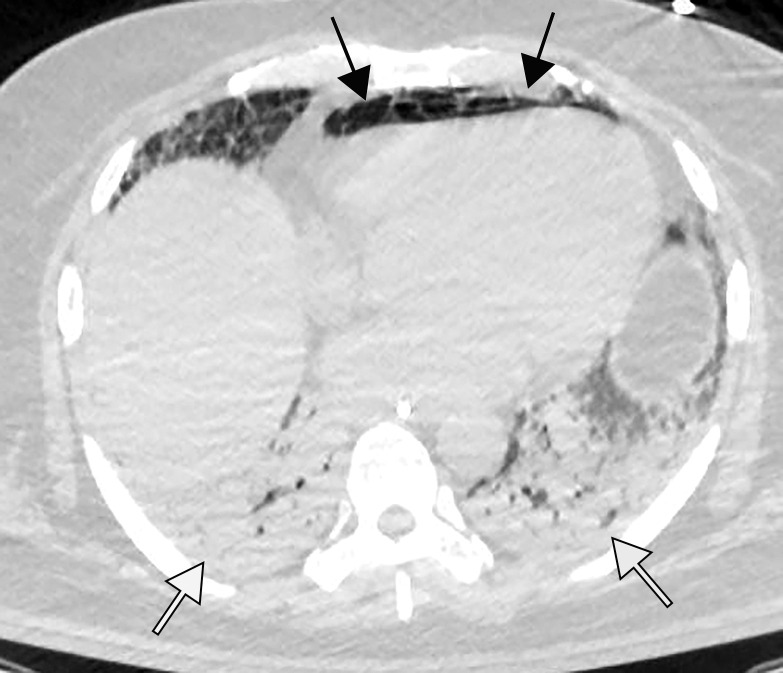
Pneumopericardium with air dissecting into the peritoneum, mimicking bowel perforation, in a 64-year-old man who underwent intubation for COVID-19 pneumonia. (a) Axial nonenhanced chest CT image shows pneumopericardium (black arrows) and bibasilar airspace consolidations (white arrows), compatible with COVID-19 pneumonia. (b) Axial nonenhanced CT image of the abdomen and pelvis shows pneumopericardium and free air under the diaphragm, anterior to the liver (black arrows), without findings of bowel wall ischemia to suggest a perforation (not shown). Note the bibasilar consolidations (white arrows).

Pneumopericardium with air dissecting into the peritoneum, mimicking bowel perforation, in a 64-year-old man who underwent intubation for COVID-19 pneumonia. (a) Axial nonenhanced chest CT image shows pneumopericardium (black arrows) and bibasilar airspace consolidations (white arrows), compatible with COVID-19 pneumonia. (b) Axial nonenhanced CT image of the abdomen and pelvis shows pneumopericardium and free air under the diaphragm, anterior to the liver (black arrows), without findings of bowel wall ischemia to suggest a perforation (not shown). Note the bibasilar consolidations (white arrows).