. 2020 Nov 2;40(7):1866–1892. doi: 10.1148/rg.2020200195

Figure 5c.

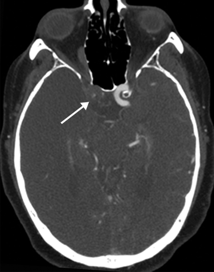
Long segment right internal carotid artery (ICA) occlusion with associated bilateral large middle cerebral artery territory infarcts in a 59-year-old man who underwent intubation due to COVID-19 who presented with sudden onset of confusion. The serum analysis results showed a high d-dimer level of 5.6 mg/mL. (a) Axial nonenhanced head CT image shows large regions of hypoattenuation (arrows) in the bilateral middle cerebral artery territories, reflecting cytotoxic edema secondary to acute bilateral middle cerebral artery territorial infarctions without hemorrhagic transformation. The findings are indicative of cardioembolic phenomena (given various vascular distributions), hypoxic-ischemic injury, or ischemic vasculopathy secondary to COVID-19. (b–d) Sagittal maximum intensity projection (b) and axial head and neck CT angiographic images (c, d) show a long segment filling defect from the proximal cervical segment of the right ICA (arrow in b) to the intracranial ICA, with involvement of the right petrous (not shown) and cavernous ICA segments (arrow in c), indicative of right ICA thrombosis and occlusion. The right carotid terminus (white arrow in d) and right middle cerebral artery are completely occluded, and the M1 segment of the left middle cerebral artery is nearly completely occluded, with only a small sleeve of intravenous contrast material opacifying the artery (black arrow in d). Note that the loss of gray matter–white matter differentiation is more pronounced on the right (arrowheads in d).

Long segment right internal carotid artery (ICA) occlusion with associated bilateral large middle cerebral artery territory infarcts in a 59-year-old man who underwent intubation due to COVID-19 who presented with sudden onset of confusion. The serum analysis results showed a high d-dimer level of 5.6 mg/mL. (a) Axial nonenhanced head CT image shows large regions of hypoattenuation (arrows) in the bilateral middle cerebral artery territories, reflecting cytotoxic edema secondary to acute bilateral middle cerebral artery territorial infarctions without hemorrhagic transformation. The findings are indicative of cardioembolic phenomena (given various vascular distributions), hypoxic-ischemic injury, or ischemic vasculopathy secondary to COVID-19. (b–d) Sagittal maximum intensity projection (b) and axial head and neck CT angiographic images (c, d) show a long segment filling defect from the proximal cervical segment of the right ICA (arrow in b) to the intracranial ICA, with involvement of the right petrous (not shown) and cavernous ICA segments (arrow in c), indicative of right ICA thrombosis and occlusion. The right carotid terminus (white arrow in d) and right middle cerebral artery are completely occluded, and the M1 segment of the left middle cerebral artery is nearly completely occluded, with only a small sleeve of intravenous contrast material opacifying the artery (black arrow in d). Note that the loss of gray matter–white matter differentiation is more pronounced on the right (arrowheads in d).