

Abstract
Diagnosis of parasitic diseases that involve tissue-stage larvae is challenging, and serology remains the most effective antemortem test for detecting these infections. Baylisascaris procyonis, the raccoon roundworm, is a zoonotic ascarid. Raccoons are the usual definitive host, and humans may be infected as accidental hosts. More than 150 species of birds and mammals may act as paratenic hosts, and rodents play an important role in the transmission and maintenance of this parasite in nature. Migratory larvae in paratenic host tissues can produce ocular disease and severe to fatal neurologic disease, but not all infected hosts develop signs. A sensitive and specific Western blot (WB) assay based on a recombinant Baylisascaris-specific antigen (rBpRAG-1) has been developed for use in humans. We evaluated the use of this antigen to detect Baylisascaris spp. infections in rodent paratenic hosts. With the use of 4 species of Peromyscus mice (Peromyscus californicus, Peromyscus leucopus, Peromyscus maniculatus, Peromyscus polionotus) from a previous infection trial, we developed species-adapted WB and ELISA assays and evaluated performance compared to detection of larvae in tissue samples. These assays revealed species-level differences in seroconversion and terminal antibody concentrations, with P. leucopus developing significantly greater antibody concentrations than P. californicus and P. polionotus at all dose levels, and P. maniculatus at the low dose. Some P. californicus and P. polionotus failed to seroconvert despite the recovery of larvae from their tissues. WB and ELISA results were correlated; however, the WB demonstrated higher sensitivity than the ELISA overall (72.2% versus 63.9%, respectively). With the use of experimental samples, specificity was 100% for WB and 94.1% for ELISA. A WB was also used to test Mus and Rattus samples, and although numbers were too limited to evaluate sensitivity and specificity, all animals known to be infected by tissue digestion were WB positive, and all uninfected animals were negative. Finally, the Peromyscus-adapted WB and ELISA were used to test a set of serum samples from wild-trapped P. maniculatus and Rattus rattus. Both assays were generally sensitive, but specificity was equivocal. This emphasizes the challenge of using serology for investigation of wildlife diseases, in which hosts have unknown exposure histories. Nevertheless, serologic methods have utility in the study of Baylisascaris spp. in paratenic hosts, either wild or captive, and have advantageous attributes (non-lethal, high-throughput), but results should be interpreted carefully.
Baylisascaris procyonis, the raccoon (Procyon lotor) roundworm, is increasingly recognized as a potential cause of neurologic disease in a broad variety of paratenic host species, including humans (Kazacos, 2016). Eggs from the infected definitive hosts (raccoons, occasionally dogs, and possibly other procyonids) are shed in the feces, become infectious after ~10–14 days in the environment, and can infect >150 species of mammals and birds. Following ingestion of infectious eggs, larvae hatch and penetrate the wall of the small intestine of the paratenic host and undergo somatic migration. These migrating larvae can cause severe or fatal neural larva migrans if they invade the central nervous system or ocular larva migrans if they invade the eye. Baylisascariasis has been implicated in paratenic host species declines and local extinctions, for example, in the Allegheny Woodrat (Neotoma magister) (Page, 2013).
The pathological effects of cerebral baylisascariasis are thought to be adaptations to increase transmission, as incapacitated animals likely become easier prey for raccoons (Kazacos, 2016; Page et al., 2001). Infection prevalence can exceed 50% in some rodent populations and raccoons readily scavenge rodent carcasses (Beasley et al., 2013; Weinstein, 2017), suggesting that these small mammals contribute to the transmission and maintenance of B. procyonis. Among rodents, deer mice (Peromyscus spp.) are likely common hosts for Baylisascaris spp. because of their caching-foraging feeding strategy, which involves scavenging plant material from raccoon feces and storing for later consumption (Logiudice, 2001; Page et al., 2001). Baylisascaris procyonis likely infects wild Peromyscus wherever they overlap with raccoons; currently, infection status in these and all other paratenic hosts can only be ascertained with lethal sampling techniques because larvae are within tissues.
The current “gold standard” method for diagnosing B. procyonis in wildlife or exotic species involves digesting tissue to recover migrating larvae, visualization of larvae in tissue squashes, or molecular detection. Larvae may also be identified morphologically in cross-sections of histological samples; however, the probability of observing a migrating larva in a small section of tissue is low, especially in low-intensity infections (Kazacos, 2016). This low probability of capture in a sufficiently small tissue sample similarly renders molecular detection of larvae difficult. Further, even tissue digestions may fail to recover dead or damaged larvae and larvae from very low-level infections, even if clinical signs are present (Sapp et al., 2016b). Currently, antemortem diagnosis of Baylisascaris spp. infections in free-ranging paratenic hosts, like Peromyscus spp., is not possible; however, such assays have been recently developed for diagnosing human infections. A WB based on a recombinant excretory-secretory (ES) antigen specific to Baylisascaris (rBpRAG-1) has been developed and validated, and it is currently used in clinical diagnosis of suspected human cases (Rascoe et al., 2013). This assay is both sensitive (88%) and specific (96%), and has been used in epidemiologic studies on subclinically infected human populations (Rascoe et al., 2013; Sapp et al., 2016a; Sircar et al., 2016; Weinstein et al., 2017). Similar sera-based diagnostics would greatly expand our ability to test wildlife and might be the key to understanding the strong species-specific differences in B. procyonis-induced pathology.
Even among similarly sized rodent species, B. procyonis exposure often results in significantly different parasite loads, pathology, and survival. For example, previous infection trials on wild-caught mice found that P. leucopus are more resistant to infection than Mus musculus based on lower larval recovery and a longer survival time (Sheppard and Kazacos, 1997). Recently, we conducted a B. procyonis experimental infection trial on 4 species of captive-bred Peromyscus (P. leucopus, P. maniculatus, P. californicus, P. polionotus) (Sapp et al., 2016b) and we noted differences in survival and tolerance towards infection. A significantly longer time until onset of neurologic disease onset was noted for P. leucopus compared with the other 3 species despite no differences in the numbers of larvae recovered (Sapp et al., 2016b). Detection and quantitation of the anti-B. procyonis humoral response can provide insight into whether these differences in tolerance are attributable to species-level differences in immunity, given that humoral immunity is increasingly recognized as an important component of host defense against helminths (Harris and Gause, 2011).
Use of serology for detection of Baylisascaris spp. infections in free-ranging wildlife would minimize the need for time-consuming and labor-intensive tissue digestions, and sera could be obtained non-lethally, allowing for long-term monitoring or studies of sensitive populations that cannot be lethally sampled. A high-performance, species-adapted Western blot (WB) or ELISA would therefore aid in studies on the exposure of rodents to Baylisascaris spp. and improve our understanding of the ecological implications of B. procyonis in wild rodent populations. These are also inexpensive, high-throughput assays that may be comparable in cost to the aforementioned tissue digestions. In this study, we adapted the rBpRAG-1 WB for use on Peromyscus, Mus, and Rattus, and developed and optimized an indirect ELISA for the quantitation of anti-rBpRAG-1 IgG in experimentally infected Peromyscus. We then tested these assays on wild P. maniculatus with and without B. procyonis infection.
MATERIALS AND METHODS
Experimental infections and sample acquisition
Experimental infections of Peromyscus spp. were conducted in a previous study (Sapp et al., 2016b). Briefly, captive-bred P. leucopus, P. maniculatus, P. californicus, and P. polionotus were inoculated with 500, 50, or 10 larvated B. procyonis eggs (n = 6 animals per dose group). Upon the development of clinical disease or 45 days post-inoculation, animals were euthanized and larvae were recovered via peptic digestion and counted from the brain, muscle, and visceral organs as described in Sapp et al. (2016b). Whole blood was collected via cardiac puncture from mice immediately following euthanasia. Blood samples were centrifuged at 1,500 g for 10 min and serum was collected and stored at −20 C until processing. All procedures involving the experimentally infected rodents were reviewed and approved by the University of Georgia’s IACUC committee (A2016 10–009).
Wild rodents (Peromyscus maniculatus, Rattus rattus) from California were trapped and processed for B. procyonis as described by Weinstein (2017). Blood was collected via cardiac puncture and processed as described above. All field captures were reviewed and approved by the University of California, Santa Barbara IACUC protocol (850.1) and California DFG permit 11188.
Antibody detection
Western blotting:
Sera from rodents were tested for anti-Baylisascaris antibodies via Western blotting using a recombinant antigen currently used in the diagnostic assay for human baylisascariasis (rBpRAG-1) (Rascoe et al., 2013). The Western blotting procedure was conducted as previously described, with the following modifications: commercially produced, GST-tagged, Escherichia coli-expressed antigen (GenScript, Piscat-away, New Jersey) was used at a concentration of 0.25 μg/ml after titration to optimize signal, and a genus-specific secondary antibody (goat-anti-mouse IgG-HRP, goat-anti-rat IgG-HRP, or goat-anti-Peromyscus IgG-HRP; Kirkegaard & Perry Laboratories, Inc., Gaithersburg, Maryland) diluted 1:5,000 was used following serum incubation. Pooled sera from 8 laboratory C57BL/6J M. musculus orally inoculated with 50 larvated B. procyonis eggs and euthanized 20 days later were used as a positive control for wild-caught M. musculus. Sera from Peromyscus spp. orally inoculated with 50 or 500 B. procyonis eggs in the prior study were pooled and used as a positive control. A positive control from Rattus spp. consisted of pooled sera from wild-caught individuals with high B. procyonis larval burdens. Pooled sera from uninfected, captive Peromyscus, Mus, or Rattus were used as negative controls for each species. The presence of a single band at 63 kDa was considered a positive.
Peromyscus-adapted ELISA:
An ELISA was developed and optimized for the quantitation of anti-Baylisascaris humoral responses in infected Peromyscus only. Briefly, optimal antigen (rBpRAG-1) concentration, serum dilutions, and secondary antibody (goat-anti-Peromyscus IgG-HRP) concentrations were determined via standard checkerboard titration protocols on Immulon 2HB 96-well plates (ThermoScientific, Rochester, New York) and selected based on optimal signal-to-noise ratio with the same controls as the WB. Substrate (3, 3′, 5, 5′-tetramethylbenzi-dine [TMB]; Kirkegaard & Perry Laboratories, Inc.) reaction time was determined with the use of a kinetic ELISA to maximize the signal-to-noise ratio. Intra- and inter-plate coefficients of variation were determined via 20 independent runs (inter-), and 50 replicates (intra-).
Antibody concentrations were expressed in arbitrary units (AU) based on a standard curve of pooled, experimentally infected Peromyscus sera (from the study described in Sapp et al., 2016b) testing strongly positive (darkest bands on WB, including at least 2 members per Peromyscus species) serially diluted in uninfected Peromyscus sera to create standard curve points. This internal standard curve was calibrated by serially diluting the pooled positive into pooled negative sera from 1:10 to 1:50 and processed according to the optimized ELISA protocol below. The dilution yielding the OD450 closest to 2.0 (1:15) was assigned 100 AU. A standard curve including 100, 60, 40, 20, 10, 5, 2.5, and 0 AU points was created and used to quantify antibody levels in experimentally infected and field Peromyscus sera.
The optimized ELISA protocol was carried out as follows: microwell plates were sensitized with 100 μl sensitization buffer (0.05M Tris-HCl pH 8.0, 1M KCl, 2 mM EDTA)/well containing 1.25 μg/ml rBpRAG-1 antigen overnight at 4 C, after which plates were washed 4 times with phosphate-buffered saline (PBS)/Tween 0.3%. One hundred microliter samples of diluted sera (1:100 in PBS/0.3% Tween/5% nonfat dry milk) were applied to each well and incubated for 30 min at room temperature on a plate shaker set at half maximum speed. Plates were washed as described, and 100 μl of conjugate antibody diluted 1:1,000 in PBS/0.3% Tween was added to each well and incubated for 30 min at room temperature on a plate shaker. Plates were washed and 100 μl substrate was applied and allowed to develop for 3 min, shaking at room temperature. The reaction was stopped by the addition of 100 μl 1N H2SO4, and the plate was read immediately at A450 nm with a VersaMax Kinetic ELISA Microplate Reader with the use of SoftMax Pro v5.4 (Molecular Devices Corporation, Sunnyvale, California). Samples with antibody concentrations above the standard curve range were diluted in pooled negative sera and re-tested, and results adjusted for dilution.
Statistical analysis
Cutoff value (minimum OD for a sample to be considered positive), sensitivity (percentage of positives correctly identified), and specificity (percentage of negatives correctly identified) and associated 95% confidence intervals for the ELISA were determined with the use of the package pROC (Robin et al., 2011) for R statistical software (v. 3.1.4) (R Core Team, 2014) with 2,000 stratified bootstrap replicates. For statistical analysis, antibody concentrations were log transformed (log[AU+1]), and species-level differences in serologic responses within dose levels were determined with the use of pairwise t-tests with Bonferroni correction for multiple comparisons. The overall relationship between antibody concentration, species, and the total number of larvae recovered was assessed via multiple regression. Agreement between WB and ELISA results was calculated with the use of Cohen’s kappa (κ). All statistical analysis was carried out in R statistical software (R Core Team, 2014).
RESULTS
Experimental rodents
Western blotting:
Serum samples from experimentally infected Mus and Peromyscus produced a positive band of the expected size with the BpRAG-1 WB assay. With the use of an antigen concentration of 0.25 μg/ml, the optimal secondary antibody dilutions were 1:2,000 for anti-Mus and 1:3,000 for anti-Peromyscus. Sera from these genera were diluted 1:50 for Western blotting. Under these conditions, assays in all uninfected rodents were negative. All inoculated Mus produced positive WB reactions; results varied by species and dose in inoculated Peromyscus (Table I). At the highest and medium egg doses (500 and 50 eggs, respectively), all P. leucopus and P. maniculatus samples produced positive reactions on WB (Table I). However, not every P. californicus or P. polionotus individual had a detectable signal at any dose (Fig. 1; Table I). At the lowest dose (10 eggs), none of the P. californicus and only a single P. polionotus sample showed evidence of seroconversion. Generally, the WB signal was stronger (i.e., darker bands) for P. leucopus and P. maniculatus. Seroconversion was evident as early as 9 days post-inoculation (dpi). All of the sentinel negative controls were consistently negative on the WB. Based on samples collected from experimentally inoculated Peromyscus spp. (inoculated vs. not inoculated regardless of larvae recovery), the sensitivity and specificity for the optimized Peromyscus-adapted WB were 72.2% (95% CI: 60.4–82.1%) and 100% (95% CI: 63.1–100%), respectively. Based on the recovery of larvae, sensitivity was 83.9% (95% CI: 71.7–92.3%) and specificity was 82.6% (95% CI: 61.2–95.1%) (Table II).
Table I.
Number of Peromyscus mice in each dose and species group testing positive on the rBpRAG-1-based Western blot (WB) and ELISA. AU = arbitrary units.
| Dose and species | No. WB positive/no. infected | No. ELISA positive*/no. infected | Mean antibody concentration (AU) | Mean antibody concentration (log AU+1) (SD) |
|---|---|---|---|---|
| High dose (500 eggs) | ||||
| Peromyscus leucopus | 6/6 | 6/6 | 4,108 | 3.34 (0.54) |
| Peromyscus maniculatus | 6/6 | 6/6 | 360.1 | 2.50 (0.27) |
| Peromyscus californicus | 5/6 | 5/6 | 285.5 | 1.93 (1.04) |
| Peromyscus polionotus | 6/6 | 6/6 | 138.2 | 2.03 (0.36) |
| Medium dose (50 eggs) | ||||
| P. leucopus | 6/6 | 6/6 | 210.3 | 2.13 (0.51) |
| P. maniculatus | 6/6 | 5/6 | 64.11 | 1.29 (0.80) |
| P. californicus | 4/6 | 2/6 | 25.73 | 0.76 (0.89) |
| P. polionotus | 5/6 | 3/6 | 13.04 | 0.41 (0.75) |
| Low dose (10 eggs) | ||||
| P. leucopus | 5/6 | 5/6 | 335.0 | 1.96 (0.94) |
| P. maniculatus | 4/6 | 3/6 | 9.88 | 0.87 (0.37) |
| P. californicus | 0/6 | 0/6 | 0.615 | 0.09 (0.14) |
| P. polionotus | 1/6 | 1/6 | 2.83 | 0.22 (0.48) |
With the use of a cutoff value of >8.27 AU for positivity.
Figure 1.
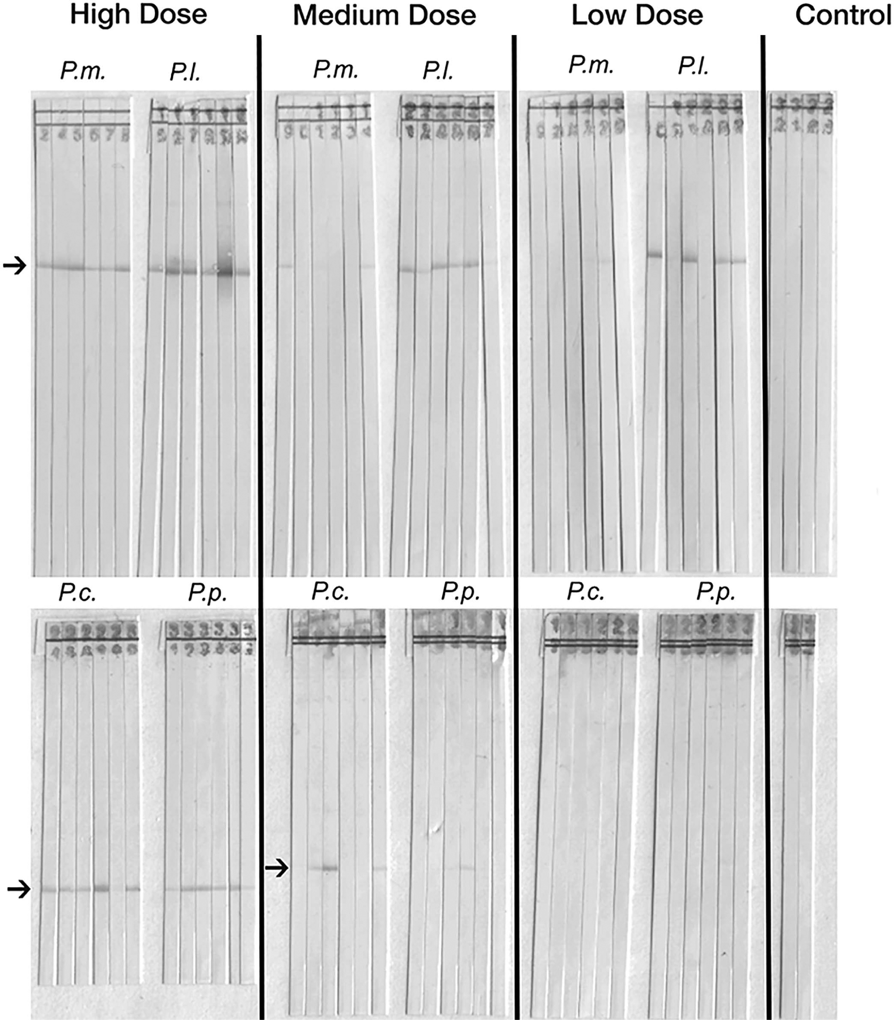
Western blot strips (BpRAG-1) exposed to sera of individuals of Peromyscus spp. experimentally infected with high, medium, or low dosages of Baylisascaris procyonis eggs. A band at 63 kDA (arrow) is considered a positive reaction. (P.m. = Peromyscus maniculatus; P.l. = Peromyscus leucopus; P.c. = Peromyscus californicus; P.p. = Peromyscus polionotus). The control column presents strips exposed to sera from unexposed controls.
Table II.
Concordance of rBpRAG-1-based Western blot (WB) results for experimentally infected Peromyscus spp., versus infection status and larval (L3) recovery (n = 80).
| Test | WB+ | WB− |
|---|---|---|
| Inoculated | 52 | 20 |
| Not inoculated | 0 | 8 |
| L3+ | 48 | 9 |
| L3− | 4 | 19 |
ELISA:
Serum samples from all experimentally infected Peromyscus were tested for anti-BpRAG-1-IgG with the use of the optimized ELISA protocol described above. For negative controls, 8 uninfected Peromyscus (~2 per species) from the experimental trials were used, as well as sera from 8 uninoculated P. maniculatus purchased from the supplier (Peromyscus Genetic Stock Center, Columbia, South Carolina). The sensitivity and specificity of this assay were 63.9 (52.8–73.6%) and 94.1% (82.3–100%), respectively, and the area under the curve was 0.815 (Fig. 2). The optimal minimum threshold value for a positive result was 8.27 AU. The inter-plate coefficient of variation (CV) was 6.82 and the intra-plate variability was 7.18. Overall, the agreement between the WB and ELISA was very good (Cohen’s κ = 0.843) (Table III).
Figure 2.

Receiver-operating characteristic curve for the Peromyscus-adapted rBpRAG-1 IgG ELISA. Area under curve = 0.815.
Table III.
Concordance between rBpRAG-1-based Western blot (WB) and ELISA results for experimentally infected Peromyscus spp. (n = 82). Cohen’s κ = 0.843.
| Test | WB+ | WB− |
|---|---|---|
| ELISA+ | 48 | 0 |
| ELISA− | 6 | 28 |
Significant associations between antibody concentration and species were detected when stratified by exposure dose (Table I). At the high and medium dose, P. leucopus had significantly greater antibody concentrations than P. californicus (high dose: P = 0.0057; medium dose: P = 0.0285) and P. polionotus (high dose: P = 0.0110; medium dose: P = 0.0045). However, antibody concentrations between P. leucopus and P. maniculatus did not differ significantly at these dose levels. At the low dose, P. leucopus had significantly greater antibody concentrations compared with the other 3 species (P. californicus: P < 0.0001; P. maniculatus: P = 0.0187; P. polionotus: P = 0.0002) (Fig. 3). There was a linear positive correlation between the total number of larvae recovered and antibody response (r = 0.679) (Fig. 4). In the linear model including species and the total number of larvae as predictors of antibody concentration, both factors were significant (P < 0.0001) and together explained 73.2% (r2) of variation in antibody concentration observed.
Figure 3.

Anti-BpRAG-1 IgG concentrations (in arbitrary units; AU) among experimentally infected Peromyscus spp. given either 500 (high dose, top); 50 (medium dose, middle), or 10 (low dose, bottom) larvated Baylisascaris procyonis eggs. *P < 0.05; **P < 0.01; ***P < 0.001 (pairwise t-test with Bonferroni correction).
Figure 4.

Linear model showing the relationship between the total numbers of Baylisascaris procyonis larvae recovered and anti-BpRAG-1 IgG concentrations (in arbitrary units; AU) at the time of euthanasia in experimentally infected Peromyscus spp. Cutoff value for positivity (8.27 AU, or 2.11 log[AU]) is indicated by the dotted line. Symbol represents species (diamond = Peromyscus peromyscus californicus; square = Peromyscus leucopus; circle = Peromyscus maniculatus; triangle = Peromyscus polionotus) and shading represents dose group (black = 500 eggs; gray = 50 eggs; white = 10 eggs).
Field samples
Serum samples from wild P. maniculatus (n = 28) from California were tested with the use of WB and ELISA. Based on previous necropsies and tissue examination, 8 of these mice were positive for Baylisascaris sp. larvae and burdens ranged from to 17 larvae. All but 1 of these samples were positive on both WB and ELISA (Table IV). The single discordant sample, which had 4 B. procyonis larvae, tested positive on WB but negative on ELISA, but was very close to the cutoff value (7.89 AU) (Table V). The number of larvae recovered and antibody concentration were positively and linearly correlated (r = 0.886) and total larvae recovered explained 71.6% (adjusted r2) of variation in antibody concentration. Data from wild P. maniculatus where no larvae were recovered were equivocal. Of 20 larvae-negative mice, 9 and produced a positive reaction on WB and ELISA, respectively; of these larvae-negative samples were positive on both serologic platforms. Samples from 3 wild R. rattus (infected with intensities of 287–793 larvae) also tested strongly positive on the WB with the use of sera diluted 1:100 and secondary antibody diluted to 1:5,000. Pooled sera from uninfected laboratory rats used as a negative control tested negative under these conditions. An ELISA was not developed for Mus or Rattus because of the low numbers of samples for full development and validation.
Table IV.
Concordance of rBpRAG-1-based Western blot (WB) and ELISA results for wild-caught Peromyscus maniculatus versus larval recovery (L3) (n = 28).
| Test | L3+ | L3− |
|---|---|---|
| WB+ | 8 | 9 |
| WB− | 0 | 11 |
| ELISA+ | 6 | 11 |
| ELISA− | 2 | 9 |
| WB+ | WB− | |
| ELISA+ | 12 | 5 |
| ELISA− | 5 | 6 |
Table V.
Serologic test results of wild rodents in which Baylisascaris sp. larvae were recovered. Abbreviations: WB = Western blot; AU = arbitrary units; ND = not done.
| Species | Total L3* recovered | WB result | ELISA result (AU) |
|---|---|---|---|
| Peromyscus maniculatus | 17 | Positive | Positive (>2,000) |
| P. maniculatus | 6 | Positive | Positive (1,641) |
| P. maniculatus | 5 | Positive | Positive (282.8) |
| P. maniculatus | 4 | Positive | Equivocal (7.89) |
| P. maniculatus | 2 | Positive | Positive (40.0) |
| P. maniculatus | 2 | Positive | Positive (106.2) |
| P. maniculatus | 1 | Positive | Positive (487.4) |
| P. maniculatus | 1 | Positive | Positive (23.8) |
| Rattus sp. | 793 | Positive | ND |
| Rattus sp. | 123 | Positive | ND |
| Rattus sp. | 89 | Positive | ND |
L3 = L3 larvae.
DISCUSSION
Serologic assays are a promising new tool for the diagnosis of Baylisascaris larva migrans in rodents. In experimentally infected Peromyscus spp., our rBpRAG-1 WB generally had high specificity and sensitivity. However, for animals inoculated with the lowest dose of eggs (10 eggs), the sensitivity and specificity varied based on how “positive” was defined (i.e., inoculation status vs. detection of larvae) as well as exposure dose with higher exposures and worm burdens having increased sensitivity. The most discordant results were observed in the low-dose group (10 eggs), which in some cases some mice had positive WB but no larvae recovered, whereas others were infected but WB negative. This suggests both that there is a minimum level of exposure needed to develop an infection and detectable antibodies and that some mice might be able to clear infection prior to sampling. Thus, although these assays might be accurate for heavily infected individuals, sensitivity and specificity are reduced in animals exposed to fewer than 10 eggs. Animals exposed to low doses may not have been successfully infected, may have cleared larvae prior to sampling, or the recovery method may simply not be sensitive enough to recover very low numbers of larvae present. For this reason, we chose to report sensitivity and specificity for both inoculation status and recovery of larvae status separately. We also observed differences in seroconversion by Peromyscus species. Several P. californicus and P. polionotus individuals failed to seroconvert despite some of them being inoculated with high numbers of eggs and had similar total numbers of larvae recovered from these hosts compared to the other Peromyscus spp. Although the mechanism is not clear, these 2 species are native to areas in which B. procyonis is believed to be historically absent, and therefore have a shorter evolutionary history with the parasite, and perhaps have undergone less selection for an effective response against infection (Sapp et al., 2016b). This demonstrates that assay performance estimates can be variable, even among congeneric species, and is an important consideration in interpreting sensitivity and specificity characteristics.
The ELISA had somewhat inferior performance characteristics compared to the WB, but concordance was favorable, which is often expected in comparing these platforms (because of alterations in epitope conformation during antigen coating, lower detection threshold and ability to separate cross-reactive fractions on WB, etc.) (Jitsukawa et al., 1989; Cortes et al., 2006; Frey et al., 2009; Fillaux and Magnaval, 2013). Despite lower sensitivity and specificity, the ELISA has a utility, as it provides a quantitative result compared with the qualitative WB and is a more rapid test for large sample sizes.
The most important finding with the quantitative ELISA output was the species-level differences among the experimentally infected Peromyscus spp. Peromyscus leucopus had significantly greater mean anti-BpRAG-1 IgG concentrations than P. californicus and P. polionotus at the high and medium dose, and all other species at the low dose. It is plausible that the anti-BpRAG-1 IgG response serves to slow or prevent larva migration through somatic tissue, and therefore delay entry of B. procyonis into the central nervous system. In addition to a significantly longer time until neurologic disease onset, we found P. leucopus had significantly higher numbers of larvae in visceral organs in our previous study (Sapp et al., 2016b). Survival time (i.e., length of infection) did not have a significant association with antibody concentration after adjustment for dose (data not shown), suggesting these observed differences are a result of the differential host responses among species and not due to longer survival time and exposure to antigens. Evidence for this exists in other ascarid species as well. For example, laboratory mice inoculated with a recombinant Ascaris suum ES product mounted a strong IgG response, and following challenge had a 54% reduction in the number of larvae recovered from lungs (Tsuji et al., 2003). Although that study did not attempt to recover larvae from other organs in the carcass, it seems likely that larvae become trapped in the livers of these immunized mice. This blocking of liver-lung larval migration could possibly be analogous to B. procyonis larva migration from viscera to the CNS.
The rBpRAG-1 antibody concentrations, as measured by ELISA, were dose-dependent, which is similar to data from studies on Toxocara. A very similar pattern was observed for ES IgG in laboratory mice inoculated with near-equivalent, graded doses of Toxocara canis eggs (500, 50, and 5 eggs) (Rodrigues e Fonseca et al., 2017). In that study, antibodies were still detectable by 170 days post-infection. Another study on laboratory mice also revealed dose-dependent patterns following small, graded doses of T. canis and T. cati eggs, with titers reaching a plateau after ~50 days (Havasiová-Reiterová et al., 1995). Although our experimental rodent study was not long-term, antibodies were detected out to 45 dpi.
This study has some important limitations. First, we used a single antigen target, BpRAG-1, which is a well-characterized Baylisascaris-specific ES antigen (Dangoudoubiyam et al., 2010). Although ES antigens are frequently used in the diagnosis of helminthic disease because of their immunogenicity, the role of anti-ES antibodies in immunity to larvae survival or migration is complex, and it is not known if antibodies to BpRAG-1 represent a protective response. However, the longer survival time and higher anti-BpRAG-1 IgG concentrations observed in P. leucopus versus other species suggest that a more robust immune response may confer a tolerant phenotype toward infection, or at least serve as an indicator of effective control of the parasite (even if not directly larvicidal) (Sapp et al., 2016b). Furthermore, immunization with ES antigens can confer some degree of protection and/or resistance in laboratory mice experimentally infected with other ascarids, including T. canis, T. vitulorum, and A. suum, and T. canis monoclonal anti-ES IgG binds directly to the cuticular surface of larvae (Nicholas et al., 1984; Bowman et al., 1987a; Abo-Shehada et al., 1991; Tsuji et al., 2003). Secreted proteins also induce protective responses in mice infected with nonascarid, tissue-dwelling helminths, such as Trichinella spiralis (Silberstein and Despommier, 1984). However, generalizations about ES antigens are difficult because of the high diversity of proteins secreted by helminths, which will all have variable impacts on the host. Evaluation of additional ES antigen targets, immunization or challenge studies, and analysis of other immune effectors, are necessary for further insight into this question.
Another limitation is that we did not collect serial blood samples from the experimentally exposed Peromyscus. Thus, we were only able to assess terminal antibody concentrations from the serum collected at the time of euthanasia, and cannot assess immune response kinetics. However, it is interesting to note that strongly positive IgG reactions were observed on WB and ELISA in Peromyscus euthanized as early as 9 days post-inoculation, and the sample with the greatest antibody concentration (14,700 AU) was from a P. leucopus euthanized at 14 dpi. This contrasts with findings from T. canis studies in laboratory mice, in which a significant IgG response in infected animals was not evident until ≥2 wk post-inoculation (Bowman et al., 1987b; Fan et al., 2003; Pinelli et al., 2007). Antisera from B. procyonis- and B. melis-infected laboratory mice that were euthanized at 11 dpi because of neurologic disease also reacted strongly on immunoblots with a crude B. procyonis ES antigen fraction (Boyce et al., 1988). Perhaps B. procyonis antigens are more immunogenic and Peromyscus spp. may be able to mount a response more rapidly than laboratory mice. We were only able to extend our study to 45 dpi, so we also cannot assess antibody persistence or changes over time. In T. canis-infected laboratory mice, anti-ES IgG peaked at 5–6 wk post-inoculation and remained at that level until the end of the 26-wk study, so it is possible that the antibody concentrations observed in surviving Peromyscus that were euthanized at the end of our study indicate maximum values (Bowman et al., 1987b).
Although assay performance was favorable among experimental rodents, based on our results on wild rodents, it seems using either the WB or ELISA on field samples may yield results that are hard to interpret. On our sample of wild-trapped P. maniculatus, all but 1 animal positive for Baylisascaris larvae had a positive result on WB or ELISA, and the WB+/ELISA− animal had an AU value very close to the cutoff. However, there were also serologic-positive wild rodents from which no larvae were recovered, which is difficult to interpret, as it is impossible to distinguish between past infection that has cleared (true positive) or cross-reactivity with other helminth fauna of wild rodents (false positive). No wild P. maniculatus were larvae positive and negative on both serologic assays, but the possibility for this situation exists as demonstrated in our findings on low-level infections (especially in P. polionotus and P. californicus) and may further complicate interpretation. Even with these limitations, these assays still may provide a sensitive method of detecting infections in wild rodents versus larval digestion or tissue squashes, although more validation is needed for reliable interpretation of serologic results. Too few Mus or Rattus samples were available for further validation of the respective species-adapted WB and development of an ELISA; however, the WB data indicated it should work on these rodent species as well.
Ultimately, serologic detection of infections in free-ranging wildlife has limitations and challenges. However, serology does have advantages that can warrant use in some situations (e.g., nonlethal, high-throughput, detection of exposure in a population vs. active infection or disease). Also, although these serologic assays cannot precisely estimate true prevalence and intensity, substantial differences in seroprevalence values across different habitat types, geographic regions, and paratenic host communities may provide some insight into B. procyonis epidemiology (i.e., magnitude of transmission). However, application of novel assays in wildlife should be interpreted appropriately. Currently, serologic testing is the only antemortem method for diagnosing Baylisascaris larva migrans in paratenic hosts. However, further efforts to improve and refine serological assays are warranted, as Baylisascaris procyonis now presents a serious disease risk to wildlife across the northern hemisphere.
ACKNOWLEDGMENTS
We would again like to thank the University of Georgia College of Veterinary Medicine summer research grant program for providing funding for experimental animals, and the staff of UGA Animal Resources for husbandry assistance. Additional support was provided by the wildlife management agencies of the Southeastern Cooperative Wildlife Disease Study member states through the Federal Aid to Wildlife Restoration Act (50 Stat. 917) and by a U.S Department of the Interior Cooperative Agreement. The findings and conclusions in this report are those of the authors and do not necessarily represent the official position of the Centers for Disease Control and Prevention.
LITERATURE CITED
- Abo-Shehada MN, al-Zubaidy BA, and Herbert IV. 1991. Acquired immunity to Toxocara canis infection in mice. Veterinary Parasitology 38: 289–298. [DOI] [PubMed] [Google Scholar]
- Beasley JC, Eagan TS, Page LK, Hennessy CA, and Rhodes OE. 2013. Baylisascaris procyonis infection in white-footed mice: Predicting patterns of infection from landscape habitat attributes. Journal of Parasitology 99: 743–747. [DOI] [PubMed] [Google Scholar]
- Bowman D, Mika-Grieve M, and Grieve RB. 1987a. Toxocara canis: Monoclonal antibodies to larval excretory-secretory antigens that bind with genus and species specificity to the cuticular surface of infective larvae. Experimental Parasitology 64: 458–465. [DOI] [PubMed] [Google Scholar]
- Bowman D, Mika-Grieve M, and Grieve RB. 1987b. Circulating excretory-secretory antigen levels and specific antibody responses in mice infected with Toxocara canis. American Journal of Tropical Medicine and Hygiene 36: 75–82. [DOI] [PubMed] [Google Scholar]
- Boyce WM, Branstetter BA, and Kazacos KR. 1988. Comparative analysis of larval excretory-secretory antigens of Baylisascaris procyonis, Toxocara canis and Ascaris suum by Western blotting and enzyme immunoassay. International Journal for Parasitology 18: 109–113. [DOI] [PubMed] [Google Scholar]
- Cortes HCE, Nunes S, Reis Y, Staubli D, Vidal R, Sager H, Leitão A, and Gottstein B. 2006. Immunodiagnosis of Besnoitia besnoiti infection by ELISA and Western blot. Veterinary Parasitology 141: 216–225. [DOI] [PubMed] [Google Scholar]
- Dangoudoubiyam S, Vemulapalli R, Hancock K, and Kazacos KR. 2010. Molecular cloning of an immunogenic protein of Baylisascaris procyonis and expression in Escherichia coli for use in developing improved serodiagnostic assays. Clinical Vaccine Immunology 17: 1933–1939. [DOI] [PMC free article] [PubMed] [Google Scholar]
- Fan CK, Lin YH, Du WY, and Su KE. 2003. Infectivity and pathogenicity of 14-month-cultured embryonated eggs of Toxocara canis in mice. Veterinary Parasitology 113: 145–155. [DOI] [PubMed] [Google Scholar]
- Fillaux J, and Magnaval JF. 2013. Laboratory diagnosis of human toxocariasis. Veterinary Parasitology 193: 327–336. [DOI] [PubMed] [Google Scholar]
- Frey CF, Schuppers ME, Nöckler K, Marinculić A, Pozio E, Kihm U, and Gottstein B. 2009. Validation of a Western blot for the detection of anti-Trichinella spp. antibodies in domestic pigs. Parasitology Research 104: 1269–1277. [DOI] [PubMed] [Google Scholar]
- Harris N, and Gause W. 2011. B cell function in the immune response to helminths. Trends in Immunology 32: 80–88. [DOI] [PMC free article] [PubMed] [Google Scholar]
- Havasiová-Reiterová K, Tomašovičová O, and Dubinský P. 1995. Effect of various doses of infective Toxocara canis and Toxocara cati eggs on the humoral response and distribution of larvae in mice. Parasitology Research 81: 13–17. [DOI] [PubMed] [Google Scholar]
- Jitsukawa T, Nakajima S, Sugawara I, and Watanabe H. 1989. Increased coating efficiency of antigens and preservation of original antigenic structure after coating in ELISA. Journal of Immunological Methods 116: 251–257. [DOI] [PubMed] [Google Scholar]
- Kazacos KR 2016. Baylisascaris larva migrans: U.S. Geological Survey Circular 1412, Abbott RC and van Riper C III(eds.),Madison, Wisconsin, 122 p. doi: 10.3133/cir1412. [DOI] [Google Scholar]
- Logiudice K 2001. Latrine foraging strategies of two small mammals: Implications for the transmission of Baylisascaris procyonis. American Midland Naturalist 146: 369–378. [Google Scholar]
- Nicholas WL, Stewart AC, and Mitchell GF. 1984. Antibody responses to Toxocara canis using sera from parasite-infected mice and protection from toxocariasis by immunisation with ES antigens. Australian Journal of Experimental Biology and Medical Science 62: 619–626. [DOI] [PubMed] [Google Scholar]
- Page KL 2013. Parasites and the conservation of small populations: The case of Baylisascaris procyonis. International Journal for Parasitology: Parasites and Wildlife 2: 203–210. [DOI] [PMC free article] [PubMed] [Google Scholar]
- Page KL, Swihart RK, and Kazacos KR. 2001. Foraging among feces: Food availability affects parasitism of Peromyscus leucopus by Baylisascaris procyonis. Journal of Mam-malogy 82: 993–1002. [Google Scholar]
- Pinelli E, Brandes S, Dormans J, Fonville M, Hamilton CM, and Van der Giessen J. 2007. Toxocara canis: Effect of inoculum size on pulmonary pathology and cytokine expression in BALB/c mice. Experimental Parasitology 115: 76–82. [DOI] [PubMed] [Google Scholar]
- R Core Team. 2014. R: A language and environment for statistical computing. R Foundation for Statistical Computing, Vienna, Austria: Available at: https://www.r-project.org/. Accessed 1 March 2018. [Google Scholar]
- Rascoe LN, Santamaria C, Handali S, Dangoudoubiyam S, Kazacos KR, Wilkins PP, and Ndao M. 2013. Interlaboratory optimization and evaluation of a serological assay for diagnosis of human baylisascariasis. Clinical Vaccine Immunology 20: 1758–1763. [DOI] [PMC free article] [PubMed] [Google Scholar]
- Robin X, Turck N, Hainard A, Tiberti N, Lisacek F, Sanchez JC, and Müller M. 2011. pROC: An open-source package for R and S+ to analyze and compare ROC curves. BMC Bioinformatics 12: 77. doi: 10.1186/1471-2105-12-77. [DOI] [PMC free article] [PubMed] [Google Scholar]
- Rodrigues e Fonseca G, dos Santos SV, Chieffi PP, de Paula FM, Gryschek RCB, and Lescano SAZ. 2017. Experimental toxocariasis in BALB/c mice: Relationship between parasite inoculum and the IgG immune response. Memórias do Instituto Oswaldo Cruz 112: 382–386. [DOI] [PMC free article] [PubMed] [Google Scholar]
- Sapp SGH, Rascoe LN, Wilkins PP, Handali S, Gray EB, Eberhard M, Woodhall DM, Montgomery SP, Bailey KL, Lankau EW, et al. 2016a. Baylisascaris procyonis roundworm seroprevalence among wildlife rehabilitators, United States and Canada, 2012–2015. Emerging Infectious Diseases 22: 2012–2015. [DOI] [PMC free article] [PubMed] [Google Scholar]
- Sapp SGH, Weinstein SB, McMahan CS, and Yabsley MJ. 2016b. Variable infection dynamics in four Peromyscus species following experimental inoculation with Baylisascaris procyonis. Journal of Parasitology 102: 538–544. [DOI] [PubMed] [Google Scholar]
- Sheppard CH, and Kazacos KR. 1997. Susceptibility of Peromyscus leucopus and Mus musculus to infection with Baylisascaris procyonis. Journal of Parasitology 83: 1104–1111. [PubMed] [Google Scholar]
- Sircar AD, Abanyie F, Blumberg D, Chin-Hong P, Coulter KS, Cunningham D, Huskins WC, Langelier C, Reid M, Scott BJ, et al. 2016. Raccoon roundworm infection associated with central nervous system disease and ocular disease—Six states, 2013–2015. Morbidity and Mortality Weekly Report 65: 930–933. [DOI] [PubMed] [Google Scholar]
- Tsuji N, Suzuki K, Kasuga-Aoki H, Isobe T, Arakawa T, and Matsumoto Y. 2003. Mice intranasally immunized with a recombinant 16-kilodalton antigen from roundworm Ascaris parasites are protected against larval migration of Ascaris suum. Infection and Immunity 71: 5314–5323. [DOI] [PMC free article] [PubMed] [Google Scholar]
- Weinstein SB 2017. Introduced rats and an endemic roundworm: Does Rattus rattus contribute to Baylisascaris procyonis transmission in California? Journal of Parasitology 103: 677–682. [DOI] [PubMed] [Google Scholar]
- Weinstein SB, Lake CM, Chastain HM, Fisk D, Handali S, Kahn PL, Montgomery SP, Wilkins PP, Kuris AM, and Lafferty KD. 2017. Seroprevalence of Baylisascaris procyonis infection among humans, Santa Barbara County, California, USA, 2014–2016. Emerging Infectious Diseases 23: 2014–2016. [DOI] [PMC free article] [PubMed] [Google Scholar]