Abstract
The COVID-19 infection has particularly affected older adults. Clinical observations in this population highlight major respiratory impairment associated with the development or aggravation of the patient’s frailty state. Mr. P is a 93-year-old frail patient, hospitalized after a COVID-19 infection. The assessment process of this patient has been supported by an innovative multi-systemic tool developed in view of the COVID-19 clinical consequences and a systemic evaluation of motor functions by the Frail’BESTest. This process allowed a mixed clinical picture associated with significant respiratory distress (linked with acute respiratory distress syndrome) and an evident motor frailty. The care plan was developed accordingly, and four assessments were done in the same manner until Mr. P returned home. This case report allows us to see a holistic COVID-19 clinical picture, showing the different axes of clinical reasoning to enhance the rehabilitation process. Furthermore, this case report illustrates the importance of rehabilitation in the COVID-19 context.
Keywords: COVID-19 - Geriatric rehabilitation - Frailty - Clinical reasoning, Screening
Introduction
The COVID-19 pandemic targets aged adults, especially when they carry comorbidities [1]. In elderly adults, frailty corresponds to the clinical consequences of physiological function decline, involving pathological aging. Frailty is characterized by a loss of functional supplies, leading to a high risk of falls, institutionalization, and sometimes death [2]. Aged adults who survive COVID-19 could present several frailty criteria following respiratory distress and may sometimes need to spend several days in an intensive care unit. There are multiple clinical pictures associating respiratory and vascular consequences, bed rest effects, and medication effects in a psychological context of anxiety [3, 4].
Case Description
The case report of Mr. P aims to underline the mixed clinical picture of Acute Respiratory Distress Syndrome (ARDS) and motor frailty. This case seems highly generalizable in the pandemic context where high age and associated diseases are major causing factors of death.
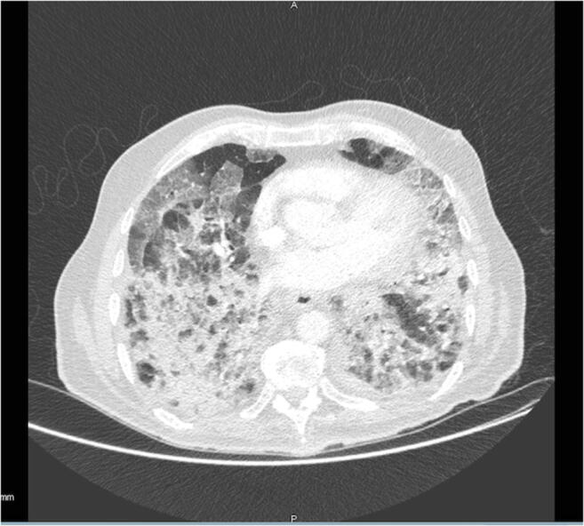
On March 3, 2020, Mr. P is hospitalized for a COVID-19 infection, confirmed by a thoracic scan showing the typical ground-glass opacities (Fig. 1). His pulmonary function decreased, entailing oxygen supplementation of 15 L/min. His clinical evolution confirms oxygen dependency between 12 and 15 L/min with high concentration mask. Mr. P is transferred to the rehabilitation unit on April 4, 2020. Mr. P lives alone in his house with a bedroom on the first floor. He is able to walk without technical support, both inside and outside. During the examination, Mr. P clearly expresses that he wants to go back at home once he regained his previous functional level. A significant fear of falling is observed, measured at 15/28 with the short FES [5]. Mr. P also indicates that he fell twice in 2019, when walking in his garden, and explains that he had difficulties getting up from the floor.
Fig. 1.

Thoracic scan showing the typical ground-glass opacities
About medical treatment, the therapeutic drugs related to the COVID-19 affection were
Plaquenil 400 mg in the morning and evening on March 31, 2020. Then the plaquenil was prescribed only in the morning for 9 days.
Methylprednisolone (corticoid) 20 mg on April 6, 2020 and 100 mg on April 7, 2020.
Tolicizumab 500 mg on April 6, 2020 and 500 mg on April 9, 2020.
Augmentin 1 g 3 times a day (morning, noon, and evening) from April 26, 2020 to May 3, 2020.
Mr. P also has an anti-coagulant treatment: Lovenox 7 ml (7000 UI). This drug is administered subcutaneously every 24 h starting March 31, 2020. From April 8, 2020, the drug is administered over 12 h.
Regarding comorbidities, Mr. P’s treatment included the following:
Furosemide (40 mg) by direct intravenous injection every 24 h (in the morning) on April 7–8, 2020. This drug was introduced due to edema in the lower limbs.
Kardegic 75.
Perindopril (2 mg) morning and evening then decreased to only in the morning from April 20 onward.
In addition, two indexes of comorbidity, the Cumulative Illness Rating Scale (CIRS) (Table 1) and Charlson Comorbidity Index (Table 2), were carried out.
Table 1.
Cumulative Illness Rating Scale (CIRS)
| System | No problem (= 0 points) | Light problem (= 1 point) | Moderate problem (= 2 points) | Severe problem (= 3 points) | Very severe problem (= 4 points) |
|---|---|---|---|---|---|
| Cardiac | 1 | ||||
| High blood pressure | 1 | ||||
| Vascular and hematopoietic | 0 | ||||
| Respiratory | 0 | ||||
| Eyes, ears, nose, throat, and larynx | 1 | ||||
| Upper gastrointestinal | 0 | ||||
| Lower gastrointestinal | 0 | ||||
| Liver, pancreas, and biliary | 0 | ||||
| Renal | 0 | ||||
| Genitourinary | 1 | ||||
| Musculoskeletal and skin | 0 | ||||
| Neurologic | 0 | ||||
| Endocrine and breast | 0 | ||||
| Psychiatric illness | 0 |
Cumulative Illness Rating Scale score = 4
Table 2.
Charlson comorbidity index
| Age | > 80 years (= 4 points) | |
|---|---|---|
| Item | Yes | No |
|
Myocardial infarction History of definite or probable MI (EKG changes and/or enzyme changes) |
0 | |
|
Congestive heart failure Exertional or paroxysmal nocturnal dyspnea and has responded to digitalis, diuretics, or afterload reducing agents |
0 | |
|
Peripheral vascular disease Intermittent claudication or past bypass for chronic arterial insufficiency, history of gangrene or acute arterial insufficiency, or untreated thoracic or abdominal aneurysm (≥ 6 cm) |
0 | |
| Cerebrovascular accident or transient ischemic attack | 0 | |
|
Dementia Chronic cognitive deficit |
0 | |
| Chronic obstructive pulmonary disease | 0 | |
| Connective tissue disease | 0 | |
|
Peptic ulcer disease Any history of treatment for ulcer disease or history of ulcer bleeding |
0 | |
|
Liver disease Severe = cirrhosis and portal hypertension with variceal bleeding history, moderate = cirrhosis and portal hypertension but no variceal bleeding history, mild = chronic hepatitis (or cirrhosis without portal hypertension) |
0 | |
| Diabetes mellitus | 0 | |
| Hemiplegia | 0 | |
|
Moderate to severe chronic kidney disease Severe = on dialysis, status post kidney transplant, uremia, moderate = creatinine > 3 mg/dL (0.27 mmol/L) |
0 | |
| Solid tumor | 0 | |
| Leukemia | 0 | |
| Lymphoma | 0 | |
| AIDS | 0 | |
Charlson comorbidity index score = 4
Diagnostic Assessment
We conducted the evaluation using a two-fold analysis. First, we evaluated the deficiencies linked with the COVID-19 infection and the associated ARDS, using a specific COVID-19 aggregation of scales. Next, we targeted the patient’s motor function using the Frail’BESTest [6] so as to guide the clinical reasoning.
Level 1: the Specific COVID-19 Evaluation
Faced with the heterogeneity of COVID-19 clinical pictures, we tried to propose a global summary that includes several clinical examinations. The available literature [7, 8] notes a typical pulmonary deficiency associated with several new clinical scripts (ARDS, psychomotor disadaptation syndrome, acquired pneumopathy, acquired neuromyopathy, etc.). The scales were aggregated using a transdisciplinary rehabilitation approach to screen all the important aspects resulting from the infection. Tests were retained for their usability, reliability, and validity.
Social and behavioral functions are evaluated with the RAMSAY score [9] and the RASS scale [10].
- Some items from the Hamilton scale (HDRS) are used to measure the psychological and emotional states [11].
- In accordance with the loss of mass frequently described [8], the body mass index is noted. Swallowing function is evaluated with simple tests.
- A binary analysis of statesthesia and tact are proposed, in addition to the other senses.
- Regarding sensitivity, a double assessment including statesthesia and touch [25] is done.
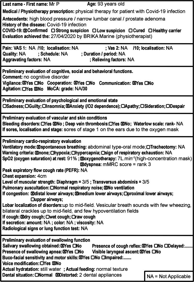
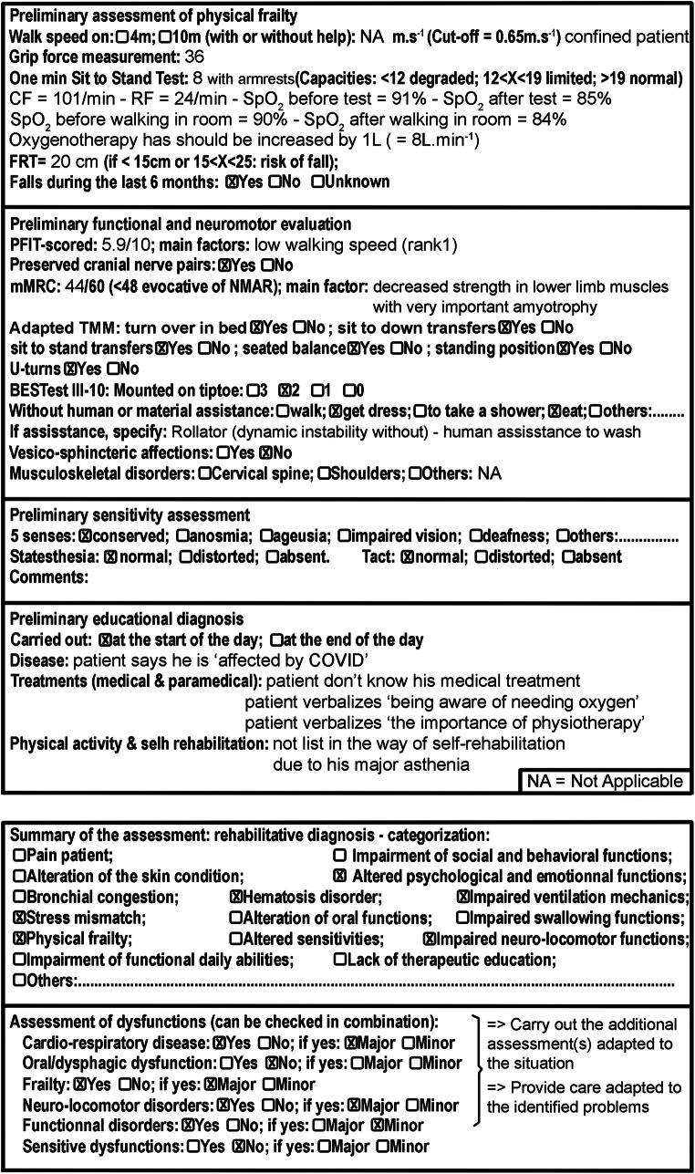
The evaluation synthesis of Mr. P is featured in Fig. 2 a and b.
Fig. 2.
a Part one of specific COVID-19 evaluation, b part two of specific COVID-19 evaluation, and c part three of specific COVID-19 evaluation
Level 2: Reasoning with the Frail’BESTest
The Frail’BESTest has been developed to make it possible to include frail older adults in systemic evaluations [6]. Therapists can therefore directly manage therapeutic intervention for different types of balance deficiencies. Overall, six sub-systems have been addressed: (1) anticipations, (2) reactions, (3) locomotion, (4) sensorial orientation, (5) biomechanical constraints, and (6) asymmetric gait.
Diagnosis
Mr. P, a 93-year-old patient, presents with a respiratory dysfunction linked to a COVID-19 infection (saturation at 91% with 7 L of oxygen supplementation), subsequent effort incapacity, and postural-motor deficiencies. Motor automatisms are impaired, and several articular and muscular constraints remain. Mr. P seems enlisted in a frailty process, leading to increased dependency, an impossibility to return home, and relative social isolation.
Therapeutic Intervention
The protocol was carried out in accordance with legal and international requirements (Declaration of Helsinki, 1964). Mr. P was informed about the published project and gave his written consent before the evaluation.
Mr. P followed a rehabilitation program which primarily included physical therapy and nutritional monitoring. He received one session of physical therapy per day. This session lasted on average of 30 min. Considering the physiotherapeutic diagnosis of Mr. P, as well as the age-specific lung physiology of the patient [26], some cardiopulmonary rehabilitation exercises allowing both the maintenance of ventilator functions and the improvement of hematosis can be proposed. During all of these exercises, precautions, red flags, and stop criteria indicated in the HAS Quick Response [27] should be followed.
The next paragraph will show the aims and exercise samples that have been proposed to Mr. P. We would like to improve both the transverse abdominis and the diaphragm via active, functional, and resistive treatments including threshold systems, hypopressive exercise, and functional ventilation during movement [28–30]. In order to limit physiological impairment, some exercises including thoracic movement with the arms, chest, and spine mobility are introduced during global therapy in both directions: inhale and exhale [31, 32]. In order to improve oxygenation and prevent congestion, ventilation should be harmonized throughout the lung territories, and mucociliary clearance should be promoted. Thus, the treatment involves high-volume ventilation-type work that includes tele-inspiratory holds, while avoiding specific collapses associated with senescence. For example, EDIC, ITLA (Inspiratory Technical for Lifting Atelectasis), Elpr, and ACBT-type exercises with open glottis are proposed [33, 34]. Concerning rehabilitation with effort, it is necessary to increase the ventilatory threshold in order to improve muscular function and decrease dyspnea. This will also improve hematosis and oxidative metabolism. An early and progressive cardiopulmonary rehabilitation program is established and based on the Borg scale [35, 36]. For Mr. P, it includes optimal loading, aerobic work measured by paliers, as well as endurance. This program mainly uses functional exercises such as treadmill walking (between 60 and 80% of the TM6 or the chair-test or top toes test) [37]. It also seems important to prevent dysphagia in the medium term and to optimize the use of the functions of the nose (to warm, filter, and humidify the air). So, nasal ventilation and correction of the tongue position is essential. For example, mindless nasal ventilation and the tongue palate position is monitored, and lingual resistive exercise and sensitive work are proposed. In a final perspective on the patient’s autonomy, throughout the rehabilitation process, education on the perception of effort, use of the Borg scale, the patient’s self-assessment of his respiratory capacities, and the criteria of alerts are all carried out [38].
On the other hand, in connection with the systemic evaluation of the balance function and motor frailty of Mr. P, several sensory-motor exercises are proposed. To improve the efficiency of postural-movement coordination, self-paced perturbations of balance were worked on with speed and variability [39]. For example, Mr. P had to reach a colored target on the ground as quickly as possible once the physiotherapist indicated the color he has to hit. To reactivate postural adaptations and fall avoidance reactions, Mr. P performs exercises that work on extrinsic imbalances (unpredictable balance perturbation) [40]. For example, Mr. P had to react to manual pushes from the physiotherapist. In order to improve muscular power, functional muscular exercises were performed in a closed chain and under a time constraint [41]. For example, Mr. P had to go up and down a step to the beat of a metronome. In order to regain physiological ankle mobility and enhance the rolling of feet when walking, active mobilization exercises were carried out during physiotherapy sessions and also by the patient independently in his room [42]. To reduce podal dependency, Mr. P performed static and dynamic balance exercises on different ground textures (e.g., standing on foam, walking on a mat, walking outside in the grass). Finally, exercises incorporating the work of spatial and temporal parameters of walking and changes of direction were carried out. These exercises were aimed at improving walking kinetics and would contribute to the evolution of help with technical walking.
Follow-up and Outcomes
The four assessments performed by the specific COVID-19 evaluation showed an overall improvement of the patient in several functions. In terms of psychological and emotional state, the anxiety with regard to oxygen dependence disappeared. Indeed, during the initial assessment, the patient had 7 L of oxygen in the high-concentration face mask. During the final evaluation, he had only 1 L of oxygen left in the nasal cannula. Pulmonary auscultation, which initially revealed a lack of ventilation associated with congestion of the middle and distal airways, also improved. Final auscultation was evaluated without particularities. The assessment of cognitive and behavioral functions remained unchanged over the course of the four assessments. Initial clinical observations did not show impairment of these functions. The initial preliminary assessment of the vascular and cutaneous system had shown the presence of a stage 1 pressure ulcer (as per the National Pressure Ulcer Advisory Panel Stage Classification) behind the ears due to the oxygen mask. Upon final evaluation, the pressure ulcer was no longer present. No vascular disorders occurred during the hospitalization. Moreover, there was no significant change in swallowing function, as Mr. P did not present any swallowing problems.
Changes to the scores of quantitative outcomes of the different functions are summarized in Table 3.
Table 3.
A summary table on the different evaluations with specific COVID-19 evaluation
| Section | Item | Evaluation 1 | Evaluation 2 | Evaluation 3 | Evaluation 4 |
|---|---|---|---|---|---|
| Date | 04/27/2020 | 05/20/2020 | 05/29/2020 | 06/08/2020 | |
| Cardio-respiratory | Oxygenotherapy (L/min) | 7 | 2 | 1 | 1 |
| Dyspnea: mMRC score | Rank 3 | Rank 3 | Rank 3 | Rank 3 | |
| Dyspnea: Borg score | 7/10 | 6/10 | 6/10 | 4/10 | |
| SpO2 (oxygen saturation) at rest | 91% | 92% | 92% | 94% | |
| SpO2 after walking | 84% | 86% | 88% | 86% | |
| Respiratory frequency | 24 | 22 | 20 | 19 | |
| Chest expansion/ventilatory asymmetry (cm) | 4 | 5 | 5 | 6 | |
| Level of muscular strength | Diaphragm = 3/5 | Diaphragm = 3/5 | Diaphragm = 3/5 | Diaphragm = 4/5 | |
| Transversus abdominis = 3/5 | Transversus abdominis = 3/5 | Transversus abdominis = 3/5 | Transversus abdominis = 4/5 | ||
| Frailty | Gait speed (m/s) | NE | 0.33 with rollator | 0.4 with rollator | 0.57 with rollator |
| Grip strength (kg) | 26 | 30 | 30 | 32 | |
| 1 min sit-to-stand test | 8 | 12 | 12 | 15 | |
| FRT (cm) | 20 | 20 | 21 | 23 | |
| Functional and neuromotor | PFIT-scored | 5.9/10 | 5.9/10 | 6.4/10 | 7.1/10 |
| mMRC | 44/60 | 50/60 | 56/60 | 56/60 | |
| BESTest III-10: Mounted on tiptoe | Score 2 | Score 2 | Score 2 | Score 2 |
The four Frail’BESTest assessments show an improvement in the score of some subsystems. The results are summarized in Table 4.
Table 4.
A summary table on the different evaluations with Frail’BESTest
| Frail’BESTest | Initial evaluation | Intermediate evaluation (number one) | Intermediate evaluation (number two) | Final evaluation |
|---|---|---|---|---|
| Date | 04/27/2020 | 05/20/2020 | 05/29/2020 | 06/08/2020 |
| System A: anticipations | 3 | 3 | 3 | 4 |
| System B: reactions | 0 | 0 | 0 | 1 |
| System C: locomotion | NE | 1 | 2 | 2 |
| System D: sensory orientation | 2 | 2 | 2 | 2 |
| System E: biomechanical constraints | 2 | 3 | 3 | 4 |
| System F: gait symmetry | 4 | 4 | 4 | 4 |
| Total score | 11 | 13 | 14 | 18 |
| Gait speed (m/s) | NE | 0.33 with rollator | 0.4 with rollator | 0.57 with rollator |
Discussion and Conclusions
This case allows us to underline the global approach that is necessary in a geriatric rehabilitation context associated with the COVID-19 infection. Although the long-term follow-up is not yet available, it seems important to continue the clinical pictures description associated with this virus in order to better organize rehabilitation strategies. Indeed, the rehabilitation process represents the other challenge of the pandemic situation in several countries characterized by a high proportion of frail patients [43]. In our opinion, it is important to understand that the issue is not only to rescue a patient from their acute respiratory problem, but more so to prevent the functional dependency associated with the infection’s consequences, especially in intensive care units where chronic diseases are frequently acquired.
Mr. P was probably lucky to return home with a high level of independency. His age and relative frailty were, at the beginning, considered to be bad prognosis factors. As is common in geriatric rehabilitation, age is not only a question of numbers. In the same manner, frailty should not be questioned as an independence level, but more in terms of functional reserves. Mr. P presented sufficient functional reserves, although he was certainly frail upon arriving at the hospital.
Several papers have described the physiotherapy associated with the COVID-19 infection since the pandemic began. A lot of them describe adult patients, often aged up to 65 years, considering respiratory or pulmonary rehabilitation. Strength recommendations are available to manage these COVID-19 patients [8]. However, age and frailty are key factors to consider when targeting the needs of these patients, and all of our health systems should be adapted to the second wave of the pandemic situation: the rehabilitation wave [44].
Although a higher vulnerability of geriatric patients has been observed, the literature on aged COVID-19 patients has remained very scarce. A few studies have already adequately targeted these patients and described interesting clinical pictures and associated medical treatments [45]. However, to our knowledge, this is the first case report highlighting the rehabilitation process with an aged COVID-19 patient who needs to be seen also as a respiratory case and as a frail patient.
Acknowledgments
The authors thank Dr. Julie Caissutti for her precious help.
Compliance with Ethical Standards
Conflict of Interest
The authors declare that they have no conflict of interest.
Ethical Approval
The protocol performed in this case report is in accordance with the ethical standards of the institutional and/or national research committee and with the 1964 Helsinki declaration and its later amendments or comparable ethical standards.
Informed Consent
Mr. P was informed about the publication of the project and gave his consent.
Footnotes
This article is part of the Topical Collection on COVID-19
Publisher’s Note
Springer Nature remains neutral with regard to jurisdictional claims in published maps and institutional affiliations.
References
- 1.Promislow DEL. A Geroscience perspective on COVID-19 mortality. J Gerontol A Biol Sci Med Sci. 2020;75(9):e30–e33. doi: 10.1093/gerona/glaa094. [DOI] [PMC free article] [PubMed] [Google Scholar]
- 2.Silva-Obregón JA, Quintana-Díaz M, Saboya-Sánchez S, Marian-Crespo C, Romera-Ortega MÁ, Chamorro-Jambrina C, Estrella-Alonso A, Andrés-Esteban EM. Frailty as a predictor of short- and long-term mortality in critically ill older medical patients. J Crit Care. 2020;55:79–85. doi: 10.1016/j.jcrc.2019.10.018. [DOI] [PubMed] [Google Scholar]
- 3.Simpson R, Robinson L. Rehabilitation after critical illness in people with COVID-19 infection. Am J Phys Med Rehabil. 2020;99(6):470–474. doi: 10.1097/PHM.0000000000001443. [DOI] [PMC free article] [PubMed] [Google Scholar]
- 4.Ceravolo MG, de Sire A, Andrenelli E, Negrini F, Negrini S. Systematic rapid “living” review on rehabilitation needs due to COVID-19: update to March 31st, 2020. Eur J Phys Rehabil Med. 2020;56(3):347–353. doi: 10.23736/S1973-9087.20.06329-7. [DOI] [PubMed] [Google Scholar]
- 5.Hauer KA, Kempen GIJM, Schwenk M, Yardley L, Beyer N, Todd C, Oster P, Zijlstra GAR. Validity and sensitivity to change of the falls efficacy scales international to assess fear of falling in older adults with and without cognitive impairment. Gerontology. 2011;57(5):462–472. doi: 10.1159/000320054. [DOI] [PubMed] [Google Scholar]
- 6.Kubicki A, Brika M, Coquisart L, Basile G, Laroche D, Mourey F. The Frail’BESTest. An adaptation of the “Balance Evaluation System Test” for frail older adults. Description, internal consistency and inter-rater reliability. CIA. 2020;15:1249–1262 [DOI] [PMC free article] [PubMed]
- 7.Lescure F-X, Bouadma L, Nguyen D, Parisey M, Wicky PH, Behillil S, Gaymard A, Bouscambert-Duchamp M, Donati F, le Hingrat Q, Enouf V, Houhou-Fidouh N, Valette M, Mailles A, Lucet JC, Mentre F, Duval X, Descamps D, Malvy D, Timsit JF, Lina B, van-der-Werf S, Yazdanpanah Y. Clinical and virological data of the first cases of COVID-19 in Europe: a case series. Lancet Infect Dis. 2020;20(6):697–706. doi: 10.1016/S1473-3099(20)30200-0. [DOI] [PMC free article] [PubMed] [Google Scholar]
- 8.Thomas P, Baldwin C, Bissett B, Boden I, Gosselink R, Granger CL, Hodgson C, Jones AYM, Kho ME, Moses R, Ntoumenopoulos G, Parry SM, Patman S, van der Lee L. Physiotherapy management for COVID-19 in the acute hospital setting: clinical practice recommendations. Aust J Phys. 2020;66(2):73–82. doi: 10.1016/j.jphys.2020.03.011. [DOI] [PMC free article] [PubMed] [Google Scholar]
- 9.Carrasco G. Instruments for monitoring intensive care unit sedation. Crit Care. 2000;4(4):217–225. doi: 10.1186/cc697. [DOI] [PMC free article] [PubMed] [Google Scholar]
- 10.Ely EW, Truman B, Shintani A, Thomason JWW, Wheeler AP, Gordon S, Francis J, Speroff T, Gautam S, Margolin R, Sessler CN, Dittus RS, Bernard GR. Monitoring sedation status over time in ICU patients: reliability and validity of the Richmond agitation-sedation scale (RASS) JAMA. 2003;289(22):2983–2991. doi: 10.1001/jama.289.22.2983. [DOI] [PubMed] [Google Scholar]
- 11.Carrozzino D, Patierno C, Fava GA, Guidi J. The Hamilton rating scales for depression: a critical review of clinimetric properties of different versions. Psychother Psychosom. 2020;89(3):133–150. doi: 10.1159/000506879. [DOI] [PubMed] [Google Scholar]
- 12.Munari AB, Gulart AA, Dos Santos K, Venâncio RS, Karloh M, Mayer AF. Modified Medical Research Council dyspnea scale in GOLD classification better reflects physical activities of daily living. Respir Care. 2018;63(1):77–85. doi: 10.4187/respcare.05636. [DOI] [PubMed] [Google Scholar]
- 13.Borg GA. Psychophysical bases of perceived exertion. Med Sci Sports Exerc. 1982;14(5):377–381. doi: 10.1249/00005768-198205000-00012. [DOI] [PubMed] [Google Scholar]
- 14.Bianchi C, Baiardi P, Khirani S, Cantarella G. Cough peak flow as a predictor of pulmonary morbidity in patients with dysphagia. Am J Phys Med Rehabil. 2012;91(9):783–788. doi: 10.1097/PHM.0b013e3182556701. [DOI] [PubMed] [Google Scholar]
- 15.Kim J, Davenport P, Sapienza C. Effect of expiratory muscle strength training on elderly cough function. Arch Gerontol Geriatr. 2009;48(3):361–366. doi: 10.1016/j.archger.2008.03.006. [DOI] [PubMed] [Google Scholar]
- 16.Middleton A, Fritz SL, Lusardi M. Walking speed: the functional vital sign. J Aging Phys Act. 2015;23(2):314–322. doi: 10.1123/japa.2013-0236. [DOI] [PMC free article] [PubMed] [Google Scholar]
- 17.Puhan MA, Siebeling L, Zoller M, Muggensturm P, ter Riet G. Simple functional performance tests and mortality in COPD. Eur Respir J. 2013;42(4):956–963. doi: 10.1183/09031936.00131612. [DOI] [PMC free article] [PubMed] [Google Scholar]
- 18.Scena S, Steindler R, Ceci M, Zuccaro SM, Carmeli E. Computerized functional reach test to measure balance stability in elderly patients with neurological disorders. J Clin Med Res. 2016;8(10):715–720. doi: 10.14740/jocmr2652w. [DOI] [PMC free article] [PubMed] [Google Scholar]
- 19.Dodds RM, Syddall HE, Cooper R, Benzeval M, Deary IJ, Dennison EM, der G, Gale CR, Inskip HM, Jagger C, Kirkwood TB, Lawlor DA, Robinson SM, Starr JM, Steptoe A, Tilling K, Kuh D, Cooper C, Sayer AA. Grip strength across the life course: normative data from twelve British studies. PLoS One. 2014;9(12):e113637. doi: 10.1371/journal.pone.0113637. [DOI] [PMC free article] [PubMed] [Google Scholar]
- 20.Denehy L, de Morton NA, Skinner EH, Edbrooke L, Haines K, Warrillow S, Berney S. A physical function test for use in the intensive care unit: validity, responsiveness, and predictive utility of the physical function ICU test (scored) Phys Ther. 2013;93(12):1636–1645. doi: 10.2522/ptj.20120310. [DOI] [PubMed] [Google Scholar]
- 21.Jolley SE, Bunnell AE, Hough CL. ICU-acquired weakness. Chest. 2016;150(5):1129–1140. doi: 10.1016/j.chest.2016.03.045. [DOI] [PMC free article] [PubMed] [Google Scholar]
- 22.Hermans G, Clerckx B, Vanhullebusch T, Segers J, Vanpee G, Robbeets C, Casaer MP, Wouters P, Gosselink R, van den Berghe G. Interobserver agreement of Medical Research Council sum-score and handgrip strength in the intensive care unit. Muscle Nerve. 2012;45(1):18–25. doi: 10.1002/mus.22219. [DOI] [PubMed] [Google Scholar]
- 23.Mourey F, Camus A, d’Athis P, Blanchon MA, Martin-Hunyadi C, Rekeneire N, Pfitzenmeyer P. Mini motor test: a clinical test for rehabilitation of patients showing psychomotor disadaptation syndrome (PDS) Arch Gerontol Geriatr. 2005;40(2):201–211. doi: 10.1016/j.archger.2004.08.004. [DOI] [PubMed] [Google Scholar]
- 24.Horak FB, Wrisley DM, Frank J. The balance evaluation systems test (BESTest) to differentiate balance deficits. Phys Ther. 2009;89(5):484–498. doi: 10.2522/ptj.20080071. [DOI] [PMC free article] [PubMed] [Google Scholar]
- 25.Baig AM. Neurological manifestations in COVID-19 caused by SARS-CoV-2. CNS Neurosci Ther. 2020;26(5):499–501. doi: 10.1111/cns.13372. [DOI] [PMC free article] [PubMed] [Google Scholar]
- 26.Skloot GS. The effects of aging on lung structure and function. Clin Geriatr Med. 2017;33(4):447–457. doi: 10.1016/j.cger.2017.06.001. [DOI] [PubMed] [Google Scholar]
- 27.Prise en charge des patients post-COVID-19 en médecine physique et de réadaptation (MPR), en soins de suite et de réadaptation (SSR), et retour à domicile. Haute Autorité de Santé. Accessed July 27, 2020. https://www.has-sante.fr/jcms/p_3179826/fr/prise-en-charge-des-patients-post-covid-19-en-medecine-physique-et-de-readaptation-mpr-en-soins-de-suite-et-de-readaptation-ssr-et-retour-a-domicile.
- 28.Babcock MA, Pegelow DF, Johnson BD, Dempsey JA. Aerobic fitness effects on exercise-induced low-frequency diaphragm fatigue. J Appl Physiol. 1996;81(5):2156–2164. doi: 10.1152/jappl.1996.81.5.2156. [DOI] [PubMed] [Google Scholar]
- 29.Fulton I, McEvoy M, Pieterse J, Williams M, Thoirs K, Petkov J. Transversus abdominis: changes in thickness during the unsupported upper limb exercise test in older adults. Physiother Theory Pract. 2009;25(8):523–532. doi: 10.3109/09593980802665023. [DOI] [PubMed] [Google Scholar]
- 30.O’Kroy JA, Coast R. Effects of flow and resistive training on respiratory muscle endurance and strength. RES. 1993;60(5):279–283. doi: 10.1159/000196216. [DOI] [PubMed] [Google Scholar]
- 31.Han JW, Kim YM. Effect of breathing exercises combined with dynamic upper extremity exercises on the pulmonary function of young adults. J Back Musculoskelet Rehabil. 2018;31(2):405–409. doi: 10.3233/BMR-170823. [DOI] [PubMed] [Google Scholar]
- 32.Petrofsky JS, Cuneo M, Dial R, Pawley AK, Hill J. Core strengthening and balance in the geriatric population. J Appl Res. 2005;5(3):11. [Google Scholar]
- 33.Lewis LK, Williams MT, Olds TS. The active cycle of breathing technique: a systematic review and meta-analysis. Respir Med. 2012;106(2):155–172. doi: 10.1016/j.rmed.2011.10.014. [DOI] [PubMed] [Google Scholar]
- 34.Pryor JA. Physiotherapy for airway clearance in adults. Eur Respir J. 1999;14(6):1418–1424. doi: 10.1183/09031936.99.14614189. [DOI] [PubMed] [Google Scholar]
- 35.Svedahl K, MacIntosh BR. Anaerobic threshold: the concept and methods of measurement. Can J Appl Physiol. 2003;28(2):299–323. doi: 10.1139/h03-023. [DOI] [PubMed] [Google Scholar]
- 36.Alexander JL, Phillips WT, Wagner CL. The effect of strength training on functional fitness in older patients with chronic lung disease enrolled in pulmonary rehabilitation. Rehabil Nurs. 2008;33(3):91–97. doi: 10.1002/j.2048-7940.2008.tb00211.x. [DOI] [PubMed] [Google Scholar]
- 37.Opasich C, Patrignani A, Mazza A, Gualco A, Cobelli F, Pinna GD. An elderly-centered, personalized, physiotherapy program early after cardiac surgery. Eur J Cardiovasc Prev Rehabil. 2010;17(5):582–587. doi: 10.1097/HJR.0b013e3283394977. [DOI] [PubMed] [Google Scholar]
- 38.O’Donnell DE, Chau LK, Webb KA. Qualitative aspects of exertional dyspnea in patients with interstitial lung disease. J Appl Physiol. 1998;84(6):2000–2009. doi: 10.1152/jappl.1998.84.6.2000. [DOI] [PubMed] [Google Scholar]
- 39.Jagdhane S, Kanekar N, Aruin AS. The effect of a four-week balance training program on anticipatory postural adjustments in older adults: a pilot feasibility study. Curr Aging Sci. 2016;9(4):295–300. doi: 10.2174/1874609809666160413113443. [DOI] [PubMed] [Google Scholar]
- 40.Mansfield A, Peters AL, Liu BA, Maki BE. Effect of a perturbation-based balance training program on compensatory stepping and grasping reactions in older adults: a randomized controlled trial. Phys Ther. 2010;90(4):476–491. doi: 10.2522/ptj.20090070. [DOI] [PubMed] [Google Scholar]
- 41.Foldvari M, Clark M, Laviolette LC, Bernstein MA, Kaliton D, Castaneda C, Pu CT, Hausdorff JM, Fielding RA, Singh MAF. Association of muscle power with functional status in community-dwelling elderly women. J Gerontol A Biol Sci Med Sci. 2000;55(4):M192–M199. doi: 10.1093/gerona/55.4.m192. [DOI] [PubMed] [Google Scholar]
- 42.Smajla D, García-Ramos A, Tomazin K, Strojnik V. Selective effect of static stretching, concentric contractions, and a one-leg balance task on ankle motion sense in young and older adults. Gait Posture. 2019;71:1–6. doi: 10.1016/j.gaitpost.2019.04.006. [DOI] [PubMed] [Google Scholar]
- 43.Özkeskin M, Elibol N, Bakırhan S. Risk of COVID-19 disease in the elderly population and physiotherapy. Physiotherapy. 2020;108:76–77. doi: 10.1016/j.physio.2020.05.003. [DOI] [PMC free article] [PubMed] [Google Scholar]
- 44.De Biase S, Cook L, Skelton DA, Witham M, Ten Hove R. The COVID-19 rehabilitation pandemic. Age Ageing. 2020;49(5):696–700. doi: 10.1093/ageing/afaa118. [DOI] [PMC free article] [PubMed] [Google Scholar]
- 45.Bentivegna E, Luciani M, Spuntarelli V, Speranza ML, Guerritore L, Sentimentale A, Martelletti P. Extremely severe case of COVID-19 pneumonia recovered despite bad prognostic indicators: a didactic report. SN Compr Clin Med. 2020;2(8):1204–1207. doi: 10.1007/s42399-020-00383-0. [DOI] [PMC free article] [PubMed] [Google Scholar]


