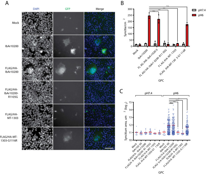
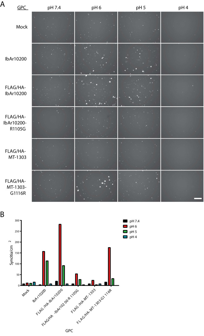
Figure 4. MT-1303 G1116 GPC variant impairs membrane fusion.
(A) Fluorescent images of individual Huh7 syncytia in the GFP cell-cell fusion reporter assay. Cells expressing GPC and T7 polymerase were exposed to pH 6 DMEM media before co-culture with Huh7 cells transfected with a reporter plasmid containing GFP under control of the T7 promoter. Cell nuclei were stained with DAPI. Scale bar represents 100 μm. (B) Quantification of syncytia density. Error bars represent standard deviation of the mean of three biological replicate experiments. ****p<0.0001 and n.s. = not significant according to the two-tailed Student’s t-test with n = 3. (C) Quantification of syncytium area. For each condition, the areas of all of syncytia identified across three biological replicate experiments are shown. *p<0.05, ***p<0.001, ****p<0.0001, and n.s. = not significant according to the Mann-Whitney U-test with n = 3.


