Abstract
This study aimed to evaluate the transection of superficial digital flexor tendon (SDFT) and deep digital flexor tendon (DDFT) in calves with severe metacarpophalangeal flexural deformities (MPFD). The study comprised 17 forelimbs of 10 calves that were diagnosed at the Animal Medical Centre, Rakuno Gakuen University. The calves were treated via transection of the SDFT and DDFT with retention of the suspensory ligament, followed by external fixation according to a post-surgical gait test. The post-procedural prognosis was determined at 14 days post-surgery. Of the 17 limbs, 14 (82%) achieved non-lameness and a good prognosis. Surgical complications were not observed in any treated calves. The transection of SDFT and DDFT is an effective first-line surgical option for calves with severe MPFD.
Keywords: bovine, deformity, external fixation, orthopedic surgery
In cattle and horses, congenital flexural limb deformities can cause structural abnormalities in the affected limbs that lead to a restricted range of motion and lameness [1,2,3,4, 6, 9, 10]. In cattle, metacarpophalangeal flexural deformities (MPFDs) in the forelimbs are the most common type of congenital limb deformity [8, 10, 13, 15, 16]. Calves with severe MPFD may require surgical treatment. These surgical procedures generally treat the limbs by sequentially transecting the superficial digital flexor tendons (SDFTs), deep digital flexor tendons (DDFTs) and suspensory ligament until the deformity is resolved [4, 19, 20]. The number of transected tendons is determined intraoperatively. Generally, transection of the SDFT and DDFT is not sufficient to improve flexion in calves with severe MPFD [4, 11]. In this study, MPFD calves were treated via the transection of SDFT and DDFT while retaining the suspensory ligament. Subsequent external fixation was performed according to post-surgical gait tests. The calves were observed their outcome until 14 days post-surgery.
This study was conducted according to the guidelines of the Experimental Animal Research Committee of Rakuno Gakuen University. Ten MPFD-affected calves (age: 11 ± 11 days old, body weight: 39 ± 8 kg, breed: Holstein-Friesian) hospitalised at the Animal Medical Centrer, Rakuno Gakuen University. None of the calves had a scar or surgical line on the forelimbs, although Calf J had a limb cast at the time of admission.
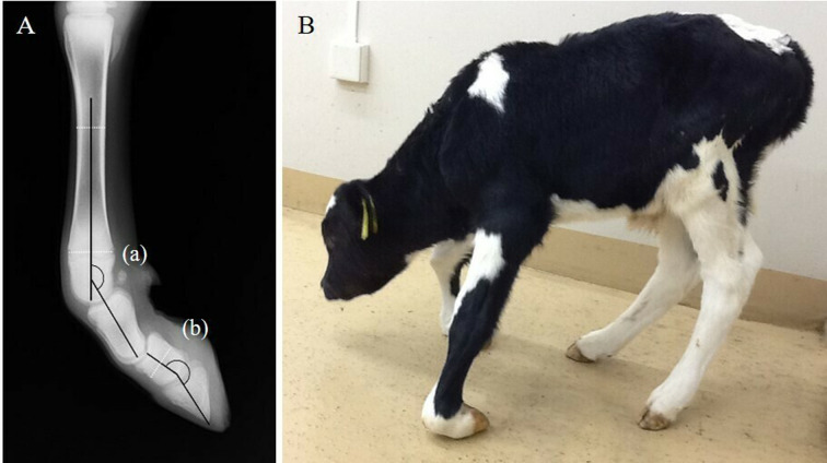
Calves were diagnosed with severe MPFD via inspection, palpation and radiographs. Severe MPFD was classified according to previous reports in which the affected animals were forced to walk on the dorsal aspect of the pastern, fetlock or carpus [2, 16]. Radiographs of affected limbs were taken as described in a previous report [12] and analysed using ImageJ (V.1.48; National Institutes of Health, Bethesda, MD, USA). The lateral angles of the metacarpophalangeal joint (MPJ) and distal interphalangeal joint (DIPJ) were measured using the axis lines of the metacarpal bone and each phalanx (Fig. 1). The preoperatively measured lateromedial angles of MPJ and DIPJ are shown in Table 1. In this study, 17 severe MPFD forelimbs were diagnosed and examined, including bilateral forelimbs from seven calves and unilateral forelimbs from three calves. The mean ± standard deviation joint angles measured on radiographs of all 17 limbs of 10 calves were as follows: MPJ, 151.1° ± 8.8° and DIPJ 204.1° ± 3.8°. The maximum MPJ value of 168.1°, which corresponded to the mildest case of MPFD, was measured in the right forelimb of Calf C; the minimum value of 135.0° was measured in the left forelimb of Calf I and corresponded to the most severe case of MPFD. The maximum and minimum values of DIPJ were 211.8° in the right forelimb of Calf A and 197.1° in the right forelimb of Calf H. According to a previous report [12], the mean joint angles in the forelimbs of normal calves were as follows: MPJ, 175.9° ± 4.6° (95% confidence interval: 174.5–177.4) and DIPJ, 211.9° ± 4.3° (95% confidence interval: 210.7–213.2). The MPJ values of all 17 limbs in the present study were smaller than that in a normal calf limb. Accordingly, the 17 limbs selected for this study appeared to be appropriate.
Fig. 1.
Pre-surgical radiograph and image of Calf F. A) Radiograph of the left forelimb. (a) Metacarpophalangeal joint (MPJ) was 151.7°. (b) Distal interphalangeal joint (DIPJ) was 204.1°. B) Picture of Calf F from left lateral side. The calf was not able to walk on the foot and was forced to walk on the dorsal aspects of MPJs or carpal joints.
Table 1. Clinical information and the results of radiographs, gait tests and prognosis of 17 forelimbs in 10 Holstein-Friesian calves with metacarpophalangeal flexural deformities.
| Limb No. | Animals ID | Affected limb | Age (days) |
Sex | Bwa)
(kg) |
MPJb)
(°) |
DIPJc)
(°) |
External fixation | Prognosis | |
|---|---|---|---|---|---|---|---|---|---|---|
| 2 days | 7 days | 14 days | ||||||||
| 1 | A | RFd) | 7 | Mf) | 28 | 147.7 | 211.8 | - | - | Good |
| 2 | B | Lfe) | 9 | M | 32.5 | 161.0 | 204.9 | - | - | Good |
| 3 | C | LF | 6 | M | 49.5 | 148.6 | 203.0 | - | - | Good |
| 4 | RF | 168.1 | 203.2 | - | - | Good | ||||
| 5 | D | LF | 20 | M | Unknown | 158.6 | 203.4 | Applied | - | Good |
| 6 | RF | 156.7 | 198.4 | Applied | - | Good | ||||
| 7 | E | LF | 8 | Fg) | 33 | 153.9 | 200.6 | Applied | - | Good |
| 8 | RF | 135.6 | 206.9 | Applied | - | Good | ||||
| 9 | F | LF | 6 | M | 38 | 151.7 | 204.1 | Applied | - | Good |
| 10 | RF | 152.2 | 206.3 | Applied | - | Good | ||||
| 11 | G | LF | 40 | M | 50.5 | 147.7 | 206.2 | Applied | - | Good |
| 12 | H | LF | 10 | M | 42 | 155.4 | 202.9 | Applied | - | Good |
| 13 | RF | 150.1 | 197.1 | Applied | - | Guarded | ||||
| 14 | I | LF | 2 | M | 34.5 | 135.0 | 200.2 | Applied | Applied | Good |
| 15 | RF | 137.6 | 204.6 | Applied | Applied | Good | ||||
| 16 | J | LF | 14 | M | 42.5 | 156.8 | 207.5 | Applied | Applied | Poor |
| 17 | RF | 151.7 | 209.0 | Applied | Applied | Poor | ||||
| Mean ± standard deviation | 151.1 ± 8.8 | 204.1 ± 3.8 | ||||||||
a) BW, body weight; b) MPJ, metacarpophalangeal joint; c) DIPJ, distal interphalangeal joint; d) RF, right forelimb; e) LF, left forelimb; f) M, male; g) F, female.
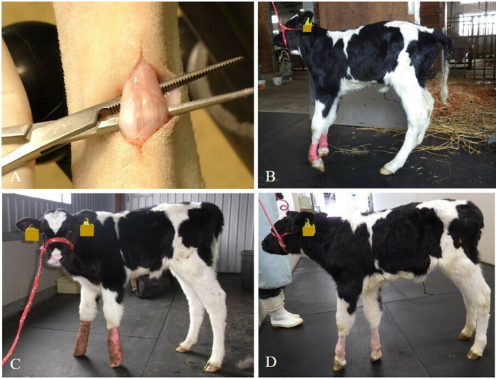
Surgical procedures were performed according to previous reports, with minor modifications [4, 11, 16, 19]. The calves were injected intravenously with 0.1 mg/kg xylazine (2% Celactal; Bayer, Osaka, Japan) as a sedative and subcutaneously with 2.5 ml lidocaine (2% Xylocaine; AstraZeneca, Osaka, Japan) in the incision line as local anesthesia. A longitudinal incision with an approximate length of 5 cm was made on the skin over the middle metacarpal at the palmar side. Subsequently, the SDFT and DDFT were confirmed and separated (Fig. 2A). The SDFT and DDFT were both resected, and a 1.5-cm length of each was removed while paying close attention to the median nerve and median blood vessels. The surgical duration was 15–30 min per limb; hence, the treatment of a calf with bilaterally affected forelimbs could be completed within 60 min. This surgical procedure was simple, and the surgical duration was short. Suture skin lines were applied using sterilized gauze and an elastic self-adhesive bandage. The calves received an antibiotic prophylaxis comprising procaine penicillin for 1–3 days after surgery, and an optional dosage of 1 mg/kg of flunixin meglumine (Furunikishin-cyu, Meiji, Tokyo, Japan) administered intravenously if pain symptoms were evident. No signs of locally infected lesions, such as heat, swelling or drainage, were observed in any calf.
Fig. 2.
Intra-operative and post-surgical images of Calf D from left lateral side. A) The picture of (a) superficial digital flexor tendon and (b) deep digital flexor tendon exposed intraoperatively. B) Two days post-surgery. The calf showed difficulty to walk on the foot and was applied with moulded polyvinyl chloride (PVC) pipes as external fixation. C) Seven days post-surgery. The calf showed no lameness with the applied PVC pipes. D) Fourteen days post-surgery. Although the calf showed mild metacarpophalangeal flexural deformities (MPFD) at the left forelimb, it showed no lameness without PVC pipe. With growth, left forelimb with mild MPFD was improved without any treatment.
The calves were subjected to a gait test on a rubber floor on days 2, 7 and 14 post-surgery. After each gait test, calves were classified as exhibiting lameness or non-lameness. Lameness with an insufficient improvement in MPFD was defined as follows: an inability to place weight on the affected toe, slow movement and a back line that dropped toward the cranial side. Non-lameness with a sufficient improvement in MPFD was defined as follows: the ability to place weight on the affected toe and to walk long strides while maintaining the back line. At 2 days post-surgery, calves that exhibited non-lameness did not receive further treatment, whereas those that exhibited lameness were treated by the application of a polyvinyl chloride (PVC) pipe to the affected limbs. At 7 days post-surgery, the PVC pipe was removed if lameness had resolved. Otherwise, fiberglass tape was used for limb cast fixation. At 14 days post-surgery, all calves underwent a final gait test without external fixations. The calves that had achieved non-lameness had a good prognosis. Conversely, the calves that exhibited lameness with mild to moderate MPFD had a guarded to poor prognosis. All 10 calves were subsequently observed until reaching an outcome such as euthanasia, death or slaughter.
Table 1 presents the prognosis based on the gait test performed 14 days post-surgery. At 2 days post-surgery, four limbs of three calves had achieved non-lameness and did not require further monitoring. Conversely, PVC pipes were applied to 13 limbs of seven calves because of lameness (Fig. 2). At 7 days post-surgery, these seven calves underwent a repeat gait test. At that time, nine limbs of five calves exhibited non-lameness, resulting in the removal of the PVC pipe at 7–14 days post-surgery. Four limbs of two calves (Calves I and J) continued to exhibit lameness and were treated with casts. At 14 days post-surgery, all 10 calves underwent a final gait test after the external fixation was removed. Fourteen limbs of nine calves exhibited non-lameness (Fig. 2), while three limbs of two calves (Calves H and J) exhibited lameness with a guarded to poor prognosis. The cumulative results indicate that 14 of 17 (82%) forelimbs with severe MPFD had a good prognosis. In all treated calves, surgical complications such as hyper-extension of treated limbs or deformation of the healthy limbs due to excess loading were not recognized at any point during the observation period before the final outcomes. These results indicated that the surgical treatment was effective for MPFD and enabled a full return of the calves to normal activity.
In this study, only the flexor tendons in limbs with severe MPFD were transected, and the suspensory ligaments were retained. Generally, the transection of only the SDFT and DDFT is considered insufficient to improve flexion in a limb with severe MPFD [4, 7, 11, 14]. In our study, however, post-operative management enabled an improvement of flexion in nearly all the MPFD-affected limbs. External fixation was easily applied after surgery because the flexion of the metacarpophalangeal joint was mainly affected by the previously transected flexor tendons. Calves that exhibited an improvement in lameness after surgery also exhibited a gradual improvement in MPFD.
Regarding post-surgical management, the transected SDFT, DDFT and suspensory ligaments of the MPFD calves arose from a destabilized carpus joint; consequently, full-limb cast fixation over the carpus joint was needed for more than 30 days post-surgery [4, 11, 17, 18]. Compared with previous reports, the post-surgical management period in our study was shorter and safer because the suspensory ligaments were retained. In young calves, these ligaments are flexible and rich in muscular substances [5]; therefore, the ligaments were maintained extensively against external fixation. Moreover, the tissues in the surrounding tendons recovered rapidly after surgery because the calves were young, and the load on the affected limbs was relatively light. This suggests that the transection of the SDFT and DDFT in a calf with MPFD would be unlikely to cause hyper-extension of the treated limb.
The clinical outcomes were evaluated for 14 days after surgery. Although this duration is short, it emphasized that the therapeutic effect was obtained fairly rapidly. As shown in the results, Calves H and J had a guarded to poor prognosis. The left forelimb of Calf H had the most severe DIPJ angle (197.1°). Although this calf could walk normally with external fixation, it exhibited lameness with mild MPFD after the external fixation was removed on day 14 post-surgery. This suggests that external fixation primarily corrects a deformity of the MPJ, whereas a deformity of the DIPJ is more difficult to correct. In Calf H, the external fixation was continued, and non-lameness was achieved at 21 days post-surgery. In contrast, the left forelimb of Calf I had the most severe MPJ angle (135.0°). Nevertheless, Calf I recovered to normal walking with a PVC pipe and cast fixation. In Calf J, the pre-surgical MPJ and DIPJ angles in the bilateral forelimbs were higher than the average values, indicating less severe flexural deformities in this calf relative to the other calves in the present study. However, Calf J exhibited lameness with moderate MPFD at 14 days post-surgery and had presented with cast fixation on the bilateral forelimbs at the time of hospital admission. This fixation might have influenced the post-surgical recovery of the joint deformities. Possibly, casting with wrist flexion might have caused joint contractures and thus prolonged the treatment period.
In the present study, 13 of 17 severe MPFD limbs required post-surgical external fixation. The MPJ and DIPJ angles of these 13 limbs exhibited a tendency toward more severe flexor deformities when compared with the angles of the other 4 limbs. In the calves of the present study, abnormal radiographic findings such as bone malformations and bone fractures were not observed. Hence, the flexor tendons and suspensory ligaments were the morphological features of concern in severely MPFD-affected limbs. External fixation after the transection of SDFT and DDFT effectively repaired limbs with severe MPFD, and particularly those with very severe deformities. We confirmed that the duration of applied external fixation after surgical treatment was shorter than those used in conventional methods [4, 11, 19]. However, it was difficult to define the criteria for surgery and post-surgical treatment in this study.
In summary, calves with severe MPFD were diagnosed according to clinical examinations and radiographs. The calves were then treated surgically via the transection of SDFT and DDFT while retaining the suspensory ligaments. After surgery, the calves received external fixations as needed. Consequently, 82% of the MPFD-affected limbs achieved a good prognosis within 14 days post-surgery, indicating a high recovery rate from severe MPFD. We conclude that positive surgical treatment involving SDFT and DDFT transection is a useful treatment for calves with severe MPFD.
REFERENCES
- 1.Adams S. B., Santschi E. M.2000. Management of congenital and acquired flexural limb deformities. Proceedings Am. Assoc. Eq. Pract. 46: 117–125. [Google Scholar]
- 2.Anderson D. E., Desrochers A., St Jean G.2008. Management of tendon disorders in cattle. Vet. Clin. North Am. Food Anim. Pract. 24: 551–566, viii. doi: 10.1016/j.cvfa.2008.07.008 [DOI] [PubMed] [Google Scholar]
- 3.Auer J. A.2006. Diagnosis and treatment of flexural deformities in foals. Clin. Tech. Equine Pract. 5: 282–295. doi: 10.1053/j.ctep.2006.09.003 [DOI] [Google Scholar]
- 4.Ducharme N. G.2017. Surgery of flexural and hyperextension deformities. pp. 519–523. In: Farm Animal Surgery, 2nd ed. (Ducharme, N. G, Desrochers, A. and Freeman, D. eds.), Elsevier, St. Louis. [Google Scholar]
- 5.Dyce K., Sack W., Wensing C. J. G.2009. The forelimb of the ruminant. pp. 728–741. In: Textbook of Veterinary Anatomy, 4th ed. (Dyce, K., Sack, W. and Wensing, C. J. G. eds.), Elsevier, St. Louis. [Google Scholar]
- 6.Gaughan E. M.2017. Flexural limb deformities of the carpus and fetlock in foals. Vet. Clin. North Am. Equine Pract. 33: 331–342. doi: 10.1016/j.cveq.2017.03.004 [DOI] [PubMed] [Google Scholar]
- 7.Gangwar A. K., Devi K. S., Singh A. K., Patel N. K. G., Srivastava S.2014. Congenital anomalies and their surgical correction in ruminants. Adv. Anim. Vet. Sci. 2: 369–376. doi: 10.14737/journal.aavs/2014/2.7.369.376 [DOI] [Google Scholar]
- 8.Hartley W. J., Wanner R. A.1974. Bovine congenital arthrogryposis in New South Wales. Aust. Vet. J. 50: 185–188. doi: 10.1111/j.1751-0813.1974.tb02361.x [DOI] [PubMed] [Google Scholar]
- 9.Kidd J. A., Barr A. R. S.2002. Flexural deformities in foals. Equine Vet. Educ. 14: 311–321. doi: 10.1111/j.2042-3292.2002.tb00197.x [DOI] [Google Scholar]
- 10.Leipold H. W., Hiraga T., Dennis S. M.1993. Congenital defects of the bovine musculoskeletal system and joints. Vet. Clin. North Am. Food Anim. Pract. 9: 93–104. doi: 10.1016/S0749-0720(15)30674-5 [DOI] [PubMed] [Google Scholar]
- 11.Pentecost R., Niehaus A. J., Anderson D. E.2016. Surgical management of fractures and tendons. Vet. Clin. North Am. Food Anim. Pract. 32: 797–811. doi: 10.1016/j.cvfa.2016.05.012 [DOI] [PubMed] [Google Scholar]
- 12.Sato A., Ishii O., Tajima M.2018. Radiographic analysis of the angle in the lateromedial projection of the metacarpophalangeal joint and the distal interphalangeal joint in metacarpophalangeal flexural deformities in calves. Vet. Rec. Open 5: e000271. doi: 10.1136/vetreco-2017-000271 [DOI] [PMC free article] [PubMed] [Google Scholar]
- 13.Shupe J. L., Binns W., James L. F., Keeler R. F.1967. Lupine, a cause of crooked calf disease. J. Am. Vet. Med. Assoc. 151: 198–203. [PubMed] [Google Scholar]
- 14.Simon S., William S. M. B., Rao G. D., Sivashanker R., Kumar S.2010. Congenital Malformations in ruminants and its surgical management. Vet. World 3: 118–119. [Google Scholar]
- 15.Sprake P.2015. Diseases of the bones, joints and connective tissues. p. 1106. In: Large Animal Internal Medicine, 5th ed. (Smith B. P. ed.), Elsevier, St. Louis. [Google Scholar]
- 16.Steiner A., Anderson D. E., Desrochers A.2014. Diseases of the tendons and tendon sheaths. Vet. Clin. North Am. Food Anim. Pract. 30: 157–175, vi. doi: 10.1016/j.cvfa.2013.11.002 [DOI] [PubMed] [Google Scholar]
- 17.Van Huffel X., DeMoor A.1987. Congenital multiple arthrogryposis of the forelimb in calves. Comp. Cont. Educ. Pract. 9: 333–339. [Google Scholar]
- 18.Verschooten F., De Moor A., Desmet P., Watte R., Gunst O.1969. Surgical treatment of congenital arthrogryposis of the carpal joint, associated with contraction of the flexor tendons in calves. Vet. Rec. 85: 140–142, passim. doi: 10.1136/vr.85.6.140 [DOI] [PubMed] [Google Scholar]
- 19.Weaver A.2005. Contracted flexor tendons. pp. 240–242. In: Bovine Surgery and Lameness, 2nd ed. (Weaver, A.D., St. Jean, G. and Steiner, A. eds.), Blackwell, Oxford. [Google Scholar]
- 20.Yardimci C., Ozak A., Nisbet O.2012. Correction of severe congenital flexural carpal deformities with semicircular external skeletal fixation system in calves. Vet. Comp. Orthop. Traumatol. 25: 518–523. doi: 10.3415/VCOT-11-11-0161 [DOI] [PubMed] [Google Scholar]


