Abstract
目的
研究放大镜和显微镜对口腔修复医生贴面牙体预备时体位的影响,从人体工程学角度对放大镜和显微镜的临床应用价值进行评价。
方法
从北京大学口腔医院修复科选择20名口腔修复医生进行前瞻性、单盲、自身对照试验。研究过程中不告知受试者研究的试验假设和真实目的,每人依次在常规视野下(空白对照组)、2.5倍头戴式放大镜下(放大镜组)和8倍医用显微镜下(显微镜组)在仿头模内完成右上中切牙开窗型贴面牙体预备,试验过程中拍摄医生的侧面和正面体位照片。贴面牙体预备完成后,由医生本人利用视觉模拟评分法对自身体位进行主观评分,由两名专家利用侧面和正面体位照片按照“改良口腔医生体位评分表”对医生的体位进行专家评分。
结果
空白对照组、放大镜组和显微镜组的主观评分分别为4.55±1.96、7.90±1.12、9.00±0.92,三组之间差异有统计学意义(P < 0.05);专家评分分别为16.38±1.52、15.15±1.30、13.60±0.88,三组之间差异有统计学意义(P < 0.05)。三组的专家评分在躯干前后向位置(1.33±0.41、1.03±0.11、1.00±0.00)、头颈前后向位置(2.75±0.38、2.13±0.36、1.23±0.38)、肘关节位置(1.38±0.43、1.40±0.45、1.13±0.22)和肩部高度(1.43±0.41、1.23±0.34、1.13±0.28)这4项评分上的差异有统计学意义(P < 0.05),其中,在头颈前后向位置和肘关节位置方面,放大镜组与显微镜组之间的差异有统计学意义(P < 0.05)。
结论
放大镜和显微镜均能改善口腔修复医生牙体预备时的体位,其中显微镜的效果更好,从人体工程学的角度二者均具有较高的临床应用价值。
Keywords: 放大镜, 显微镜, 口腔医生, 体位, 人体工程
Abstract
Objective
To assess the effects of loupes and microscope on the posture of prosthodontists when preparing the laminate veneer, and to assess the clinical value of loupes and microscope from the ergonomic aspects.
Methods
Twenty young prosthodontists from Department of Prosthodontics, Peking University School and Hospital of Stomatology were recruited into this study, which was a prospective, single blind, self-control trials. The research hypothesis was concealed and the participants were deceived about the precise purpose of the study to counterbalance the lack of direct blinding. The prosthodontists prepared laminate veneers of open window type in the artificial dental model, under routine visual field (control group), 2.5× headwear loupes (loupes group), and 8× operating microscope (microscopic group) by turning. The participants were photographed from profile view and front view. Thereafter, the subjective assessment was performed by themselves using the visual analogue score (VAS). The expert assessment was performed by two professors using modified-dental operator posture assessment instrument on the basis of photographs of the profile view and front view.
Results
The subjective assessment scores for the control group, loupes group and microscopic group were 4.55±1.96, 7.90±1.12, and 9.00±0.92, respectively. There was significant difference between the three groups' subjective scores (P < 0.05). The expert assessment scores for the control group, loupes group and microscopic group were 16.38±1.52, 15.15±1.30, and 13.60±0.88, respectively. There was significant difference between the three groups' expert assessment scores (P < 0.05). Specifically, the three groups' expert assessment scores were significantly different (P < 0.05) in trunk position (front to back) (1.33±0.41, 1.03±0.11, 1.00±0.00), head and neck position (front to back) (2.75±0.38, 2.13±0.36, 1.23±0.38), elbows level (1.38±0.43, 1.40±0.45, 1.13±0.22), and shoulders level (1.43±0.41, 1.23±0.34, 1.13±0.28). Thereinto, the microscopic group was better than loupes group in head and neck position (front to back) and elbows level (P < 0.05).
Conclusion
Loupes and microscope improve the posture of the prosthodontist when preparing the laminate veneer, in which the microscope is better than loupes. Therefore, the magnification devices have clinical value from the ergonomic aspects.
Keywords: Loupes, Microscopes, Dentists, Posture, Human engineering
口腔医生工作时经常长时间保持同一体位,因此罹患肌肉骨骼疾病(musculoskeletal disorders,MSDs)的风险很高,且逐年上升[1-2]。口腔医生中肌肉骨骼疼痛的发生率高达64%~93%,其中最常见的疼痛部位是背部(36%~60%)和颈部(20%~85%)[3]。
人体工程学是研究“人-机-环境”系统中人、机和环境三大要素之间的关系,为系统中人的效能和健康问题提供理论与方法的学科。应用人体工程学原则设计的口腔医学诊疗设备将有助于保持自然的体位[4],而符合人体工程学的工作体位将有助于预防MSDs,这对减少口腔医生的骨骼肌疼痛至关重要[2]。
放大镜是近年来在口腔医学领域广泛应用的设备之一,包括头戴式和眼镜式两类,均固定在医生的头部,可以辅助医生获得更好的视野。由于放大镜具有特定的焦距,戴用放大镜时需要使医生的眼睛与术区保持特定的距离,可防止医生的头颈和背部过于弯曲。有研究表明,使用放大镜可以帮助口腔医生获得人体工程学益处,并减少MSDs的发生[5-6],但是放大镜对口腔修复医生的人体工程学意义尚未见报道。
除了放大镜以外,医用显微镜在口腔医学领域的应用也越来越多。显微镜同样具有特定范围的焦距,但是与放大镜不同,显微镜固定在独立的台式支架上,而非医生的头部,其位置不受医生的主观因素影响。口腔修复医生能否利用显微镜获得人体工程学益处尚不得而知。
本研究的主要目的是探讨放大镜和显微镜对口腔修复医生贴面牙体预备时体位的影响,从人体工程学角度对二者的临床应用价值进行评价。
1. 资料与方法
1.1. 研究对象
从北京大学口腔医院口腔修复科60名年轻医生当中随机选择20人参加本项研究,纳入标准:(1)年龄在35岁以下,(2)裸眼视力或矫正视力达到1.0,(3)已有独立完成贴面牙体预备10例以上的临床经验。
1.2. 试验方法
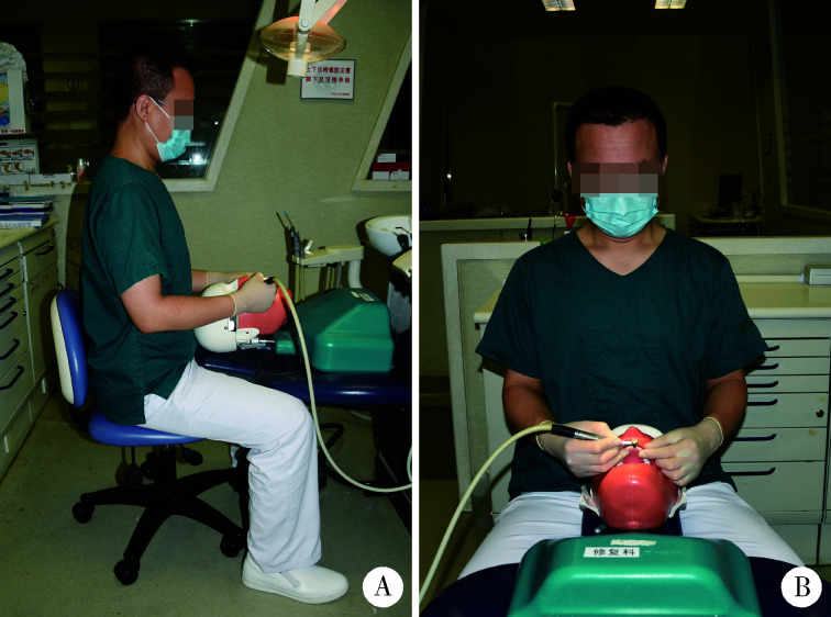
本研究采用前瞻性、单盲、自身对照的试验方法,对受试者实施盲法。20名口腔修复医生依次在常规视野下(空白对照组)、2.5倍头戴式放大镜下(放大镜组)和8倍医用显微镜下(显微镜组)在仿头模内完成右上中切牙开窗型贴面牙体预备。试验时以“评价牙体预备速度和牙体预备质量”来掩盖本试验的真实目的,试验过程中拍摄医生的侧面和正面体位照片(图 1)。牙体预备完成以后,由医生本人利用视觉模拟评分法(visual analogue scale,VAS)对自身体位进行主观评分,由第三方专家利用照片对医生体位进行专家评分。
图1.
口腔修复医生体位的侧面(A)和正面照(B)
Profile view (A) and front view (B) of the prosthodontist
1.3. 医生体位主观评分
每名医生利用VAS对自身体位进行主观评分,将体位用0~10共11个数字表示,分值越低说明体位越差,分值越高说明体位越好。
1.4. 医生体位专家评分
利用医生的侧面和正面体位照片,由两名口腔修复教研室高级职称医师采用改良口腔医生体位评分表(modified-dental operator posture assessment instrument,M-DOPAI)[2]对医生体位进行专家评价。M-DOPAI包含12个评分项,每项分为3个等级:良好为1分、一般为2分、较差为3分,其中8项分值为1~3分、4项分值为1~2分,总分最低为12分、最高为32分,分值越低说明体位越符合人体工程学要求,分值越高说明体位越背离人体工程学要求。利用侧面体位照片评价臀部高度、躯干前后向位置、头颈前后向位置、上臂与躯干的相对位置关系、肩部是否放松以及腕关节有无屈伸。利用正面体位照片评价躯干有无倾斜或旋转、头颈部有无倾斜或旋转、肘关节位置、肩部高度和腕关节有无屈伸。两名评价者首先回顾理想的人体工程学体位、浏览全部照片并了解可能的体位变化之后再进行独立评分,以二者的平均分为最终专家评分。
1.5. 仪器及设备
使用的仪器设备包括:放大镜(速迈公司,中国)、医用显微镜(Leica公司,德国)、仿头模及人工牙颌模型(Nissin公司,日本)、牙体预备用钻针(Coltene公司,瑞士)、拍照手机(华为公司,中国)等。
1.6. 统计学分析
计量数据以“均数±标准差”表示,使用SPSS Statistics 20.0软件(IBM公司,美国)对数据进行统计分析,对主观评分和专家评分进行两因素方差分析,并通过Tukey HSD方法进行组间多重比较,以P < 0.05为差异有统计学意义。
2. 结果
2.1. 体位的主观评分
表 1结果显示,空白对照组、放大镜组和显微镜组的主观评分分别为4.55±1.96、7.90±1.12、9.00±0.92,三组间差异有统计学意义(P < 0.05);多重比较结果显示,每两组之间的差异均有统计学意义(P < 0.05)。
表1.
体位的主观评分与专家评分结果
Subjective assessment and expert assessment of the posture
| Group | Subjective evaluation (VAS scores*, x±s) | Expert assessment evaluation (x±s) | ||||||||||||
| Total scores* | Hips | Trunk (front to back)* | Trunk (side to side) | Trunk (rotation) | Head and neck (front to back)* | Head and neck (side to side) | Head and neck (rotation) | Upper arms | Elbows* | Shoulder (relaxed or slumped) | Shoulder level* | Wrists (flexion or extension) | ||
| For each column, groups identified by different superscript letters were significantly different (P < 0.05). *, there was significant difference between groups (P < 0.05). VAS, visual analogue score. | ||||||||||||||
| Control group | 4.55±1.96a | 16.38±1.52a | 1.58±0.44a | 1.33±0.41a | 1.05±0.15a | 1.13±0.22a | 2.75±0.38a | 1.03±0.11a | 1.00±0.00 | 1.70±0.55a | 1.38±0.43a | 1.03±0.11a | 1.43±0.41a | 1.00±0.00 |
| Loupes group | 7.90±1.12b | 15.15±1.30b | 1.58±0.44a | 1.03±0.11b | 1.05±0.15a | 1.08±0.18a | 2.13±0.36b | 1.05±0.15a | 1.00±0.00 | 1.63±0.51a | 1.40±0.45a | 1.00±0.00a | 1.23±0.34a, b | 1.00±0.00 |
| Microscopic group | 9.00±0.92c | 13.60±0.88c | 1.55±0.46a | 1.00±0.00b | 1.00±0.00a | 1.00±0.00a | 1.23±0.38c | 1.00±0.00a | 1.00±0.00 | 1.58±0.49a | 1.13±0.22b | 1.00±0.00a | 1.13±0.28b | 1.00±0.00 |
| P value | <0.001 | <0.001 | 0.727 | <0.001 | 0.270 | 0.091 | <0.001 | 0.377 | 0.180 | 0.001 | 0.377 | 0.004 | ||
2.2. 体位的专家评分
表 1结果显示,空白对照组、放大镜组和显微镜组的专家评分分别为16.38±1.52、15.15±1.30、13.60±0.88,三组间差异有统计学意义(P < 0.05);多重比较结果显示,每两组之间的差异均有统计学意义(P < 0.05)。
对M-DOPAI包含的12个评分项分别进行分析,空白对照组、放大镜组和显微镜组的专家评分在躯干前后向位置、头颈前后向位置、肘关节位置和肩部高度这4项上的差异有统计学意义(P < 0.05);在臀部高度、躯干有无倾斜、躯干有无旋转、头颈有无倾斜、头颈有无旋转、上臂与躯干的相对位置关系、肩部是否放松和腕关节有无屈伸这8项上的差异无统计学意义(P>0.05)。
多重比较显示:在头颈前后向位置上,空白对照组、放大镜组和显微镜组每两组间的差异均有统计学意义(P < 0.05)。在躯干前后向位置上,放大镜组与显微镜组间的差异无统计学意义(P>0.05),二者与空白对照组间的差异有统计学意义(P < 0.05)。在肘关节位置上,空白对照组和放大镜组间的差异无统计学意义(P>0.05),二者与显微镜组间的差异有统计学意义(P < 0.05)。在肩部高度上,空白对照组与显微镜组间的差异有统计学意义(P < 0.05),二者与放大镜组间的差异无统计学意义(P>0.05)。
3. 讨论
本研究的主观评分和专家评分结果表明,放大镜和显微镜均可改善口腔修复医生贴面牙体预备时的体位。从人体工程学的角度来看,二者均具有临床应用价值,尤其是显微镜对医生体位的改善效果更明显、人体工程学价值更高。
以往有学者观察了口腔医生的工作体位,在4 h的观察期内,有85%的时间口腔医生的颈部前倾30°以上,超过50%的时间躯干弯曲超过30°、肩部抬升到躯干侧方30°以上[7],这种长期的不良体位直接与MSDs的高发生率相关[8-9],因此,保持符合人体工程学的自然体位对减少MSDs有重要意义。然而,不符合人体工程学的最大原因在于视野不好[10],应用口腔显微设备(包括放大镜和显微镜)的最初目的就是提高视野的清晰度和分辨率。以往的研究表明,在模拟临床情况下,与常规视野相比,使用放大镜和显微镜均可以提高牙体预备精度,其中显微镜的效果优于放大镜[11]。另外,显微设备都有特定的焦距范围,更好的视野和特定的焦距可以减少过度的头颈部以及躯干部的弯曲,保持符合人体工程学的自然体位,减少口腔医生MSDs的发生。
以往的研究表明,口腔医生使用放大镜时可以显著改善体位[5, 12],可减少肩颈部、上后背部和手腕部疼痛的发生概率[13]。本研究的主观评分和专家评分结果显示,口腔修复医生使用显微设备时可以显著改善体位,放大镜可以改善头颈前后向位置和躯干前后向位置,使之更符合人体工程学的要求。使用显微镜时,除了可以在头颈前后向位置和躯干前后向位置上获益外,还可以改善肘关节位置和肩部高度,减少异常体位的出现。此外,与放大镜相比,显微镜可以更显著地减少头部前倾,而头部长时间过于前倾与肩颈部疼痛增加有关[14]。因此,就口腔修复医生而言,显微镜比放大镜的人体工程学意义更大。
在医生的臀部高度、躯干与头颈部有无倾斜或旋转、上臂位置及腕关节屈伸方面,口腔显微设备没有显著影响,这是因为大多数医生已经掌握了基本的人体工程学原则,可以通过调整医生座椅的高度、仿头模的三维位置以及医患的相对位置来获得常规视野上述各方面的合理体位,而不依赖于视觉辅助设备。
由于受试者知晓试验设计后可能对试验结果产生影响,因此,在研究中可以隐藏真实的研究目的[15]。本研究中,以“评价牙体预备速度和牙体预备质量”来掩盖试验的真实目的,一定程度上可以抵消不能直接应用盲法所带来的偏倚,进而提高研究结果的可信度。
本研究的缺点在于仅评价了放大镜和显微镜对口腔修复年轻医生体位的影响,未评价其对预防或减少MSDs的作用,且受试者未包含不同工作经验和年限的医生,此外,使用显微设备有无长期副作用仍有待长期追踪随访。
综上,放大镜和显微镜均可改善口腔修复医生贴面牙体预备时的体位,其中使用显微镜时更符合人体工程学需求,推荐使用放大镜和显微镜作为人体工程学解决方案,改善口腔修复医生的体位,以期减少口腔医生MSDs的患病率。
Funding Statement
中华口腔医学会青年科研基金(CSA-R2018-01)和北京大学口腔医院教学改革项目基金(2017-PT-01)
Supported by the Chinese Stomatological Association Research Fund (CSA-R2018-01) and the Program for Educational Reform of Peking University School and Hospital of Stomatology (2017-PT-01)
Contributor Information
杨 洋 (Yang YANG), Email: Yyangpkuss@163.com.
谭 建国 (Jian-guo TAN), Email: tanwume@vip.sina.com.
References
- 1.Al-Mohrej OA, AlShaalan NS, Al-Bani WM, et al. Prevalence of musculoskeletal pain of the neck, upper extremities and lower back among dental practitioners working in Riyadh, Saudi Arabia: a cross-sectional study. BMJ Open. 2016;6(6):e011100. doi: 10.1136/bmjopen-2016-011100. [DOI] [PMC free article] [PubMed] [Google Scholar]
- 2.Partido BB, Wright BM. Self-assessment of ergonomics amongst dental students utilising photography: RCT. Eur J Dent Educ. 2018;22(4):223–233. doi: 10.1111/eje.12335. [DOI] [PubMed] [Google Scholar]
- 3.Hayes M, Cockrell D, Smith DR. A systematic review of musculoskeletal disorders among dental professionals. Int J Dent Hyg. 2009;7(3):159–165. doi: 10.1111/j.1601-5037.2009.00395.x. [DOI] [PubMed] [Google Scholar]
- 4.陈 曦, 边 专, 聂 敏. 人体工程学原则在口腔医学中的应用. 国外医学·口腔医学分册. 2006;33(2):134–135, 138. doi: 10.3969/j.issn.1673-5749.2006.02.017. [DOI] [Google Scholar]
- 5.Maillet JP, Millar AM, Burke JM, et al. Effect of magnification loupes on dental hygiene student posture. J Dent Educ. 2008;72(1):33–44. doi: 10.1002/j.0022-0337.2008.72.1.tb04450.x. [DOI] [PubMed] [Google Scholar]
- 6.Hayes MJ, Osmotherly PG, Taylor JA, et al. The effect of wearing loupes on upper extremity musculoskeletal disorders among dental hygienists. Int J Dent Hyg. 2014;12(3):174–179. doi: 10.1111/idh.12048. [DOI] [PubMed] [Google Scholar]
- 7.Marklin RW, Cherney K. Working postures of dentists and dental hygienists. J Calif Dent Assoc. 2005;33(2):133–136. [PubMed] [Google Scholar]
- 8.Fals Martínez J, González Martínez F, Orozco Páez J, et al. Musculoskeletal alterations associated factors physical and environmental in dental students. Rev Bras Epidemiol. 2012;15(4):884–895. doi: 10.1590/S1415-790X2012000400018. [DOI] [PubMed] [Google Scholar]
- 9.Memarpour M, Badakhsh S, Khosroshahi SS, et al. Work-related musculoskeletal disorders among Iranian dentists. Work. 2013;45(4):465–474. doi: 10.3233/WOR-2012-1468. [DOI] [PubMed] [Google Scholar]
- 10.Garcia P, Gottardello ACA, Wajngarten D, et al. Ergonomics in dentistry: experiences of the practice by dental students. Eur J Dent Educ. 2017;21(3):175–179. doi: 10.1111/eje.12197. [DOI] [PubMed] [Google Scholar]
- 11.Eichenberger M, Biner N, Amato M, et al. Effect of magnification on the precision of tooth preparation in dentistry. Oper Dent. 2018;43(5):501–507. doi: 10.2341/17-169-C. [DOI] [PubMed] [Google Scholar]
- 12.Branson BG, Bray KK, Gadbury-Amyot C, et al. Effect of magnification lenses on student operator posture. J Dent Educ. 2004;68(3):384–389. doi: 10.1002/j.0022-0337.2004.68.3.tb03755.x. [DOI] [PubMed] [Google Scholar]
- 13.Hayes MJ, Taylor JA, Smith DR. Predictors of work-related musculoskeletal disorders among dental hygienists. Int J Dent Hyg. 2012;10(4):265–269. doi: 10.1111/j.1601-5037.2011.00536.x. [DOI] [PubMed] [Google Scholar]
- 14.Ariens GA, Bongers PM, Douwes M, et al. Are neck flexion, neck rotation, and sitting at work risk factors for neck pain? Results of a prospective cohort study. Occup Environ Med. 2001;58(3):200–207. doi: 10.1136/oem.58.3.200. [DOI] [PMC free article] [PubMed] [Google Scholar]
- 15.Wilson AT. Counterfactual consent and the use of deception in research. Bioethics. 2015;29(7):470–477. doi: 10.1111/bioe.12142. [DOI] [PubMed] [Google Scholar]


