



Abstract
Exosome-encapsulated microRNAs (miRNAs) have been identified as potential cancer biomarkers and pro-tumorigenic mediators for several cancers. However, the miRNA profiling in BCa-Exo (exosomes from plasma of patients with bladder cancer) has not yet been explored. Hence, the aim of this study was to analyze the miRNA profiling in BCa-Exo and to explore the function and mechanism of the selected miR-4644 in BCa progression. Of the 8 differentially expressed miRNAs in BCa-Exo relative to NC-Exo (exosomes from plasma of normal control subjects), hsa-miR-4644 was the only upregulated (fold change >2.0, P<0.05) miRNA, which was further confirmed to be upregulated in plasma of BCa patients and BCa cell lines. Further in vitro assays demonstrated that miR-4644 mimic promoted, whereas miR-4644 inhibitor suppressed BCa cell proliferation and invasion. miR-4644 negatively regulated expression of UBIAD1 (UbiA prenyltransferase domain-containing protein 1) by directly binding to its 3’-UTR region. UBIAD1 overexpression effectively abrogated the promoting effects of miR-4644 mimic on BCa proliferation, migration, and invasion. Additionally, intratumoral injection of miR-4644 antagomir downregulated miR-4644 expression in tumors and suppressed tumorigenesis in mouse xenografts. Collectively, miR-4644 promotes BCa progression by targeting UBIAD1. miR-4644 may be an important therapeutic target for BCa treatment.
Keywords: Exosome, miR-4644, UBIAD1, bladder cancer
Introduction
Bladder cancer (BCa) is one of the most prevalent malignancies of the urinary system, accounting for approximately 3% of total cancer-related death [1]. Despite the improvement in the treatment, the 5-year survival rate is still dissatisfied. Hence, it is indispensable for us to find novel diagnostic indicators and therapeutic targets to improve the clinical efficacy of treatment for BCa.
Exosomes are nanoscale membrane vesicles that contain bioactive molecules such as lipids, proteins, mRNAs, and microRNAs (miRNAs) and have been regarded as a crucial means of intercellular communication [2]. Exosome-encapsulated miRNAs have been identified as potential cancer biomarkers and pro-tumorigenic mediators during carcinogenesis [3-5]. To date, many studies have highlighted the important role of miRNAs in BCa diagnosis and progression [6-8]. However, the miRNA profiling in BCa-Exo (exosomes from plasma of patients with BCa) has not yet been explored. In the present study, we performed a miRNA microarray to analyze the miRNA profile of BCa-Exo and NC-Exo (exosomes from plasma of normal controls) and found that hsa-miR-4644 was the only upregulated miRNA in BCa-Exo. A previous study suggested that miR-4644 in salivary exosome could be used as a potential biomarker for pancreatobiliary tract cancer [9]. However, the functional role and the underlying mechanism of miR-4644 in BCa have not been explored.
Our bioinformatics analysis (Targetscan) revealed that UbiA prenyltransferase domain-containing protein 1 (UBIAD1, also referred to as TERE1) was a putative target of miR-4644 by harboring two putative miR-4644 binding sites in the 3’-UTR of its mRNA. UBIAD1 has been shown to inhibit the proliferation of BCa cells [10]. Thus, in the present study, we examined the functional role of miR-4644 in regulating BCa cell behaviors and elucidated whether UBIAD1 was involved in this process. Our study provides new information regarding miR-4644 as a novel therapeutic target for BCa treatment.
Materials and methods
Patients and sample collection
Peripheral blood was withdrawn from patients with pathologically confirmed BCa and age-matched healthy volunteers as normal control (NC). The patients who underwent chemotherapy or radiotherapy before blood sampling were excluded. The clinicopathological characteristics of bladder patients, including age, gender, and cancer stage is shown in Table 1. There were 57 patients, with 12 in Ta, 13 in T1, 15 in T2, and 10 in T3, and 7 in T4. Peripheral blood was collected into tubes and centrifugated at 1,000× g for 10 min to obtain plasma. The obtained plasma was stored at -80°C prior to exosome isolation. This study was approved by the Research Ethics Committee of the First Affiliated Hospital of Zhengzhou University and all tested individuals signed an informed consent form.
Table 1.
Clinicopathological characteristics of bladder patients (n=57)
| Parameters | Value |
|---|---|
| Age (y, mean ± SD) | 62.1±4.02 |
| Sex ratio (male/female) | 45/12 |
| Stage | |
| Ta | 12 |
| T1 | 13 |
| T2 | 15 |
| T3 | 17 |
Isolation and characterization of plasma-derived exosomes
Exosomes were isolated from plasma of NC subjects or BCa patients using differential centrifugation, size-exclusion chromatography on Sephadex G50 columns, and ultracentrifugation, as previously described [11,12]. Approximately 10 mL of plasma was collected for exosome isolation. The exosome pellet was resuspended in PBS and protein concentration was measured using Bradford protein assay. Isolated exosomes were immediately used for subsequent experiments or stored at 4°C.
For identification, the morphologic characteristics of exosomes were observed by transmission electron microscopy (TEM) as previously described [13]. The particle size distribution of exosomes was determined by Nanosight. The protein levels of exosomal surface markers CD9, CD63, and Tsg101 were measured by western blot.
miRNA microarray analysis
miRNA microarray analysis was performed by Shanghai Biotechnology Corporation (Shanghai, China). miRNA expression profiles in the collected BCa-Exo and NC-Exo were analyzed using the Human miRNA microarray kit (Agilent Technologies, Spain), and the differentially expressed miRNAs were screened out based on the filtration criteria of fold change >1.0 or <-1.0 and P<0.05.
Cell lines and culture
The immortalized human bladder epithelial cell line SV-HUC-1 (ATCC, Manassas, VA, USA) and human BCa cell lines (T24, J82, 5637, and UMUC3; ATCC) were cultured in complete DMEM (Gibco) containing 10% FBS (Gibco), 100 U/mL penicillin and 0.1 mg/mL streptomycin in a humidified 95% air and 5% CO2 atmosphere at 37°C.
Cell transfection
The UBIAD1 overexpression vector (pcDNA3.1-UBIAD1), an empty pcDNA3.1 vector, miR-4644 mimic, mimic NC, miR-4644 inhibitor, inhibitor NC, UBIAD1 siRNA (si-UBIAD1) and scramble control siRNA (si-Ctrl) were purchased from GenePharma (Shanghai, China). BCa cells at 80-90% confluence were transfected with the indicated constructs, miRNAs, and siRNAs using LipofectamineTM 2000 (Invitrogen).
The sequences were as follows: miR-4644 mimic (forward, 5’-UGGAGAGAGAAAAGAGACAGAAG-3’; reverse, 5’-CUUCUGUCUCUUUUCUCUCUCCA-3’), miR-4644 inhibitor (5’-CUUCUGUCUCUUUUCUCUCUCCA-3’), negative control mimic (mimic NC; forward, 5’-UCACAACCUCCUAGAAAGAGUAGA-3’, reverse, 5’-UCUACUCUUUCUAGGAGGUUGUGA-3’), inhibitor NC (5’-UCUACUCUUUCUAGGAGGUUGUGA-3’), si-UBIAD1 (5’-GUAAUUUGGUCAACACUUATT-3’), si-NC (5’-UUCUCCGAACGUGUCACGUTT-3’).
Quantitative real-time PCR
Total RNA was extracted from plasma or BCa cells using Trizol reagent (Invitrogen). TaqMan MicroRNA Reverse Transcription Kit (Thermo Fisher Scientific) was used to reverse-transcribe RNA to cDNA. qRT-PCR was performed using the Taqman Universal PCR Master Mix (Thermo Fisher Scientific). For internal control, cel-miR-39 was applied for the detection of miRNAs in plasma samples, while U6 was used as the internal control of miRNAs in BCa cells. The UBIAD1 mRNA level was normalized to that of GAPDH.
Western blot
Proteins were extracted from isolated exosome with M-PER buffer (Pierce Biotechnology) or from BCa cells using RIPA lysis buffer (Beyotime). Proteins were separated by electrophoresis through 12% SDS-polyacrylamide gels and then transferred to nitrocellulose membranes. Finally, an ECL kit (Perkin Elmer, Waltham, MA, USA) was used to visualize protein bands. The primary antibodies and secondary antibodies were purchased from Abcam (Cambridge, MA).
RNA pull-down assay
The biotin-labeled miR-4644 and random pull-down probe sequences as NC were designed and synthesized. T24 cells were washed twice with pre-cooled PBS solution, and the cells were lysed in 1 mL 0.1% NP40 lysate containing protease inhibitor and centrifuged at 12000× g for 5 min to extract supernatant. Biotinylated RNAs were then incubated with streptavidin agarose-treated magnetic beads (Life Technologies, Thermo Fisher Scientific) and the cell lysates at room temperature for 1 h. The precipitated UBIAD1 mRNA level was detected by qRT-PCR.
Cell proliferation assay
Following transfection with the designated plasmids or siRNAs, cells were plated in 96-well plates for the indicated time. Then 20 μL of MTT (Sigma-Aldrich, 5 mg/mL) was added into each well. After 4 h of incubation, the medium was replaced with 150 μL dimethyl sulfoxide. After 10 min, the optical density (OD) at 490 nm was measured by a spectrophotometer.
Transwell migration and invasion assay
Cell invasion was assessed by 24-well Transwell plates (8-μm pore size, Corning, NY, USA) pre-coated with Matrigel (BD Biosciences, San Jose, CA, USA) as previously described [14]. The migration assay was conducted the same way as the invasion assay described above, except that the upper chambers were coated without Matrigel.
Dual-luciferase reporter assay
The putative miR-4644 binding site at the 3’-UTR of UBIAD1 was cloned into a pMIR-REPORT vector (Ambion; Thermo Fisher Scientific). For luciferase reporter assay, 293T cells were co-transfected with hsa-miR-4644 or mimic NC, wild-type or mutant luciferase constructs, and pRL-TK (internal control; Promega, Madison, WI, USA) using Lipofectamine 2000 (Invitrogen). The luciferase activity 48 h post-transfection was analyzed using the dual-luciferase reporter assay system (Promega).
In vivo xenograft experiments
BALB/c nude mice (six-week-old; male) were randomly divided into the following 3 groups: control, antagomir-NC, and miR-4644 antagomir, with 6 mice in each group. T24 cells (3×10⁶ cell/mouse) were injected subcutaneously into the right flank of BALB/c nude mice in their backs. After 1 week, 25 nM of miR-4644 antagomir (RiboBio Co., Guangzhou, China; diluted in 100 μL PBS) and 25 nM of antagomir-NC (diluted in 100 μL PBS) were injected intratumorally three times per week for 2 weeks. Mice in the control group were treated with PBS instead. Tumor diameters were measured every week. The tumor volume was calculated. After 6 weeks, the mice were sacrificed, and tumors were excised; the weight of the tumor was measured. All experimental procedures were performed in strict accordance with the guidelines for the Care and Use of Laboratory Animals of the National Institutes of Health.
Statistical analysis
All statistical analyses were performed using SPSS version 20.0. The differences between two groups were analyzed using Student’s t-test. The differences between three or more groups were analyzed by one-way ANOVA. P-value <0.05 was considered statistically significant.
Results
Characterization of NC-Exo and BCa-Exo
TEM revealed that the vesicles from the plasma of NC subjects or BCa patients exhibited a “round-shaped” appearance (Figure 1A). Furthermore, data from NanoSight measurements showed that these vesicles were uniform in size (30-150 nm in diameter) (Figure 1B). Moreover, western blot analysis showed that exosomes in both groups were positive for the exosomal surface markers CD9, CD63, and Tsg101 (Figure 1C).
Figure 1.
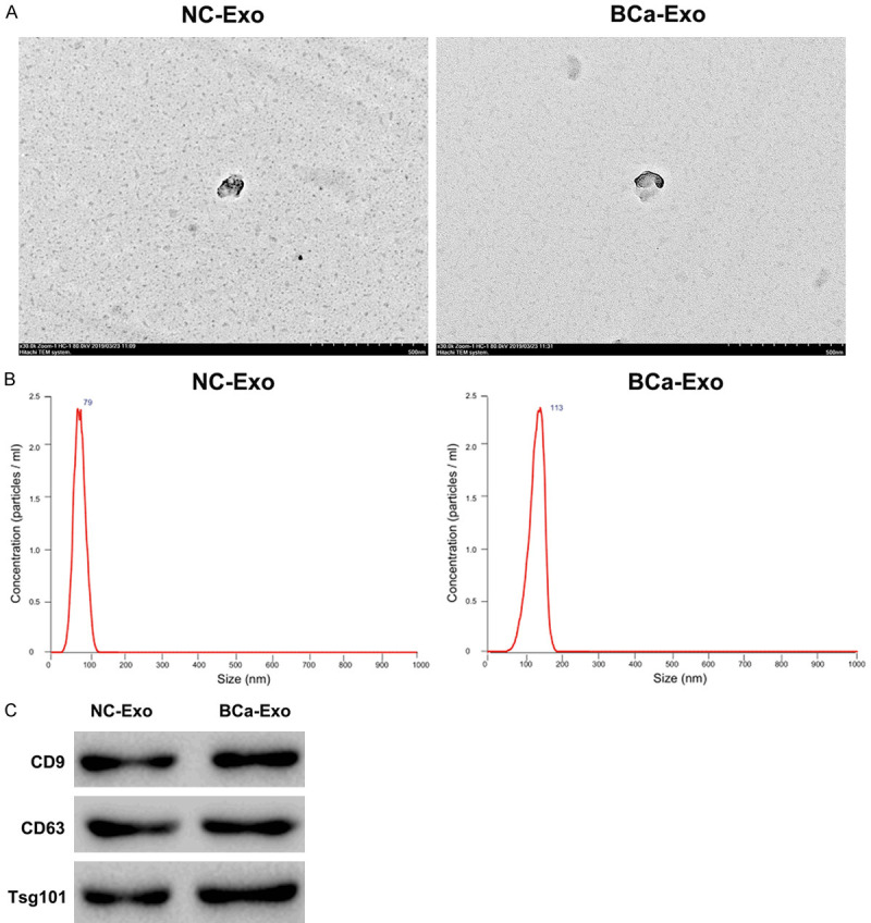
Characterization of exosomes from plasma. Exosomes were isolated from plasma of NC or BCa patients. A. The morphologic characteristics of exosomes were observed by transmission electron microscopy. Scale bar: 500 nm. B. Exosomes were evaluated by Nanosight. The plot shows a broad size distribution. C. Exosomes were positive for exosomal surface markers CD9, CD63, and Tsg101.
Expression profiles of miRNAs in BCa-Exo and expression of selected miRNAs in BCa plasma
The miRNA profile of NC-Exo and BCa-Exo was analyzed using a miRNA microarray. Volcano plot (Figure 2A) and cluster analysis (Figure 2B) revealed 8 miRNAs that were differentially expressed in BCa-Exo compared with NC-Exo, of which 1 was upregulated (hsa-miR-4644; fold change >2.0, P<0.05) and 7 miRNAs were downregulated (hsa-miR-4298, hsa-miR-4499, hsa-miR-4669, hsa-miR-6756-5p, hsa-miR-6809-5p, hsa-miR-6812-5p, and hsa-miR-7107-5p; fold change <-1.0, P<0.05). The fold change values of 8 selected miRNAs and their p-values are shown in Table 2.
Figure 2.
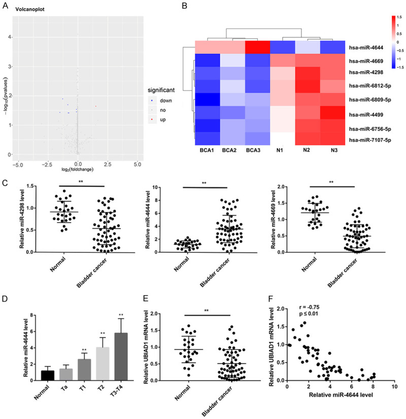
Expression profiles of miRNAs in BCa-Exo and expression of selected miRNAs in BCa plasma. Volcano plot (A) and cluster analysis (B) of differentially expressed exosomal miRNAs in plasma from NC and BCa patients. In (A), red (fold change >1.0; P<0.05) and blue point (fold change <-1.0; P<0.05) represents upregulated or downregulated miRNAs with statistical significance, respectively. In (B), red represents high relative expression levels; blue represents low relative expression levels. (C) qRT-PCR analysis of 3 differentially expressed miRNAs (hsa-miR-4298, hsa-miR-4644, and hsa-miR-4669; in miRNA microarray analysis fold change >2.0 or <-2.0; P<0.05) in plasma from BCa patients (N=57) and NC individuals (N=25). (D) qRT-PCR analysis of hsa-miR-4644 in plasma from BCa patients (N=57) and NC individuals (N=25). (E) qRT-PCR analysis of UBIAD1 mRNA level in plasma from BCa patients (N=57) and NC individuals (N=25). (F) Negative correlation between hsa-miR-4644 expression and UBIAD1 mRNA in plasma from BCa patients (N=57). **P<0.05 vs. Normal.
Table 2.
The most differentially expressed microRNAs in BCa-Exo
| Rank | miRNAs | Fold change | P value |
|---|---|---|---|
| Upregulated miRNAs | |||
| 1 | hsa-miR-4644 | 2.872710454 | 0.022668085 |
| Downregulated miRNAs | |||
| 1 | hsa-miR-4669 | -2.819818006 | 0.036963923 |
| 2 | hsa-miR-4298 | -2.385001842 | 0.012674751 |
| 3 | hsa-miR-6812-5p | -1.770953192 | 0.02010558 |
| 4 | hsa-miR-4499 | -1.740916158 | 0.0382360419794306 |
| 5 | hsa-miR-7107-5p | -1.663734565 | 0.038351644 |
| 6 | hsa-miR-6809-5p | -1.078021675 | 0.035334227 |
| 7 | hsa-miR-6756-5p | -1.073376001 | 0.029084244 |
To verify the results from miRNA array data, the top 3 miRNAs (hsa-miR-4644, hsa-miR-4669, and hsa-miR-4298; fold change >2.0 or <-2.0, P<0.05) that were significantly upregulated and downregulated were selected and analyzed in plasma from 57 BCa patients and 25 normal controls by qRT-PCR analysis. Consistent with miRNA array data, qRT-PCR further confirmed that expression of hsa-miR-4298 and hsa-miR-4669 was significantly downregulated, whereas expression of hsa-miR-4644 was notably upregulated in most plasma samples from BCa patients when compared with the Normal group (Figure 2C). Furthermore, miR-4644 expression was upregulated along with the disease progressed to the advanced stage (Figure 2D). Conversely, UBIAD1 mRNA level was significantly downregulated in most plasma samples from BCa patients (Figure 2E). Moreover, a negative correlation between miR-4644 level and UBIAD1 mRNA level in BCa plasma samples was observed (Figure 2F).
miR-4644 promotes BCa cell proliferation and invasion
We next evaluated the biological roles of the most upregulated miRNA, miR-4644, in BCa cell proliferation and invasion. qRT-PCR results showed that miR-4644 expression was significantly higher in BCa cells (T24, J82, 5637, and UMUC3) than that in SV-HUC-1 cells (Figure 3A). MTT assay demonstrated that the growth of both T24 and J82 cells was significantly facilitated following transfection with miR-4644 mimic (Figure 3B). Transwell invasion assays indicated that the number of invaded cells in T24 and J82 cells transfected with miR-4644 mimic was notably more than those transfected with mimic NC (Figure 3C). In contrast, miR-4644 inhibitor transfection significantly inhibited cell proliferation and invasion of T24 and J82 cells (Figure 3B, 3C).
Figure 3.
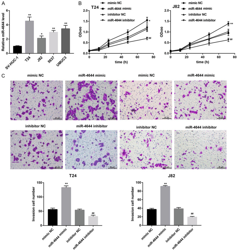
miR-4644 promotes BCa cell proliferation and invasion. (A) qRT-PCR analysis of miR-4644 in human bladder epithelial cell line SV-HUC-1 and human BCa cell lines (T24, J82, 5637, and UMUC3). Cell proliferation (B) and invasion (C) in T24 and J82 cells transfected with miR-4644 mimic, miR-4644 inhibitor, and corresponding controls were evaluated by MTT assay and Transwell invasion assay, respectively. *P<0.05, **P<0.01 vs. SV-HUC-1 or mimic NC, ##P<0.01 vs. inhibitor NC. Data are expressed as the mean ± standard deviation (n=3).
miR-4644 directly targets and downregulates UBIAD1 expression
UBIAD1 has been shown to inhibit the proliferation of BCa cells [10]. Our bioinformatics analysis (Targetscan) revealed that UBIAD1 was a putative target of miR-4644. Thus, we examined whether miR-4644 regulated BCa cell behaviors by targeting UBIAD1. In turn, RNA pull-down and a luciferase reporter assay were performed to analyze the interaction between miR-4644 and UBIAD1. Data from RNA pull-down assay confirmed the direct interaction between miR-4644 and UBIAD1 (Figure 4A). Furthermore, Data from luciferase reporter assay revealed that miR-4644 mimic notably reduced WT luciferase activity to less than 40% of levels in mimic NC-transfected cells, suggesting that UBIAD1 3’-UTR was indeed directly targeted by miR-4644. Furthermore, the activities of the Mut1 construct and the Mut2 construct were over 50% of that of the control. Moreover, Mut1+Mut2 construct disrupting both binding sites completely blocked the downregulation of luciferase activity (Figure 4B). These results indicated that miR-46446 targeting to UBIAD1 mRNA was through binding to both sites on the 3’-UTR.
Figure 4.
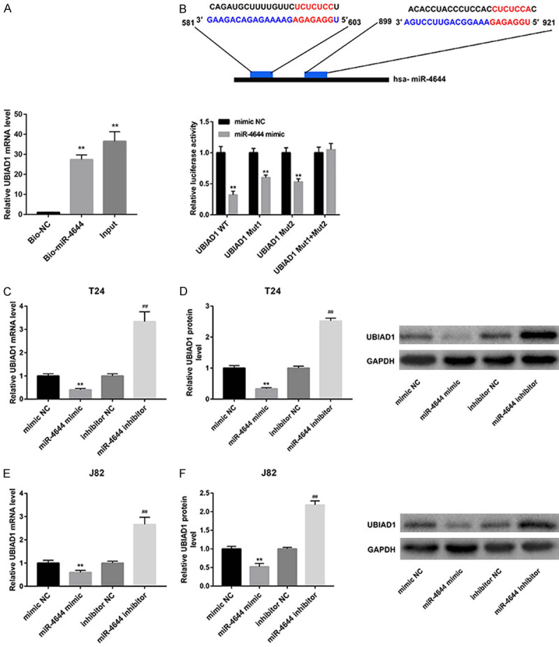
miR-4644 directly targets and downregulates UBIAD1 expression. (A) RNA pull-down was performed to analyze the interaction between miR-4644 and UBIAD1. **P<0.01 vs. Bio-NC. (B) Sequences of the two putative miR-4644 binding sites in the 3’-UTR of UBIAD1. Luciferase activity in 293T cells co-transfected with wild type (WT) or mutant (Mut1, Mut2, Mut1+Mut2) UBIAD1 3’-UTR luciferase reporter plasmids and miR-4644 mimics or mimic NC. The mRNA (C, E) and protein levels (D, F) of UBIAD1 in T24 and J82 cells following transfection with miR-4644 mimic, miR-4644 inhibitor, and corresponding controls. **P<0.01 vs. mimic NC, ##P<0.01 vs. inhibitor NC. Data are expressed as the mean ± standard deviation (n=3).
Furthermore, results from qRT-PCR (Figure 4C) and western blot (Figure 4D) demonstrated that the mRNA and protein levels of UBIAD1 in T24 cells were significantly decreased following transfection with miR-4644 mimic, whereas markedly increased upon transfection with miR-4644 inhibitor. Consistent results were observed in J82 cells (Figure 4E, 4F). These data suggested that miR-4644 directly targeted and downregulated UBIAD1 expression.
UBIAD1 inhibits BCa cell proliferation and invasion
To gain further insight into the biological role of UBIAD1 in BCa, we assessed the effect of UBIAD1 overexpression and knockdown on BCa cell proliferation and invasion. MTT assay demonstrated that UBIAD1 overexpression by pcDNA3.1-UBIAD1 transfection significantly suppressed the growth of both T24 and J82 cells (Figure 5A). Transwell invasion assays indicated that the number of invaded cells in T24 and J82 cells transfected with pcDNA3.1-UBIAD1 was notably less than those transfected with vector control (Figure 5B). In contrast, UBIAD1 knockdown significantly facilitated cell proliferation and invasion of T24 and J82 cells (Figure 5A, 5B).
Figure 5.
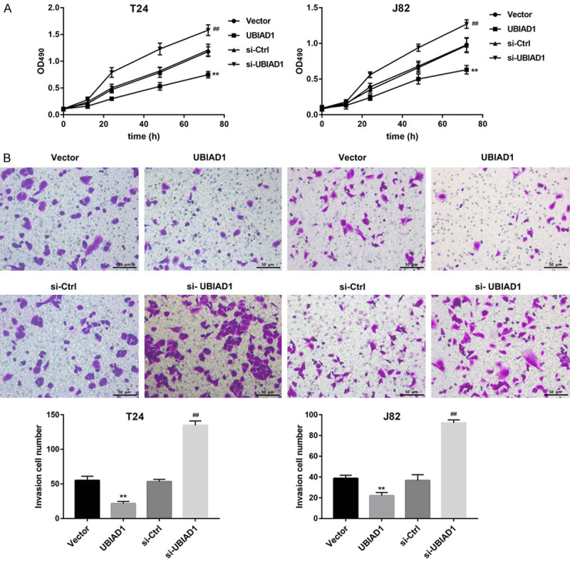
UBIAD1 inhibits BCa cell proliferation and invasion. Cell proliferation (A) and invasion (B) in T24 and J82 cells transfected with UBIAD1 overexpression vector, si-UBIAD1, and corresponding controls were evaluated by MTT assay and Transwell invasion assay, respectively. **P<0.01 vs. Vector, ##P<0.01 vs. si-Ctrl. Data are expressed as the mean ± standard deviation (n=3).
miR-4644 facilitates BCa cell proliferation, migration, and invasion by targeting UBIAD1
Finally, we tested whether UBIAD1 was involved in the miR-4644 mimic-induced BCa cell proliferation, migration, and invasion. MTT assay (Figure 6A) and Transwell assay (Figure 6B, 6C) revealed that UBIAD1 overexpression effectively attenuated the miR-4644 mimic-mediated promotion of cell proliferation, migration, and invasion in both T24 and J82 cells, indicating that miR-4644 facilitated BCa cell proliferation, migration, and invasion via inhibiting UBIAD1 expression.
Figure 6.
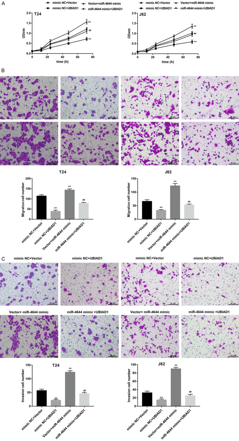
miR-4644 facilitates BCa cell proliferation and invasion via inhibiting UBIAD1. Cell proliferation (A), migration (B), and invasion (C) in T24 and J82 cells co-transfected with miR-4644 mimic/mimic NC, UBIAD1 overexpression vector/control vector were evaluated by MTT assay and Transwell assay, respectively. **P<0.01 vs. mimic NC+Vector, ##P<0.01 vs. mimic NC+UBIAD1 or Vector+miR-4644 mimic. Data are expressed as the mean ± standard deviation (n=3).
Antagomir-4644 suppressed tumorigenesis in mouse xenografts
To ascertain the functional role of miR-4644 in vivo, subcutaneous tumors formed by T24 cells in BALB/c nude mice were administered miR-4644 antagomir once every 3 days for 2 weeks. Importantly, in vivo xenograft tumor growth in nude mice was effectively inhibited by intratumor injection of miR-4644 antagomir, as evidenced by a significant decrease in tumor weight (Figure 7B) and tumor volume (Figure 7A and 7C) in the miR-4644 antagomir group relative to the antagomir-NC group. Expectedly, intratumor injection of miR-4644 antagomir significantly decreased miR-4644 expression in tumors (Figure 7D). However, the mRNA and protein levels of UBIAD1 in tumors were notably increased following intratumor injection of miR-4644 antagomir (Figure 7E and 7F).
Figure 7.

Antagomir-4644 suppresses tumorigenesis in mouse xenografts. The BALB/c nude mice were injected subcutaneously with T24 cells and then administered PBS (control), antagomir-NC or miR-4644 antagomir via intratumoral injection. After 6 weeks, the tumors were exercised. (A) Images of representative tumors excised from mice. (B) Tumor weight. (C) Tumor volume (mm3) was calculated every week. qRT-PCR analysis of miR-4644 (D), qRT-PCR analysis of UBIAD1 mRNA level (E), and western blot analysis of UBIAD1 protein level (F) in tumor tissues excised from mice in each group. **P<0.01 vs. antagomir-NC. N=6 in each group.
Discussion
To the best of our knowledge, this was the first report demonstrating that miR-4644 was upregulated in plasma from BCa patients and that miR-4644 promoted BCa progression via targeting UBIAD1.
Exosomes could serve as a source of diagnostic and prognostic biomarkers for multiple cancers [15], one of which the most exciting marker for cancer diagnosis is miRNA [4,16]. In this study, we performed miRNA microarray in exosome samples from the plasma of BCa patients to analyze differentially expressed miRNAs. Our microarray results showed that the only upregulated miRNA in BCa-Exo was hsa-miR-4644, which was further confirmed by qRT-PCR to be upregulated in plasma of BCa patients. However, more research is warranted to elucidate the potential of exosomal miR-4644 as a biomarker for predicting the clinical behavior of BCa.
Dysregulation of miRNAs is associated with the progression of BCa [1,6,7,17]. For example, Zhang et al. [7] observed that miR-154 expression was notably decreased in BCa tissues and cell lines, and acted as a tumor suppressor in BCa by directly targeting autophagy-related gene 7 (ATG7). MiR-4644 was a recently identified miRNA, whose functions have not been characterized. A previous study suggested that miR-4644 in salivary exosome could be used as a potential biomarker for pancreatobiliary tract cancer [9]. However, until recently, there were few studies concerning miR-4644. In the present study, we selected miR-4644 to examine its biological function in BCa. Our in vitro gain- and loss- of function assays suggested that miR-4644 promoted BCa cell proliferation, migration, and invasion. Furthermore, antagomir-4644 suppressed tumorigenesis in mouse xenografts. These findings demonstrated the pro-tumoral role of miR-4644 in BCa, which merits further investigation.
UBIAD1 protein has been identified as a tumor suppressor in BCa [18]. Recently, Xu et al. [10] suggested that UBIAD1 suppressed the BCa cell proliferation by regulating H-Ras intracellular trafficking via interaction with the C-terminal domain of H-Ras. Consistent with this, our findings revealed that UBIAD1 overexpression significantly inhibited, whereas UBIAD1 knockdown facilitated BCa cell proliferation and invasion. Using luciferase reporter assay, we confirmed that UBIAD1 was a target of miR-4644, which can negatively regulate UBIAD1 mRNA and protein levels. More significantly, the restoration of UBIAD1 in miR-4644-overexpressed BCa cells rescued the promoting effects of miR-4644, indicating that miR-4644 promoted BCa cell proliferation and invasion via targeted silencing of UBIAD1.
In conclusion, we demonstrated that miR-4644 was upregulated in plasma of BCa patients and its derived exosomes. Furthermore, miR-4644 promoted BCa progression by targeting UBIAD1. Our findings contribute to a better understanding of the molecular mechanisms occurring in bladder tumorigenesis and progression.
Acknowledgements
This study was supported by grants from the Key Research Projects of Henan Higher Education Institutions [21A320054].
Disclosure of conflict of interest
None.
References
- 1.Yu H, Duan P, Zhu H, Rao D. miR-613 inhibits bladder cancer proliferation and migration through targeting SphK1. Am J Transl Res. 2017;9:1213–1221. [PMC free article] [PubMed] [Google Scholar]
- 2.Colombo M, Raposo G, Thery C. Biogenesis, secretion, and intercellular interactions of exosomes and other extracellular vesicles. Annu Rev Cell Dev Biol. 2014;30:255–289. doi: 10.1146/annurev-cellbio-101512-122326. [DOI] [PubMed] [Google Scholar]
- 3.Liu Q, Peng F, Chen J. The role of exosomal micrornas in the tumor microenvironment of breast cancer. Int J Mol Sci. 2019;20:884. doi: 10.3390/ijms20163884. [DOI] [PMC free article] [PubMed] [Google Scholar]
- 4.Movahedpour A, Ahmadi N, Ghasemi Y, Savardashtaki A, Shabaninejad Z. Circulating microRNAs as potential diagnostic biomarkers and therapeutic targets in prostate cancer: current status and future perspectives. J Cell Biochem. 2019;20:16316–16329. doi: 10.1002/jcb.29053. [DOI] [PubMed] [Google Scholar]
- 5.Ma J, Xu M, Yin M, Hong J, Chen H, Gao Y, Xie C, Shen N, Gu S, Mo X. Exosomal hsa-miR199a-3p promotes proliferation and migration in neuroblastoma. Front Oncol. 2019;9:459. doi: 10.3389/fonc.2019.00459. [DOI] [PMC free article] [PubMed] [Google Scholar]
- 6.Li Q, Wang H, Peng H, Huang Q, Huyan T, Huang Q, Yang H, Shi J. MicroRNAs: key players in bladder cancer. Mol Diagn Ther. 2019;23:579–601. doi: 10.1007/s40291-019-00410-4. [DOI] [PubMed] [Google Scholar]
- 7.Zhang J, Mao S, Wang L, Zhang W, Zhang Z, Guo Y, Wu Y, Yi F, Yao X. MicroRNA154 functions as a tumor suppressor in bladder cancer by directly targeting ATG7. Oncol Rep. 2019;41:819–828. doi: 10.3892/or.2018.6879. [DOI] [PMC free article] [PubMed] [Google Scholar]
- 8.Hofbauer SL, de Martino M, Lucca I, Haitel A, Susani M, Shariat SF, Klatte T. A urinary microRNA (miR) signature for diagnosis of bladder cancer. Urol Oncol. 2018;36:531.e1–531.e8. doi: 10.1016/j.urolonc.2018.09.006. [DOI] [PubMed] [Google Scholar]
- 9.Machida T, Tomofuji T, Maruyama T, Yoneda T, Ekuni D, Azuma T, Miyai H, Mizuno H, Kato H, Tsutsumi K, Uchida D, Takaki A, Okada H, Morita M. miR1246 and miR4644 in salivary exosome as potential biomarkers for pancreatobiliary tract cancer. Oncol Rep. 2016;36:2375–2381. doi: 10.3892/or.2016.5021. [DOI] [PubMed] [Google Scholar]
- 10.Xu Z, Duan F, Lu H, Abdulkadhim Dragh M, Xia Y, Liang H, Hong L. UBIAD1 suppresses the proliferation of bladder carcinoma cells by regulating H-Ras intracellular trafficking via interaction with the C-terminal domain of H-Ras. Cell Death Dis. 2018;9:1170. doi: 10.1038/s41419-018-1215-4. [DOI] [PMC free article] [PubMed] [Google Scholar]
- 11.Hong CS, Muller L, Whiteside TL, Boyiadzis M. Plasma exosomes as markers of therapeutic response in patients with acute myeloid leukemia. Front Immunol. 2014;5:160. doi: 10.3389/fimmu.2014.00160. [DOI] [PMC free article] [PubMed] [Google Scholar]
- 12.Muller L, Hong CS, Stolz DB, Watkins SC, Whiteside TL. Isolation of biologically-active exosomes from human plasma. J Immunol Methods. 2014;411:55–65. doi: 10.1016/j.jim.2014.06.007. [DOI] [PMC free article] [PubMed] [Google Scholar]
- 13.Lee YS, Kim SY, Ko E, Lee JH, Yi HS, Yoo YJ, Je J, Suh SJ, Jung YK, Kim JH, Seo YS, Yim HJ, Jeong WI, Yeon JE, Um SH, Byun KS. Exosomes derived from palmitic acid-treated hepatocytes induce fibrotic activation of hepatic stellate cells. Sci Rep. 2017;7:3710. doi: 10.1038/s41598-017-03389-2. [DOI] [PMC free article] [PubMed] [Google Scholar]
- 14.Zhang Y, Yan J, Li C, Wang X, Dong Y, Shen X, Zhang X. LncRNA H19 induced by helicobacter pylori infection promotes gastric cancer cell growth via enhancing NF-kappaB-induced inflammation. J Inflamm (Lond) 2019;16:23. doi: 10.1186/s12950-019-0226-y. [DOI] [PMC free article] [PubMed] [Google Scholar]
- 15.Cui S, Cheng Z, Qin W, Jiang L. Exosomes as a liquid biopsy for lung cancer. Lung Cancer. 2018;116:46–54. doi: 10.1016/j.lungcan.2017.12.012. [DOI] [PubMed] [Google Scholar]
- 16.Joyce DP, Kerin MJ, Dwyer RM. Exosome-encapsulated microRNAs as circulating biomarkers for breast cancer. Int J Cancer. 2016;139:1443–1448. doi: 10.1002/ijc.30179. [DOI] [PubMed] [Google Scholar]
- 17.Su H, Jiang H, Tao T, Kang X, Zhang X, Kang D, Li S, Li C, Wang H, Yang Z, Zhang J, Li C. Hope and challenge: precision medicine in bladder cancer. Cancer Med. 2019;8:1806–1816. doi: 10.1002/cam4.1979. [DOI] [PMC free article] [PubMed] [Google Scholar]
- 18.Fredericks WJ, McGarvey T, Wang H, Lal P, Puthiyaveettil R, Tomaszewski J, Sepulveda J, Labelle E, Weiss JS, Nickerson ML, Kruth HS, Brandt W, Wessjohann LA, Malkowicz SB. The bladder tumor suppressor protein TERE1 (UBIAD1) modulates cell cholesterol: implications for tumor progression. DNA Cell Biol. 2011;30:851–864. doi: 10.1089/dna.2011.1315. [DOI] [PMC free article] [PubMed] [Google Scholar]