Abstract
BACKGROUND AND PURPOSE: While MR findings in progressive multifocal leukoencephalopathy (PML) have been described previously, usually in retrospective studies with limited sample size, what has not been well addressed is whether any are predictive of longer survival. Our participation in a large prospective clinical trial of AIDS patients with biopsy-proved PML and MR correlation allowed us to test our hypothesis that certain MR features could be found favorable to patient survival.
METHODS: The patient cohort derived from a randomized multicenter clinical trial of cytosine arabinoside for PML. Pretreatment T1- and T2-weighted noncontrast images (n = 48) and T1-weighted contrast-enhanced images (n = 45) of 48 HIV-positive patients with a PML tissue diagnosis as well as the follow-up images in 15 patients were reviewed to determine signal abnormalities, lesion location and size, and the presence or absence of mass effect, contrast enhancement, and atrophy, and to ascertain the frequency of these findings. A statistical analysis was performed to determine if any MR abnormalities, either at baseline or at follow-up, were predictive of patient survival.
RESULTS: No MR abnormalities either on univariate or multivariate analysis significantly correlated with patient survival, with the exception of mass effect, which was significantly associated with shorter survival. The mass effect, however, always minimal, was infrequent (five of 48). More severe degrees of cortical atrophy and ventricular dilatation, lesion location and size, and other MR variables were not predictive of outcome.
CONCLUSION: Except for mass effect, we found no MR findings predictive of the risk of death in patients with PML. The mass effect, however, was so infrequent and minimal that it was not a useful MR prognostic sign.
Progressive multifocal leukoencephalopathy (PML) is a fulminating opportunistic infection of the brain that occurs in approximately 4% of AIDS patients (1–6). Caused by the JC papovavirus, a DNA virus, it typically has caused death within 2½ to 4 months of diagnosis (1, 2, 7). In the setting of severely altered cellular immunity, this virus causes extensive myelin breakdown and white matter destruction, because of its targeting of the oligodendrocytes (8–11). Despite the use of different medical therapies (1, 11–21), this infection has generally remained refractory to treatment (1, 8, 14). Only a small percentage of patients with a more benign clinical course have reportedly had a prolonged survival (22–25). The lack of a successful therapy for this usually fatal infection has prompted numerous investigators to explore further the efficacy of different medical regimens (1, 11–24). The hope of finding an effective therapy has amplified the need to noninvasively diagnose PML early in its course and to assess treatment response.
While tissue diagnosis has been the mainstay for confirming the presence of PML (1, 7, 8, 11, 13, 26–28), the need for a less invasive test in these often critically ill patients has been acknowledged. Although polymerase chain reaction (PCR) testing of CSF for the JC virus has had promising results (1, 19, 28–35), the use of supplementary tests, such as MR imaging, has seemed very appealing. The combination of CSF analysis, clinical correlation, and MR findings to establish the diagnosis of PML of the brain in a much less invasive way than tissue diagnosis appears especially promising in light of the recent advent of highly active antiretroviral therapy (HAART) and the hope that it will slow the deterioration of the immune system in HIV-positive persons (1, 29, 36–39).
Although MR findings in patients with PML have been previously reported with the use of both conventional (18, 25, 40–52) and newer pulse sequences, and with specialized MR techniques, such as magnetization transfer (MT) (53, 54), fast fluid-attenuated inversion recovery (FLAIR) (55, 56), and MR proton spectroscopy (57–59), there have been limitations of sample size in these mainly retrospective studies in addition to some limitations with regard to follow-up. These limitations, as well as the promise of new therapies for PML, prompted us to undertake the current investigation. Our participation in a prospective therapeutic clinical trial afforded us the unique opportunity to study exclusively by MR imaging a sizable number of AIDS patients (n = 48), all with biopsy-proved PML of the brain. We hypothesized that with this large patient cohort with pathologically proved PML we could find MR features, such as lack of significant cortical atrophy, that were favorable for patient survival. The identification of responders might, of course, prove valuable in guiding future medical therapies. Furthermore, familiarity with MR abnormalities characteristic of PML might facilitate noninvasive diagnosis and follow-up.
Methods
The patient cohort for this analysis derived from a randomized, multicenter, open-label study, designated AIDS Clinical Trials Group (ACTG) Protocol #243 (1). The purpose of this protocol was to determine the efficacy of different treatment methods for PML (ie, a nucleoside antiretroviral regimen alone in a zidovudine and DDI combination versus in conjunction with intravenous or intrathecal cytosine arabinoside [ARA-C]). Thirteen ACTG sites, all with institutional review board study approval, enrolled patients with written informed consent from April 1994 to August 1996. These subjects, all with HIV type I infection and all with clinical and radiologic findings compatible with PML, had the diagnosis of PML confirmed by tissue obtained from stereotactic brain biopsy samples within 2 months of entry into the study. Final diagnosis was established either by conventional neuropathologic examination of this tissue or by in situ hybridization for JC virus or by both (1). PCR analysis of CSF for JC virus complemented the laboratory analysis in some subjects. The details of this Protocol #243 as well as its clinical results have recently been reported by Hall et al (1). This study, which showed no improved survival of HIV-positive patients with PML with either intravenous or with intrathecal cytarabine over antiretroviral therapy alone, was terminated early, as no ARA-C benefit was observed (1). By the last analysis of the clinical investigation (which began before the advent of HAART), 42 patients had died (37 from PML) and only seven had completed 24 weeks of therapy (1). While the clinical results of this Protocol #243 included data from 57 HIV-positive patients with PML, the current radiologic analysis is based on 48 patients in whom complete MR studies were available.
The subjects included 40 men and eight women with a mean age of 39 years (median age, 38 years; age range, 26 to 54 years). T-helper (CD4) absolute count at baseline (n = 39) ranged from 4 to 420, with a median of 65 and a mean of 89. The median survival for the 48 subjects was 11 weeks from study entry (range, 8 to 15 weeks), with a 95% confidence interval. Thirty-five of the 48 subjects died. Given that the maximum time from diagnosis of PML to study entry was 2 months, their survival estimates are consistent with previous reports of 2 to 4 months (1, 2, 7).
The pretreatment baseline noncontrast (n = 48) and contrast-enhanced (n = 45) MR images were reviewed. Follow-up MR studies, available in 15 patients, were also analyzed. Routinely, T1-weighted images were acquired in axial, coronal, sagittal planes or both with the following parameters: 500–750/20–26/1 (TR/TE/excitations); a section thickness of 5–7 mm; usually, a matrix of 256 × 192; and a flip angle of 90°. Proton density–and T2-weighted images, obtained in axial and often coronal views, were acquired routinely with the following parameters: 1900–2300/20,80/1, a flip angle of 90°, a section thickness of 5 to 7 mm, and a matrix of 200 × 256. FLAIR and MT images occasionally complemented the routine scans but were not required for study inclusion. No volumetric or signal intensity measurements were made. For the contrast studies, contrast material was administered intravenously at the recommended doses and scanning was done immediately thereafter.
These images, obtained from 13 different institutions using a variety of mid- to high-field-strength magnets, were then sent to a single neuroradiologist who analyzed them for the presence or absence of the following abnormalities: cortical atrophy (mild, moderate, or severe, as determined by visual inspection); ventricular dilatation (mild, moderate, or severe); white matter lesions; gray matter lesions; number and size of discrete white matter lesions; signal intensity of white matter lesions on T1- and T2-weighted images (low intensity, isointensity, high intensity); mass effect; meningeal disease; contrast enhancement; and interval changes from baseline pretreatment studies (better, worse, or no change; number of new lesions; and change in size or confluence of white matter lesions). White matter lesions were further analyzed to determine if they were confluent, discrete, unilateral, bilateral, supratentorial, or infratentorial. If supratentorial, their general location was designated as being in the periventricular white matter, centrum semiovale, subcortical white matter, internal capsule, external capsule, corpus callosum, or other location. Specific sites (frontal, temporal, parietal, occipital lobes) were also determined. Infratentorial white matter lesions were characterized as being in the brain stem (medulla, pons, midbrain), cerebellar peduncle, or cerebellar white matter. If gray matter involvement was present, its location in the thalamus, basal ganglia, cortical gray matter, or other site was recorded. Follow-up studies were analyzed without knowledge of the treatment arm. All MR information was recorded on data sheets with the results recorded in a numerical fashion. These data were then sent to a separate site for data entry.
Statistical analyses were performed to associate the recorded MR data with clinical data to determine characteristic MR findings in PML of the brain in HIV-positive patients at baseline and at follow-up and to determine if any of these MR findings were predictive of patient survival. The effect of MR findings on survival was investigated through the log-rank test (univariate analyses) while multiple factors were simultaneously considered in the Cox proportional hazards model (multivariate analyses). General association between factors expressed as ordered categories (eg, severity of cortical atrophy) was assessed via the Mantel-Haenszel test.
Results
The abnormalities and their frequency on the initial pretreatment cranial MR images in these 48 AIDS patients are summarized in Table 1. In 85% of patients there was either no cortical atrophy or mild cortical atrophy and in 94% there was either no ventricular dilatation or mild ventricular dilatation. White matter lesions, seen in all, were bilateral in the vast majority of patients (although asymmetrical in their severity), confluent in 94%, and discrete in 67% (the latter including from one to 30 lesions ranging in size from 2 mm to 30 mm). All patients had multiple lesions, of which all were hyperintense relative to gray matter on T2-weighted images and most were hypointense on T1-weighted images (Fig 1). While there was no patient without one or more lesions that appeared hypointense on T1-weighted images, some patients also had lesions that were isointense. Supratentorial white matter lesions (Fig 2B) predominated over infratentorial involvement (Figs 2A and 3). Only three of the 48 patients had disease isolated to the posterior fossa. Gray matter involvement (Fig 2B), when present, was a secondary finding. Mass effect, seen in only five patients, consisted of minimal cortical sulcal effacement (Fig 4) or minimal compression of the adjacent ventricle or both. No contrast uptake was evident in 44 of the 45 patients in whom contrast studies were obtained. In the single patient in whom contrast enhancement of white matter lesions was observed, it was slight and punctate in appearance. No meningeal disease was detected.
TABLE 1:
Frequency of pretreatment MR findings in 48 HIV-positive patients with biopsy proved PML
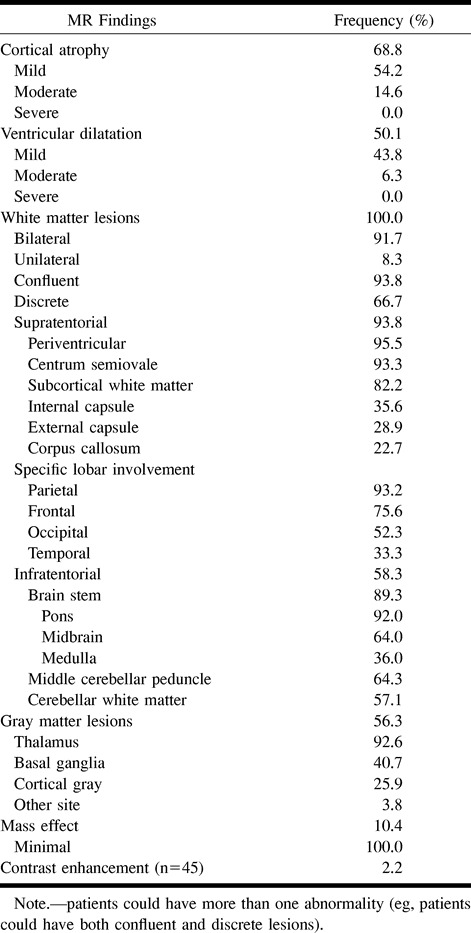
fig 1.
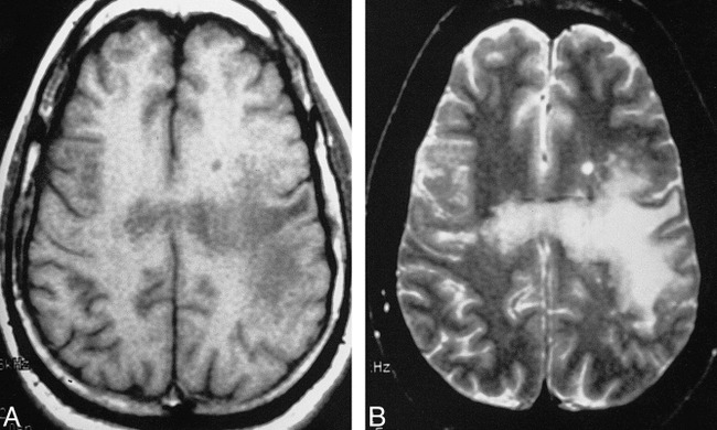
Signal characteristics of PML.
A and B, Axial MR studies show low-signal-intensity lesions on T1-weighted (533/25/1) image (A) and hyperintense lesions on T2-weighted (2000/80/1) image (B) in the centrum semiovale bilaterally, subcortical white matter, and corpus callosum. Note the asymmetrical white matter involvement, with the left cerebral hemispheric white matter more severely affected.
fig 2.
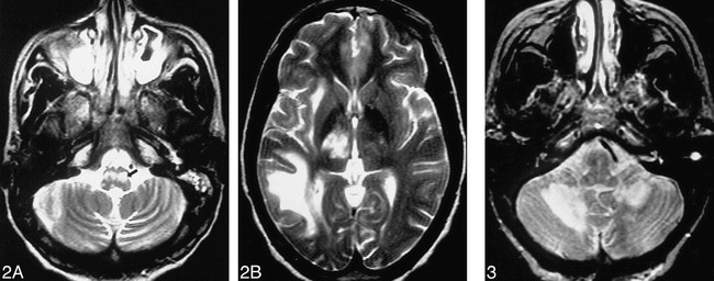
Supratentorial and posterior fossa PML.A and B, Axial T2-weighted images (3700/150/2) show involvement of the medulla (arrow, A), right temporal lobe white matter, right thalamus, and right internal and external capsules, with mild cortical atrophy and no mass effect. Note the asymmetrical involvement of PML, with the right cerebral hemisphere affected to a much greater degree than the left. (Note also the maxillary sinus and left mastoid opacification.)
fig 3. Cerebellar PML. Axial T2-weighted image (2000/80/1) shows bilateral hyperintense lesions in the cerebellar white matter (right greater than left).
fig 4.
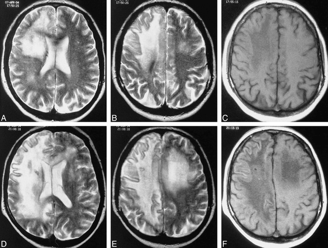
Marked progression of PML documented by serial MR studies.
A and B, Axial T2-weighted images (3500/95/1) show the right frontal lobe confluent hyperintense signal abnormalities extending from the periventricular white matter to the subcortical white matter, with much milder white matter involvement in the right parietal lobe and minimal involvement of the left cerebral hemisphere.
C, Axial T1-weighted image (600/15/1) shows corresponding low signal abnormalities in the affected white matter on the right as well as minimal mass effect on cortical sulci.
D–F, Eight weeks later, marked progression of disease is evident with extension and increasing confluence of the right frontal and parietal lobe lesions, corpus callosum involvement, and greater involvement of the left cerebral hemisphere. Also seen is an increase in white matter low signal abnormality on axial T1-weighted image (600/15/1). Patient died 7 days after this study. (Biopsy tract is also evident in the right cerebral hemisphere.)
Of these MR abnormalities, the only MR finding at baseline that correlated significantly with the risk of death was mass effect. As indicated by a P value of .02, there was a significant association between mass effect and a shorter period of survival. With regard to severity of cortical atrophy and ventricular dilatation, there was no predictive value for the risk of death. None of the other MR variables (see Methods) correlated with patient survival either; however, a trend was observed between increasing number of discrete white matter lesions and increasing survival rates (P = .08). We also noted a trend with a twofold increase for the risk of death if there was gray matter involvement in the basal ganglia. When a multivariate analysis was performed, grouping MR abnormalities in different ways, no additional MR findings predictive of patient survival were discovered.
Follow-up MR studies, available in 15 subjects, showed worsening of findings in 13 patients (Figs 4 and 5) and no significant progression of disease in two. Table 2 summarizes these changes over an interval ranging from 1 to 24 weeks (median, 10 weeks). A dramatic visual change in signal was also seen with a more strikingly low signal intensity in the white matter lesions on T1-weighted images in four patients as well as a greater number of white matter lesions appearing hypointense on T1-weighted images in any given patient. Progression of white matter abnormalities occurred in seven patients in a short 9-week span, with death following shortly thereafter in five.
Concerning treatment arms, seven patients were in the ARA-C intrathecal treatment arm, five in the ARA-C IV treatment arm, and three in the antiretroviral treatment arm. No significant differences were found between treatment arm and MR abnormalities on these follow-up studies. Furthermore, no MR findings at follow-up were found to be predictive of patient survival.
JCV PCR measurements from CSF, available only for the patients in the ARA-C intrathecal treatment arm, were negative in eight and positive in five. Of these 13 patients tested, 12 had periventricular white matter involvement on MR images (seven of whom were PCR-negative and five of whom were PCR-positive). No significant correlations were found between the type of MR findings and the results of the PCR test.
Discussion
Because of the inexorable neurologic decline that leads to coma and death in the majority of patients with PML (2, 9–11, 21, 23, 24, 60–66), the need to develop ways to diagnose and treat PML early in its course has been a pressing one. This need has been considered even more urgent in patients with PML who are infected with HIV, since the two infections appear to be synergistic (8, 35, 67, 68). Survival rates in patients with cellular immunodeficiency due to causes other than HIV have typically been longer, often ranging from 9 to 18 months (2, 9, 19, 27, 41, 64). In contrast, in HIV-infected persons, PML has been reported to have a more aggressive course (13, 35, 67). Not only has the prevalence of PML risen since the advent of AIDS but the number of patients succumbing to PML in a shorter time span has increased (5, 7, 22, 35, 69, 70), with the median survival being only 2½ to 4 months (1, 2, 7, 69). While reports of prolonged survival, both spontaneously and in conjunction with a variety of medical therapies, have been recorded (12–19, 21, 23, 25, 46, 71–74), subsequent studies have shown the inefficacy of many of these regimens (14). Nevertheless, with the introduction of HAART, and with its use now standard in the United States since 1996, the suggestion that AIDS patients with PML may live longer has recently been made, although it has yet to be extensively documented.
As part of the search for ways to predict longer patient survival and to aid in future noninvasive diagnosis and treatment algorithms, we undertook our current study. We hypothesized that if certain MR features were found that favored a delay in death, we could also search in these patients for corollary clinical and laboratory data favorable to prolonged survival. Tailoring of therapies based on any positive predictors of survival might then prove rewarding. Unfortunately, no MR findings significantly correlated in our study with the risk of death, save for mass effect, which was infrequent and always slight and, therefore, practically speaking, of little use. None of the MR variables, such as location and size of lesions, number or type of lesions, or atrophy, predicted a delay in death either on baseline or on follow-up studies. Surprising to us and negating one of our original suppositions, the finding of more marked cortical or deep atrophy at baseline did not correlate with a shortened life span. As for the trend toward a longer patient survival with an increasing number of discrete lesions, it most likely can be explained by the fact that with disease progression white matter lesions coalesce and appear more confluent. While these negative results are obviously disappointing, they are not unexpected, given the ineffectiveness of cytarabine now shown by this clinical trial (ACTG Protocol #243) and given the fact that the study began before the advent of HAART, which bolsters the immune system (36–39, 74). Armed with the knowledge that the majority of patients lived only a short time from study entry, it does not seem surprising that MR abnormalities were not predictive of a delay in death either on baseline or on follow-up studies.
Of definite benefit, however, is the MR imaging evidence of the natural history of this infection that our investigation incidentally produced. Since none of the three different drug regimens appeared to influence patient survival (1) or MR findings, our MR results can serve as a valuable reference or database for future noninvasive diagnosis and for clinical trials. The most commonly found MR abnormalities in our HIV patients with pathologically proved PML might be used in future studies to diagnose PML noninvasively when combined with JCV PCR CSF testing. With positive JCV PCR CSF results and MR findings characteristic of PML, brain biopsy might be avoided in many AIDS patients. The high specificity (95.8% according to Fong et al and 92% according to McGuire et al) (31, 32) of a positive JCV PCR CSF result for PML makes the avoidance of a brain biopsy reasonable when combined with supportive clinical and MR data. Such a course of action is, in fact, now being used in a new ACTG Protocol #363, which is a safety tolerance trial for cidofovir in AIDS patients with PML. Such less-invasive approaches have also been suggested by other investigators (19, 31–33, 35, 50), with brain biopsy being reserved for atypical cases (33). This less-invasive workup seems especially appealing in light of the ongoing refinements in JCV PCR CSF testing, which, it is hoped, will improve the test's sensitivity. Fong et al (32) reported a sensitivity of 74%; Weber et al (29), 82%. A higher sensitivity (92%) was noted by McGuire et al (31), although only eight of their 26 patients had biopsy proof of diagnosis. Limitations to this test were also found in our study: eight of 13 patients with biopsy-proved PML had negative JCV PCR CSF results. Nevertheless, despite these limitations, we agree with McGuire et al that the low likelihood of a false-positive result coupled with the clinical desire to avoid brain biopsy makes the use of this PCR test, when complemented by imaging findings, very attractive. Further test improvements will make such a less-invasive workup even more compelling.
We found that on pretreatment initial MR studies, cortical atrophy and ventricular dilatation were not predominant features of PML. While the infrequent finding of moderate or severe cortical or deep atrophy on pretreatment MR images has not been dwelt on in the imaging literature, it has been pointed out in neuropathologic reports (8, 11, 75). On macroscopic examination before sectioning, the brain in patients with PML shows either no abnormalities or only minimal atrophy (8, 75). This lack of significant cortical or deep atrophy is important, since it can be used as one of the differentiating features from HIV encephalitis on imaging studies. HIV encephalitis, one of the leading diagnostic possibilities in the differential diagnosis of non–mass-producing nonenhancing white matter lesions in the brain of AIDS patients, is typically associated in encephalopathic patients with a significant degree of cortical and deep atrophy (76–79).
An essential feature of PML, because of the targeting of the oligodendrocytes by the JC papovavirus, is that white matter lesions are typically bilateral (although asymmetrical in their degree of involvement), confluent, and multiple. These imaging findings are key diagnostic aides, since they can be used to differentiate PML from HIV encephalitis (8, 12, 14, 41, 42, 48, 50). Possibly because HIV induces a secondary demyelination and does not directly target the oligodendrocytes, the white matter changes in HIV encephalitis are usually symmetrical and diffuse.
Concerning the location of white matter lesions in PML, the periventricular white matter, centrum semiovale, and subcortical white matter were all commonly affected. White matter lesions in PML have been reported to start subcortically, the site of highest blood flow, and then to extend into the deep white matter (35, 41). This subcortical location has been highlighted in the literature and has been touted as an important differentiating feature between PML and HIV encephalitis (22, 35, 40, 42, 46, 50). The scalloping has been attributed to U fiber involvement at the gray/white matter interface of the cerebral cortex. Although the subcortical white matter was also affected in the majority of our patients and did appear scalloped, the higher prevalence of periventricular white matter involvement remains unexplained.
While a posterior location has been used as an imaging sign favorable to the diagnosis of PML, particularly when lesions have been observed in the occipital or parietal lobes, such a location has not been deemed pathognomonic of PML (40–42, 44, 47, 80). Other sites, either alone or in conjunction with posterior lesions, have been reported (15, 16, 40–42, 44, 47, 48). Our study showed that the JC papovavirus commonly infects the frontal lobe, a result worthy of emphasis, since it appears to be a new finding. The parietal lobe, however, remains the most commonly involved by PML in the HIV-positive patient. Our series also showed a higher prevalence of cerebellar, brain stem, and gray matter involvement than previously reported (42, 47, 67, 81), most likely because of our use of MR imaging rather than CT.
In our study, mass effect was infrequent and always minimal, but it did correlate with shorter survival. This lack of significant mass effect has been noted as an important differentiating feature in PML (2, 4, 7, 22, 40, 42, 82, 83). Marked mass effect suggests alternative diagnoses. Similarly, the finding of robust contrast enhancement favors other pathologic processes, since PML lesions typically do not enhance, as evidenced by our study and others (2, 7, 14–16, 30, 41, 43, 44, 46, 50, 84, 85). Contrast enhancement, however, especially when faint and peripheral, does not exclude a PML diagnosis (13, 22, 40, 44, 45, 47, 86). Recently, in fact, contrast enhancement has been considered by Berger et al (71) to be one of the factors predictive of prolonged survival in AIDS patients with PML, along with higher CD4 counts, possibly due to the patient's ability to mount a better response via an intensification of the inflammatory response around the lesions (10, 22, 23). Our study neither confirms nor refutes the value of contrast enhancement as a favorable prognostic sign; but with the advent of HAART, it may be worthy of further exploration.
Concerning the signal abnormalities on T1- and T2-weighted images, low signal intensity on T1-weighted images and hyperintense signal on T2-weighted images typified PML in our patients and those reported in the literature (7, 22, 25, 30, 41, 42, 44, 50). The low signal on T1-weighted images has been thought to be a differentiating factor from HIV encephalitis (47, 50). These typical signal abnormalities on T1- and T2-weighted images with PML have generally been attributed to demyelination (25, 41, 43). Edema as an explanation for the high signal change on T2-weighted images has been reserved as an explanation for those patients whose MR studies have rapidly worsened with changes in treatment regimens, while a decrease in interstitial water or possibly even a remyelination has been used to explain MR imaging improvements with medical therapy (12, 15, 38). The seminal work of Armand et al (53) and Dousset et al (54) with MT imaging in AIDS patients with PML gives strong credence to the supposition that the majority of signal abnormalities are due to demyelination. This may also explain why in the majority of our patients with follow-up MR studies, a further decrease in signal intensity was observed in lesions already hypointense on initial T1-weighted images and why some lesions previously isointense on T1-weighted images became hypointense at follow-up. A greater degree of myelin destruction would certainly explain this incremental lowering of signal intensity on T1-weighted images and would correlate with the progressive decrease in MT contrast values noted in the PML patients being followed up by Dousset et al (54). Increasing demyelination culminating in more extensive areas of destruction and more strikingly hypointense lesions may well correspond to a greater degree of necrosis. Corollary work with proton MR spectroscopy appears to support these theories, as choline, a marker of cellular membrane integrity, is elevated in PML patients, most likely because of cell membrane and myelin breakdown (57, 59).
Concerning follow-up MR studies, since the mean survival rate was only 2½ months in our 48 AIDS patients, it is understandable that 13 of the 15 follow-up studies showed progression of disease and that no MR finding, even on follow-up studies, was found predictive of prolonged survival. Not fully appreciated heretofore, however, has been the impact that conventional MR imaging can have on patient follow-up. Our study shows that serial MR images can depict progression of disease within 1 to 24 weeks, including a more rapid change in many patients in just 9 weeks. This suggests that increasing severity of cortical and deep atrophy, increasing confluence and extent of white matter lesions, spread of disease across the corpus callosum, and increasing hypoattenuation of T1-weighted images in white matter lesions can be used, if persistent, as indicators of a poor prognosis. The presence of generalized atrophy in the late stages of PML may well relate to white matter loss and gliosis, as previously suggested on CT scans (11, 81), while other changes may relate to direct extension of PML lesions (7, 11, 14, 16, 18, 21, 40, 44, 81). Well-demarcated low-signal-intensity lesions on T1-weighted images in patients with PML appear to indicate a very aggressive and severe stage of infection. In future investigations, findings such as increasing hypoattenuation of PML lesions on T1-weighted images might be used as a sign of disease progression, which might then prompt changes in medical therapy.
Follow-up studies might, of course, also be used to document the benefit of a particular therapy. Stabilization of MR findings, a decrease in the size of lesions, and lesion resolution have all been used in the past to suggest the effectiveness of different medical therapies for PML (12, 18, 19, 21, 25, 38). In some cases, MR improvement has lagged behind clinical improvement by 2 to 6 months, and a temporary worsening of abnormalities has even been noted, followed only later by a better MR appearance (12, 15). Most recently, improvement on MR studies has been used along with clinical improvement and a loss of detection of JC virus on PCR CSF testing to indicate an improved prognosis in patients being treated with cidofovir (87). In yet another recent report, serial MR images have been used to show stabilization in one patient and decreased PML lesional size in another patient being treated with HAART and acyclovir therapy (88). In a recent abstract, the positive impact of HAART therapy was noted: improved survival was seen in patients with PML whose most recent viral load after therapy was below the baseline (31). Nonetheless, the response in patients with PML on HAART therapy has not always been deemed beneficial (89), and further investigations of the impact of HAART are needed. The results of our study clearly indicate that conventional MR imaging can be used to detect progression of PML in an early enough time frame to effect a change in medical management. Combined with worsening of JCV PCR CSF results and progressive clinical disease, MR findings might be used as a marker for response to therapy in future clinical trials. We believe that the addition of other noninvasive techniques, such as proton MR spectroscopy and MT contrast ratios, to this type of treatment monitoring and serial assessments of viral load will only give further impetus to this noninvasive approach. Since a noninvasive approach can never reach the diagnostic accuracy of a tissue diagnosis, reliance on complementary studies, given the clinical reality of the situation, only seems reasonable and practical.
Conclusion
While our series did not uncover any MR findings predictive of patient survival in PML, our investigation did reveal MR abnormalities that might be useful to patient management both in initial noninvasive diagnosis and in follow-up. MR findings in PML do evolve quickly and might be useful as intermediate end points in drug trials. With the optimism generated by HAART and other new drug regimens, we can look forward to future investigations that might be able to identify prognostic MR indicators of prolonged survival in this heretofore fulminating infection.
TABLE 2:
Changes on follow-up MR studies in 15 HIV-positive patients with PML on treatment
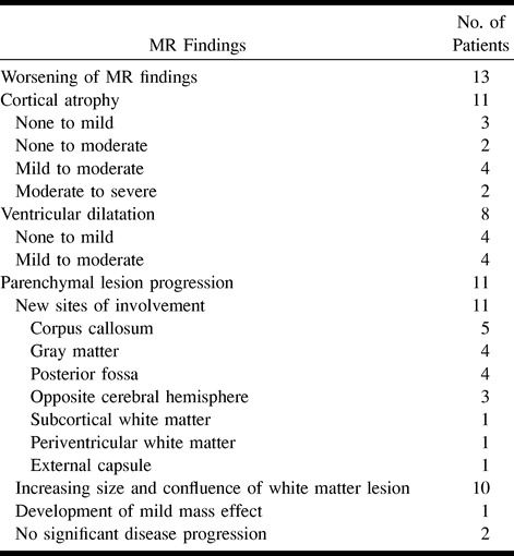
fig 5.
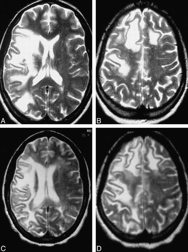
PML progression.
A–D, Axial T2-weighted images before (3700/150/2) (A and B) and at 4½-month follow-up (2500/96/1) (C and D) show worsening of PML as evidenced by increasing cortical atrophy and ventricular dilatation, as well as by further extension of bilateral white matter disease.
Acknowledgments
Gratitude is expressed to Jean Alli for her excellent secretarial assistance and to Robert Burke for his superb photographic work. Appreciation is expressed to the following sites and individuals who contributed MR studies and who were involved in this project: M. Chappel, Community Constituency Group, San Francisco, CA; H. Hollander, University of California at San Francisco School of Medicine, San Francisco, CA; S. Manmann and K. Tyler, University of Colorado Health Sciences Center, Denver, CO; M. Crawford, J. Dobkin, G. Dooneief, and K. Marker, Columbia Presbyterian Medical College, New York, NY; B. Navia, T. Flynn, M. Hirsch, and E. McCarthy, Massachusetts General Hospital and Harvard Medical School, Boston, MA; S. Remick, Albany College Medical Center, Albany, NY; J. R. Berger, University of Kentucky School of Medicine, Lexington, KY; P. E. Wetherill, Yale University School of Medicine, New Haven, CT; C. Marra, University of Washington, Seattle, WA; R. Levy, Northwestern University, Evanston, IL; and the Chicago AIDS Clinical Trials Unit.
Footnotes
Supported by grants 1 PO1 NS3228, A1–25868, RR00036–37, RR00046, RR00051, RR00722, NS26643, and A125915 from the National Institutes of Health and by grant AI38858 from the Adult AIDS Clinical Trials Group, National Institute of Allergy and Infectious Diseases.
Presented at the annual meeting of the American Society of Neuroradiology, Philadelphia, May 1998.
Address reprint requests to M. Judith Donovan Post, MD, Department of Radiology (R 308), University of Miami School of Medicine, MRI Center, 1115 NW 14th St, Miami, FL 33136.
References
- 1.Hall CD, Dafini U, Simpson D, et al. Failure of cytarabine in progressive multifocal leukoencephalopathy associated with human immunodeficiency virus infection. N Engl J Med 1998;338:1345-1351 [DOI] [PubMed] [Google Scholar]
- 2.Berger JR, Kaszovitz B, Post MJD, Dickinson G, Progressive multifocal leukoencephalopathy associated with human immunodeficiency virus infection. Ann Intern Med 1987;107:78-87 [DOI] [PubMed] [Google Scholar]
- 3.Krupp LB, Lipton RB, Swerdlow ML, Leeds NE, Llena J, Progressive multifocal leukoencephalopathy: clinical and radiographic features. Ann Neurol 1985;17:344-349 [DOI] [PubMed] [Google Scholar]
- 4.Petito CK, Cho ES, Lemann W, Navia BA, Price RW, Neuropathology of acquired immunodeficiency syndrome (AIDS): an autopsy review. J Neuropathol Exp Neurol 1986;45:635-646 [DOI] [PubMed] [Google Scholar]
- 5.Levy RM, Bredesen DE, Rosenblum ML, Neurological manifestations of the acquired immune deficiency syndrome (AIDS): experience at UCSF and review of the literature. J Neurosurg 1985;62:475-495 [DOI] [PubMed] [Google Scholar]
- 6.Berger JR, Moskovitz L, Fischl M, Kelley RE, The neurologic complications of AIDS: frequently the initial manifestation. Neurology 1984;34(Suppl 1):134-1356537847 [Google Scholar]
- 7.Karahalios D, Breit R, Dal Conto MC, Levy RM, Progressive multifocal leukoencephalopathy in patients with HIV infection: lack of impact of early diagnosis by stereotactic brain biopsy. J Acquir Immune Defic Syndr Hum Retrovirol 1992;5:1030-1038 [PubMed] [Google Scholar]
- 8.Sweeney BJ, Miller RF, Harrison MJG, Progressive multifocal leukoencephalopathy. Br J Hosp Med 1993;50:187-192 [PubMed] [Google Scholar]
- 9.Richardson EP, Jr, Progressive multifocal leukoencephalopathy 30 years later. N Engl J Med 1988;318:315-317 [DOI] [PubMed] [Google Scholar]
- 10.Walker DL, Progressive multifocal leukoencephalopathy. In: Vinken PJ, Bruyn GW, Klawans HL, eds. Demyelinating Diseases: Handbook of Clinical Neurology. Amsterdam: Elsevier; 1985; 47:503–524
- 11.Brooks BR, Walker DL, Progressive multifocal leukoencephalopathy. Neurol Clin 1984;2:299-313 [PubMed] [Google Scholar]
- 12.Portegies P, Algra PR, Hollak CEM, et al. Response to cytarabine in progressive multifocal leukoencephalopathy in AIDS. Lancet 1991;337:680-681 [DOI] [PubMed] [Google Scholar]
- 13.Conway B, Halliday WC, Brunham RC, Human immunodeficiency virus-associated progressive multifocal leukoencephalopathy: apparent response to 3-azido-3 deoxythymidine. Rev Infect Dis 1990;12:479-482 [DOI] [PubMed] [Google Scholar]
- 14.de Truchis P, Flament-Saillour M, Uetizberea J-A, Hassine D, Clair B, Inefficacy of cytarabine in progressive multifocal leukoencephalopathy in AIDS. Lancet 1993;342:622-623 [DOI] [PubMed] [Google Scholar]
- 15.Nicoli F, Chave B, Peragut JC, Gastaut JL, Efficacy of cytarabine in progressive multifocal leucoencephalopathy in AIDS. Lancet 1992;306: [DOI] [PubMed] [Google Scholar]
- 16.Steiger MJ, Tarnesby G, Gable S, McLaughlin J, Schapira AH, Successful outcome of PML with cytarabine and interferon. Ann Neurol 1993;33:407-411 [DOI] [PubMed] [Google Scholar]
- 17.Berger JR, Pall L, McArthur JG, et al. A pilot study of recombinant alpha 2A interferon in the treatment of AIDS-related progressive multifocal leukoencephalopathy (abstract). Neurology 1992;42(Suppl 3):2571734312 [Google Scholar]
- 18.Tashiro K, Doi S, Moriwaka F, Maruo Y, Nomura M, Progressive multifocal leukoencephalopathy with magnetic resonance imaging verification and therapeutic trials with interferon. J Neurol 1987;234:427-429 [DOI] [PubMed] [Google Scholar]
- 19.O'Riordan T, Daly PA, Hutchinson M, Shattock AG, Gardner SD, Progressive multifocal leukoencephalopathy: remission with cytarabine. J Infect 1990;20:51-54 [DOI] [PubMed] [Google Scholar]
- 20.Wolinsky JS, Johnson KP, Rand K, Merrigan TC, Progressive multifocal leukoencephalopathy: clinical pathological correlates and failure of a drug trial in two patients. Trans Am Neurol Assoc 1976;101:81-82 [PubMed] [Google Scholar]
- 21.Garrels K, Kucharczyk W, Wortzman G, Shandling M, Progressive multifocal leukoencephalopathy: clinical and MR response to treatment. AJNR Am J Neuroradiol 1996;17:597-600 [PMC free article] [PubMed] [Google Scholar]
- 22.Berger JR, Mucke L, Prolonged survival and partial recovery in AIDS-associated progressive multifocal leukoencephalopathy. Neurology 1988;38:1060-1065 [DOI] [PubMed] [Google Scholar]
- 23.Fong IW, Toma E, the Canadian PML Study Group The natural history of progressive multifocal leukoencephalopathy in patients with AIDS. Clin Infect Dis 1995;20:1305-1310 [DOI] [PubMed] [Google Scholar]
- 24.Hedley-White ET, Smith BP, Tyler HR, Peterson WP, Multifocal leukoencephalopathy with remission and five-year survival. J Neuropathol Exp Neurol 1966;25:107-116 [Google Scholar]
- 25.Lortholary O, Pialoux G, Dupont B, et al. Prolonged survival of a patient with AIDS and progressive multifocal leukoencephalopathy. Clin Infect Dis 1994;18:826-827 [DOI] [PubMed] [Google Scholar]
- 26.Chappel ET, Guthrie BL, Orenstein J, The role of stereotactic biopsy in the management of HIV-related focal brain lesions. Neurosurgery 1992;30:825-829 [DOI] [PubMed] [Google Scholar]
- 27.Silver SA, Arthur RR, Erozan YS, Sherman ME, McArthur JC, Uematsu S, Diagnosis of progressive multifocal leukoencephalopathy by stereotactic brain biopsy utilizing immunohistochemistry and the polymerase chain reaction. Acta Cytol 1995;39:35-44 [PubMed] [Google Scholar]
- 28.Levy RM, Rothholtz V, HIV-I-related neurologic disorders: neurosurgical implications in neuroimaging of AIDS II. Neuroimaging Clin N Am 1997;7:527-559 [PubMed] [Google Scholar]
- 29.Weber T, Turner RW, Frye B, et al. Specific diagnosis of progressive multifocal leukoencephalopathy by polymerase chain reaction. J Infect Dis 1994;169:1138-1141 [DOI] [PubMed] [Google Scholar]
- 30.Moret H, Guichad M, Matherson S, et al. Virological diagnosis of progressive multifocal leukoencephalopathy: detection of JC virus DNA in cerebrospinal fluid and brain tissue of AIDS patients. J Clin Microbiol 1993;10:3313. [DOI] [PMC free article] [PubMed] [Google Scholar]
- 31.McGuire D, Barhite S, Hollander H, et al. JC virus DNA in cerebrospinal fluid of human immunodeficiency virus-infected patients: predictive value for progressive multifocal leukoencephalopathy. Ann Neurol 1995;37:395-399 [DOI] [PubMed] [Google Scholar]
- 32.Fong IW, Britton CB, Luinstra KE, et al. Diagnostic value of detecting JC virus DNA in cerebrospinal fluid of patients with progressive multifocal leukoencephalopathy. J Clin Microbiol 1995;33:484-486 [DOI] [PMC free article] [PubMed] [Google Scholar]
- 33.Weber T, Turner RW, Frye S, et al. Progressive multifocal leukoencephalopathy diagnosed by application of JC virus-specific DNA from cerebrospinal fluid. AIDS 1994;8:49-57 [DOI] [PubMed] [Google Scholar]
- 34.Brouqui P, Bollet C, Delmont J, Bourgeade A, Diagnosis of progressive multifocal leukoencephalopathy by PCR detection of JC virus from CSF. Lancet 1992;339:1182. [DOI] [PubMed] [Google Scholar]
- 35.Major EO, Ault GS, Progressive multifocal leukoencephalopathy: clinical and laboratory observations on a viral induced demyelinating disease in the immunodeficient patient. Curr Opin Neurol 1995;8:184-190 [PubMed] [Google Scholar]
- 36.Elliott BC, Aromin I, Flanigan TP, Mileno M, Leukoencephalopathy with combined antiretroviral therapy (abstract). Intervent Conf AIDS 1996;11:222. [DOI] [PubMed] [Google Scholar]
- 37.Mileno M, Tashima K, Farrar D, et al. Resolution of AIDS-related opportunistic infections with addition of protease inhibitor treatment (abstract). In: Program and Abstracts of the Fourth Conference on Retrovirus and Opportunistic Infections. Washington, DC: January 22–26, 1997
- 38.Huang SS, Skolasky RL, Dal Pan GJ, Royal W, III, McArthur J, Survival prolongation in HIV-associated progressive multifocal leukoencephalopathy treated with alpha-interferon: an observational study. J Neurovirol 1998;4:324-332 [DOI] [PubMed] [Google Scholar]
- 39.Clifford DB, the Neurologic AIDS Research Consortium Natural history of progressive multifocal leukoencephalopathy (PML) in AIDS modified by anti-retroviral therapy (abstract). [Neuroscience of HIV infection. Basic Research and Clinical Frontiers, June 3–6, 1998; Chicago, IL] J Neurovirol 1998;7:346 [Google Scholar]
- 40.Wheeler AL, Truwit CL, Kleinschmidt-DeMasters BK, Bryne WR, Hannon RN, Progressive multifocal leukoencephalopathy: contrast enhancement on CT scans and MR images. AJR Am J Roentgenol 1993;161:1049-1051 [DOI] [PubMed] [Google Scholar]
- 41.Guilleux M-H, Steiner RE, Young IR, MR imaging in progressive multifocal leukoencephalopathy. AJNR Am J Neuroradiol 1986;7:1033-1035 [PMC free article] [PubMed] [Google Scholar]
- 42.Mark AS, Atlas SW, Progressive multifocal leukoencephalopathy in patients with AIDS: appearance on MR images. Radiology 1989;173:517-620 [DOI] [PubMed] [Google Scholar]
- 43.Olsen WL, Longo FM, Mills CM, Norman D, White matter disease in AIDS: findings at MR imaging. Radiology 1988;169:445-448 [DOI] [PubMed] [Google Scholar]
- 44.Ng S, Tse VC, Rubinstein J, Bradford E, Enzmann DR, Conley FK, Progressive multifocal leukoencephalopathy: unusual MR findings. J Comput Assist Tomogr 1995;19:302-305 [DOI] [PubMed] [Google Scholar]
- 45.Newton HB, Makley M, Slivka AP, et al. Progressive multifocal leukoencephalopathy presenting as multiple enhancing lesions on MRI: case report and literature review. J Neuroimaging 1995;5:125-128 [DOI] [PubMed] [Google Scholar]
- 46.Trotot PM, Vazeux R, Yamashita HK, et al. MRI pattern of progressive multifocal leukoencephalopathy (PML) in AIDS. J Neuroradiol 1990;17:233-254 [PubMed] [Google Scholar]
- 47.Whiteman MLH, Post MJD, Berger JR, Tate LG, Bell MD, Limonte LP, Progressive multifocal leukoencephalopathy in 47 HIV-seropositive patients: neuroimaging with clinical and pathogenic correlation. Radiology 1993;187:233-240 [DOI] [PubMed] [Google Scholar]
- 48.Balakrishnan J, Becker PS, Kumar AJ, Zinreich SJ, McArthur JC, Bryan RN, Acquired immunodeficiency syndrome: correlation of radiologic and pathologic findings in the brain. Radiographics 1990;10:201-215 [DOI] [PubMed] [Google Scholar]
- 49.Sze G, Brant-Zawadzki MN, Norman D, Newton TH, The neuroradiology of AIDS. Semin Roentgenol 1987;22:42-53 [DOI] [PubMed] [Google Scholar]
- 50.Sarrazin JL, Soulie D, Derosier C, Lescop J, Schill H, Cordoliani YS, MRI patterns of progressive multifocal leukoencephalopathy. J Neuroradiol 1995;22:172-179 [PubMed] [Google Scholar]
- 51.Lizerbram EK, Hesselink JR, Viral infections. Neuroimaging Clin N Am (Neuroimaging of AIDS I) 1997;7:261-280 [PubMed] [Google Scholar]
- 52.Scatliff JH, Kwock L, Chancellor K, Bouldin TW, Kapoor CC, Castillo M, Postmortem MR imaging of the brains of patients with AIDS. Neuroimaging Clin N Am (Neuroimaging of AIDS I) 1997;7:297-320 [PubMed] [Google Scholar]
- 53.Armand J-D, Dousset V, Viaud B, et al. HIV encephalitis and progressive multifocal leukoencephalopathy: a magnetization transfer study to differentiate pathologic processes. In: Proceedings of the 34th Annual Meeting of the American Society of Neuroradiology, Seattle, 1996. Oak Brook, IL; American Society of Neuroradiology, 1996:27
- 54.Dousset V, Armand J-P, Huot P, Viaud B, Caille J-M, Magnetization transfer imaging in AIDS-related brain diseases. Neuroimaging Clin N Am (Neuroimaging of AIDS II) 1997;7:447-460 [PubMed] [Google Scholar]
- 55.Thurnher MM, Thurnher SA, Fleischmann D, et al. Comparison of T2-weighted and fluid-attenuated inversion-recovery fast spin-echo MR sequences in intracerebral AIDS-associated disease. AJNR Am J Neuroradiol 1997;18:1601-1609 [PMC free article] [PubMed] [Google Scholar]
- 56.Post MJD, Fluid-attenuated inversion-recovery fast spin-echo MR: a clinically useful tool in the evaluation of neurologically symptomatic HIV-positive patients. AJNR Am J Neuroradiol 1997;18:1611-1616 [PMC free article] [PubMed] [Google Scholar]
- 57.Chang L, Miller B, McBride D, et al. Brain lesions in patients with AIDS: H-1 MR spectroscopy. Radiology 1995;197:527. [DOI] [PubMed] [Google Scholar]
- 58.Chang L, Ernst T, Tornatore C, et al. Metabolic abnormalities in progressive multifocal leukoencephalopathy: a proton magnetic resonance spectroscopy study. Neurology 1997;48:836-845 [DOI] [PubMed] [Google Scholar]
- 59.Chang L, Ernst T, MR spectroscopy and diffusion-weighted MR imaging in focal brain lesions in AIDS. Neuroimaging Clin N Am (Neuroimaging of AIDS II) 1997;7:409-426 [PubMed] [Google Scholar]
- 60.Miller JR, Barrett RE, Britton CB, et al. Progressive multifocal leukoencephalopathy in a male homosexual with T cell immunodeficiency. N Engl J Med 1982;307:1436-1438 [DOI] [PubMed] [Google Scholar]
- 61.Blum LM, Chambers RA, Schwartzman RJ, Streletz LJ, Progressive multifocal leukoencephalopathy in acquired immune deficiency syndrome. Arch Neurol 1985;42:137-139 [DOI] [PubMed] [Google Scholar]
- 62.Astrom K-E, Mancall EL, Richardson EP, Jr, Progressive multifocal leukoencephalopathy: a hitherto unrecognized complication of chronic lymphatic leukemia and Hodgkin's disease. Brain 1958;81:93-111 [DOI] [PubMed] [Google Scholar]
- 63.Zu Rhein GM, Chou S-M, Particles resembling papova viruses in human cerebral demyelinating disease. Science 1965;148:1477-1479 [DOI] [PubMed] [Google Scholar]
- 64.Silverman L, Rubinstein LJ, Electron microscopic observation in a case of progressive multifocal leukoencephalopathy. Acta Neuropathol (Berlin) 1965;5:215-224 [DOI] [PubMed] [Google Scholar]
- 65.Padgett BL, Walker DL, Zurhein GM, Echroade RJ, Dessel BH, Cultivation of papova-like virus from human brain with progressive multifocal leukoencephalopathy. Lancet 1971;1:1251-1260 [DOI] [PubMed] [Google Scholar]
- 66.Bernick C, Gregorios JB, Progressive multifocal leukoencephalopathy in a patient with acquired immunodeficiency syndrome. Arch Neurol 1984;41:780-783 [DOI] [PubMed] [Google Scholar]
- 67.Aksamit A, Gendelman H, Orenstein J, Pezeshkpou RG, AIDS-associated progressive multifocal leukoencephalopathy (PML): comparison to non-AIDS PML with in situ hybridization and immunohistochemistry. Neurology 1990;40:1073-1078 [DOI] [PubMed] [Google Scholar]
- 68.Vazeux R, Cumont M, Girard P, et al. Severe encephalitis resulting from co-infection with HIV and JC virus. Neurology 1990;40:944-948 [DOI] [PubMed] [Google Scholar]
- 69.Berger JR, Concha M, Progressive multifocal leukoencephalopathy: the evaluation of a disease once considered rare. J Neurovirol 1995;1:5-18 [DOI] [PubMed] [Google Scholar]
- 70.Budka H, Neuropathology of myelitis, myelopathy and spinal infections in AIDS. Neuroimaging Clin N Am (Neuroimaging of AIDS II) 1997;7:639-650 [PubMed] [Google Scholar]
- 71.Berger JR, Levy RM, Flomenhoft D, Dobbs M, Predictive factors for prolonged survival in AIDS: associated PML. J Neurovirol 1998;4:342. [DOI] [PubMed] [Google Scholar]
- 72.Wright D, Schneider A, Berger JR, Central nervous system opportunistic infections. Neuroimaging Clin N Am (Neuroimaging of AIDS II) 1997;7:513-525 [PubMed] [Google Scholar]
- 73.Henry K, Worley J, Sullivan C, et al. Documented improvement in late stage manifestations of AIDS after starting ritonavir in combination with two reverse transcriptase inhibitors (abstract). Opportun Infec 1997; Jan 22;26:130 [Google Scholar]
- 74.Palella FJ, Delaney KM, Moorman AC, et al. Declining morbidity and mortality among patients with advanced human immunodeficiency virus infection. N Engl J Med 1988;338:853-860 [DOI] [PubMed] [Google Scholar]
- 75.Esiri M, Kennedy P, Viral diseases. In: Adams JH, Duchen LW, eds. Greenfield's Neuropathology. 5th ed. London: Edward Arnsel; 1992:367–370
- 76.Post MJD, Tate LG, Quencer RM, et al. CT, MR and pathology in HIV encephalitis and meningitis. AJNR Am J Neuroradiol 1988;9:469-476 [DOI] [PubMed] [Google Scholar]
- 77.Post MJD, Berger JR, Duncan R, et al. Asymptomatic and neurologically symptomatic HIV-seropositive subjects: results of long term MR imaging and clinical follow-up. Radiology 1993;188:727-733 [DOI] [PubMed] [Google Scholar]
- 78.Chrysikopoulos HS, Press GA, Grafe MR, Hesselink JR, Wiley CA, Encephalitis caused by human immunodeficiency virus: CT and MR imaging manifestations with clinical and pathologic correlation. Radiology 1990;175:185-191 [DOI] [PubMed] [Google Scholar]
- 79.Post MJD, Sheldon JJ, Hensley GT, et al. Central nervous system disease in acquired immunodeficiency syndrome: prospective correlation using CT, MRI and pathologic studies. Radiology 1986;158:141-148 [DOI] [PubMed] [Google Scholar]
- 80.Ramsey RG, Geremia GK, CNS complications of AIDS: CT and MR findings. AJR Am J Roentgenol 1988;151:449-454 [DOI] [PubMed] [Google Scholar]
- 81.Ledoux S, Libman I, Robert F, Just N Progressive multifocal leukoencephalopathy with gray matter involvement. Can J Neurol Sci 1989;16:200-202 [DOI] [PubMed] [Google Scholar]
- 82.Cunningham ME, Kishore PRS, Rengachary SS, Preskorn S, Progressive multifocal leukoencephalopathy presenting as focal mass lesion in the brain. Surg Neurol 1997;8:448-450 [PubMed] [Google Scholar]
- 83.Vanneste JAL, Bellot SM, Stam FC, Progressive multifocal leukoencephalopathy presenting as a single mass lesion. Eur Neurol 1984;23:113-118 [DOI] [PubMed] [Google Scholar]
- 84.Carroll BA, Lane B, Norman D, Enzmann D, Diagnosis of progressive multifocal leukoencephalopathy by computed tomography. Radiology 1977;122:137-141 [DOI] [PubMed] [Google Scholar]
- 85.Tuite M, Keetonen L, Kieburtz K, Handy B, Efficacy of gadolinium in MR brain imaging of HIV-infected patients. AJNR Am J Neuroradiol 1993;14:257-263 [PMC free article] [PubMed] [Google Scholar]
- 86.Heinz ER, Drayer BP, Haeinggell CA, Painter MJ, Crumrine P, Computed tomography in white matter disease. Radiology 1979;130:371-378 [DOI] [PubMed] [Google Scholar]
- 87.Al-Sahi R, Sadler M, Davies E, Nelson MR, Gazzard BG, Progressive multifocal leukoencephalopathy (PML) treatment with cidofovir. Presented at the 12th World AIDS Conference, June 28–July 3, 1998, Geneva, Switzerland
- 88.Acosta A, Regression or stabilization of PML by highly active antiretroviral therapy (HAART) and acyclovir therapy. Presented at the 12th World AIDS Conference, June 28–July 3, 1998, Geneva, Switzerland
- 89.Fichtenbaum CJ, Tantisiviwan W, Tebas P, Clifford DB, Powderly WG, Fichtenbaum CJ, The occurrence of progressive multifocal leukoencephalopathy (PML) in HIV infected persons taking highly active antiretroviral therapy (HAART). Presented at the 12th World AIDS Conference, June 28–July 3, 1998, Geneva Switzerland


