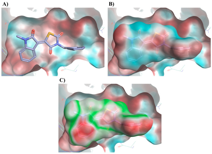
Figure 10.
Electrostatic potential (ESP) of the binding pocket (A) and of compound 20 (B). Red colour indicates a positive ESP region and blue colour negative ESP region. 20 places its large negative electrostatic area (blue) in correspondence of the positive portion of the protein (red). Protein compound 20 electrostatic complementarity (C). Green colour indicates complementarity between protein and ligand, while red indicates an electrostatic clash. Binding mode for compound 20 obtained after MD simulation.

