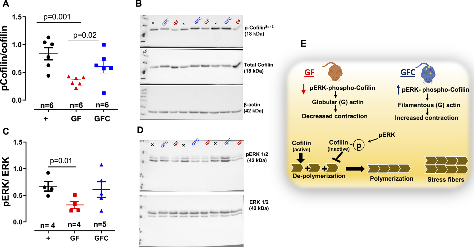
Figure 5.

Densitometric analysis and representative images of immunoblots from total and phospho cofilin (A and B) and ERK1/2 (C and D) in vascular smooth muscle cells (VSMCs) from male 7-week-old Sprague Dawley [(SD, positive control (+)] rats, SD rats that were germ-free (GF) or germ-free conventionalized (GFC). Illustration (E) shows that acquisition of microbiota in GFC rats, ERK 1/2 phosphorylates and inactivates the actin-depolymerizing protein cofilin leading to a stabilization of actin stress fibers to maintain vascular tone. Number of animals and p values are indicated in the graphs. Data are presented as mean ± SEM. One-way ANOVA or Student’s t test.
