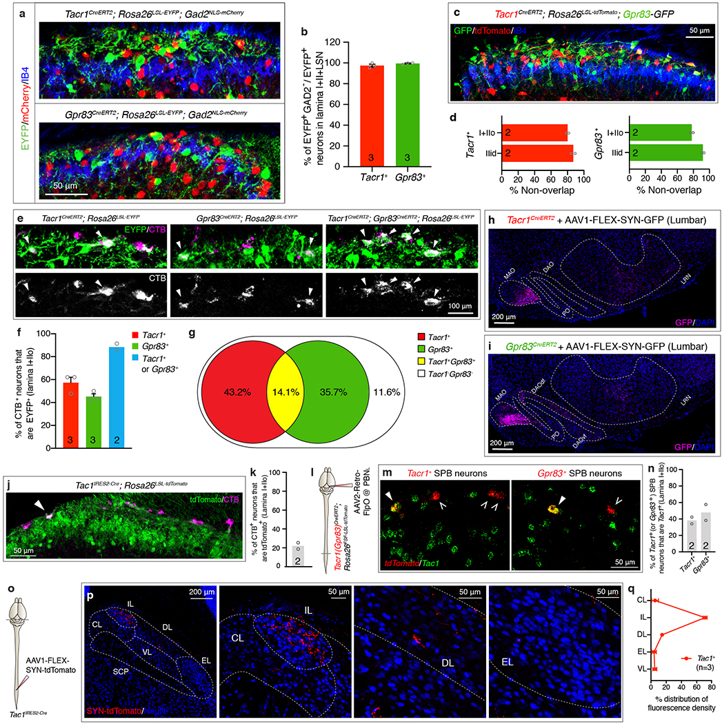
Extended Data Figure 2. Comparative analysis of the Gpr83+, Tacr1+, and Tac1+ SPB populations.

a, Distribution of EYFP-expressing Tacr1+ (top) or Gpr83+ (bottom) spinal neurons and mCherry-expressing Gad2+ neurons in the superficial lamina of the spinal cord dorsal horn. b, Quantification of % of Gad2-negative neurons in EYFP+ neurons. 97.5 ± 1.4 % of Tacr1+ neurons and 99.5 ± 0.5 % of Gpr83+ neurons were Gad2-negative. c, Distribution of tdTomato-expressing Tacr1+ neurons and GFP-expressing Gpr83+ neurons in the spinal cord dorsal horn. d, Quantification of co-expression of tdTomato and GFP. 80.2 ± 1.5% and 87.0 ± 2.5% of tdTomato-expressing Tacr1+ neurons are not positive for GFP expression in lamina I+IIo and lamina IIid, respectively. Conversely, 78.0 ± 1.8% and 92.0 ± 1.4% of GFP-expressing Gpr83+ neurons are not positive for tdTomato expression in lamina I+IIo and lamina IIid, respectively. e, Distribution of EYFP-expressing Tacr1+ neurons, Gpr83+ neurons, or both in the superficial lamina of the spinal cord dorsal horn. The SPB neurons were retrogradely labeled with CTB injected into the PBNL. Arrowheads, CTB and EYFP double-positive neurons. f, Quantification for % of Tacr1+ SPB neurons, Gpr83+ SPB neurons, and either Tacr1+ or Gpr83+ SPB neurons. g, % of Tacr1+, Gpr83+, Tacr1+ Gpr83+, and Tacr1− Gpr83− SPB neurons calculated from experiments in e, f. h, i, Coronal sections of the ventral brain stem of Tacr1CreERT2 (f) or Gpr83CreERT2 (h) mice whose lumbar spinal cords were injected with AAV1-FLEX-Synaptophysin-GFP viruses. MAO, medial accessory olivary nucleus; DAOdf, dorsal accessory olivary nucleus dorsal fold; DAOvf, dorsal accessory olivary nucleus ventral fold; PO, primary olivary nucleus. j, Distribution of tdTomato-expressing Tac1+ neurons in the superficial lamina of the spinal cord dorsal horn. The SPB neurons were retrogradely labeled with CTB injected into the PBNL. Arrowhead, CTB and tdTomato double-positive neuron. k, Quantification of % of Tac1+ SPB neurons. l, Schematic of injections of AAV2-retro-FlpO viruses into the PBNL. m, Distribution of tdTomato-expressing Tacr1+ (left) or Gpr83+ (right) SPB neurons and Tac1-expressing neurons in the spinal cord dorsal horn. tdTomato (red) and Tac1 (green) mRNA molecules were detected with gene-specific RNAscope probes. Filled arrowheads, double-positive neurons; empty arrowheads, tdTomato+ SPB neurons that do not express Tac1. n, Quantification of co-expression of tdTomato and Tac1 in lamina I+IIo. o, Schematic of lumbar injections of an AAV1-FLEX-Synaptophysin-tdTomato virus. p, Distribution of tdTomato-positive synaptic terminals of Tac1+ SPB neurons in the PBNL. q, Quantification of distribution of tdTomato-positive synaptic terminals of Tac1+ SPB neurons in the PBNL. n = number of mice (indicated in the graph). Error bars, s.e.m.
