Abstract
Background
Ischemia-reperfusion injury (IRI) is a major cause of acute kidney injury (AKI) and increases the risk of subsequently developing chronic kidney disease. Angiogenesis has been shown to play an important role in reducing renal injury after ischemia reperfusion. In this study, we investigated whether IMD could reduce renal IRI by promoting angiogenesis.
Methods
The kidneys of Wistar rats were subjected to 45 min of warm ischemia followed by 24 h of reperfusion. IMD was overexpressed in vivo using the vector pcDNA3.1-IMD transfected by an ultrasound-mediated system. The renal injury after ischemia reperfusion was assessed by detection of the serum creatinine concentration and histologic examinations of renal tissues stained by PAS and H&E. Real-time PCR and Western blotting were used to determine the mRNA and protein levels, respectively. Histological examinations were used to assess the expression of CD31, MMP2, MMP9, ET-1, VEGF and VEGFR2 in tissues.
Results
Renal function and renal histological damage were significantly ameliorated in IMD-transfected rats after ischemia reperfusion. Compared to the IRI, IMD significantly promoted angiogenesis. IMD also upregulated the protein and mRNA expression levels of VEGF and VEGFR2 and downregulated the expression level of MMP2, MMP9 and ET-1.
Conclusion
IMD could protect the kidney after renal ischemia-reperfusion injury by promoting angiogenesis and reducing the destruction of the perivascular matrix.
Keywords: intermedin, ischemia-reperfusion injury, kidney, angiogenesis, VEGF, VEGFR2, MMP2, MMP9, ET-1
Background
Ischemia-reperfusion injury (IRI) is a major cause of acute kidney injury (AKI) and increases the risk of later developing chronic kidney disease (CKD).1,2 The risk of kidney IRI mainly increases as the result of in urologic operations, such as partial nephrectomy, renal transplantation and other clinical situations.3,4 The multifactorial pathogenesis of IRI is still not clear, and no method has been clearly confirmed to be effective in preventing IRI or decreasing injury.
Intermedin/adrenomedullin-2 (IMD/ADM2), a member of the calcitonin gene-related peptide (CGRP) family, was identified in 2004 and is expressed in a variety of human tissues, such as the nervous system, heart, kidney, lung, gastrointestinal tract and thymus.5,6 Our previous studies found that it could alleviate renal IRI in vivo and in vitro. However, the mechanism is not clear.
The extracellular matrix (ECM) plays an important role in the structural integrity and morphogenesis of tissue.7 The degradation of ECM after renal IRI plays an important role in the loss of tubule function, tubule fluid back-leak and neovascularization.8
Vascular endothelial growth factor (VEGF) and its receptor (VEGFR2) play important roles in maintaining integrity and regulating ECM proliferation.
Matrix metalloproteinases (MMPs) are enzymes that belong to the subfamily of the zinc metalloprotease family, defined as matrixins, and they are responsible for the degradation of ECM.9 Ischemic AKI could induce the upregulation of MMPs that could destroy the peritubular capillaries by decomposing renal microvascular matrix.10 MMP2 and MMP9 have been shown to play important roles in the pathophysiology of renal IRI.11 Endothelin-1 (ET-1), which is mostly secreted by endothelial cells, is a potent vasoconstrictor.12 Downregulated ET-1 showed a clear function in protecting the kidney after ischemia/reperfusion injury by reducing the vasoconstriction effect.13 Our previous studies indicated that IMD and microvessel density were upregulated after renal IRI. Therefore, we mainly focused on determining whether IMD protects against renal IRI by inhibiting the expression of MMP2, MMP9, and ET-1 and increasing neovascularization.
Materials and Methods
Ultrasound-Mediated Gene Delivery into the Kidney
The eukaryotic expression plasmid pcDNA3.1-IMD containing the full-length cDNA sequence of rat IMD was created according to the methods described in our previous study.14 After excising the right kidney, the pcDNA3.1-IMD plasmid or control empty vector was transfected into the left kidney of male Wistar rats via the renal artery via an ultrasound-mediated system, as described in our previous study.14 In brief, SonoVue (sulfur hexafluoride, an echocardiographic contrast microbubble; BRACCO Imaging B.V.) was dissolved with 0.9% sodium chloride to a final volume of 5 mL (45 μg/mL). The pcDNA3.1-IMD plasmid or control empty plasmid was mixed with the above mixture at 1:1 vol/vol. The mixed solution containing 50 μg of the designated plasmid was injected into the left renal artery, and the renal artery and vein were temporarily blocked (<5 min). The left kidney was irradiated by continuous-wave ultrasound, 0.95-MHz, 5% power output for a total of 60 s with 30 s intervals. Four rats were selected and killed after 7 days to detect the transfection rate by real-time PCR and Western blot analyses.
Animals and Experimental Design
Male Wistar rats (180–200 g) were acquired from the Experimental Animal Center of Shanxi Medical University (Taiyuan, China) and maintained in a specific pathogen-free environment in our facility. All animals were fed standard chow and had free access to water. All animal experiments were performed in a humane manner and in accordance with the Institutional Animal Care Instructions. This study was conducted under experimental protocols approved by the Ethics Committee for Animals of Shanxi Medical University.
The rats were randomly divided into the following four groups: Sham, IRI, IRI + IMD and IRI + empty plasmid (n=20 in each group). The rats in the IRI+IMD and IRI + empty plasmid groups were transfected by ultrasound as described above 1 week before the induction of renal IRI. The renal IRI model was created by clamping the left renal artery with a nontraumatic vascular clamp for 45 min 1 week after the removal of the right kidney.14 Sham rats underwent similar operative procedures without occlusion of the renal artery. Experimental rats were sacrificed 24 hours after reperfusion. Blood and the left kidney were harvested for further analysis.
Reverse Transcription-Polymerase Chain Reaction and Real-Time PCR
The reverse transcription-polymerase chain reaction (RT-PCR) method was described in our previous work.15 Real-time PCR amplification was performed using the SYBR Green I system the Stratagene M3000 Sequence Detection System (Stratagene). The specific primers were used were as follows: IMD (sense: 5ʹ-CCTCACTTCGGCCTGTAGTT-3ʹ and antisense: 5ʹ-ACCCACCTCAGCCATAACTT-3ʹ), CD31 (sense: 5ʹ-CAGAGCCAGCATTGTGACCAGTC-3ʹ and antisense: 5ʹ-CAAGGCGGCAATGACCACTCC-3ʹ), MMP-2 (sense: 5ʹ-TAGTGATGGTTCCCCTCCTC-3ʹ and antisense: 5ʹ-TACTTGTTTGCCATTCCCA-3ʹ), VEGF (sense: 5ʹ-GAGGAAAGGGAAAGGGTCAA-3ʹ and antisense: 5ʹ-CGCGAGTCTGTGTTTTTGG −3ʹ), VEGFR2 (sense 5ʹ-GAATGCGGGCTCCTGACTAC-3ʹ and antisense: 5ʹ-GCACACTTCCTCTTCCTCCATAC-3ʹ), MMP9 (sense: 5ʹ-GGAAGATGCTGCTGTTCA-3ʹ and antisense: 5ʹ-CCACCTGGTTCAACTCAC-3ʹ), ET-1 (sense: 5ʹ-TTTTGAAGACCGCGCTGAG-3ʹ and antisense: 5ʹ-GGTTGCTCTGATCGCCTCTG-3ʹ), and β-actin (sense: 5ʹ-GGAAATCGTGCGTGACATTAAG-3ʹ and antisense: 5ʹ- GGACTCGTCATACTCCTGCTTG −3ʹ).
Western Blot Analysis
Western blot analysis was performed as previously described.15 The primary antibody, rabbit monoclonal anti-rat IMD antibody (Phoenix Biotech, Beijing, P.R. China), was added at a dilution of 1:3000. Then, the fluorescein-linked secondary antibody (Santa Cruz Biotechnology, Santa Cruz, CA) was added at a dilution of 1:3000. The specific bands were visualized by fluorography using an enhanced chemiluminescence kit (Pierce, Rockford, USA). The relative density was quantified using the Quantity One analysis system (Bio-Rad, California, USA).
Renal Function
Renal function was analyzed by measuring serum creatinine levels with an autoanalyzer (Beckman Instruments, Palo Alto, Calif., USA). The experiment was repeated at least three times.
Histological Examinations
After fixation with 10% paraformaldehyde, paraffin-embedded transverse kidney slices were sectioned at a thickness of 3 μm and stained with hematoxylin-eosin (H&E) and Periodic Acid-Schiff (PAS) stains. Renal tissue damage was evaluated using the scoring system described by Hussein Sheashaa.16 The scoring system included scoring of active injury, regenerative changes and chronic changes. In brief, the active injury scoring was mainly according to the degree of tubular injury; necrotic tubules were given a score of 1, 2, 3 or 4, corresponding to the presence of 1‑3, 4‑5, 6‑10 and >10 necrotic tubules. The scoring of regenerative changes involved counting the mitotic figures as the number/10 high power fields (HPF) and giving a score of 1, 2 or 3, corresponding to 1‑2, 3‑5 and >5 mitotic figures per 10 HPFs. Solid interstitial sheets of cells were given a score of 1, 2 or 3, corresponding to 1‑2, 3‑5 and >5 solid interstitial sheets of cells per HPF. Solid tubules were scored as 1, 2 or 3 corresponding to 1‑2, 3‑5 and >5 solid tubules per HPF. The scoring of chronic changes scored interstitial fibrosis as 1, 2, 3 or 4 corresponding to a fibrosed interstitium content of <25, 25‑50, 50‑75 and >75%, respectively. Tubular atrophy was scored as 1, 2 or 3, corresponding to 1‑5, 6‑10 and 7‑10 atrophied tubules per HPF.
Immunohistochemistry
The following primary antibodies were utilized: (1) goat polyclonal anti-rat CD31 antibody (R&D Systems, American), dilution 1:250; (2) rabbit polyclonal anti-rat VEGF and VEGFR2 antibody (BiossAntibodies, Beijing, P.R. China), dilution 1:250; (3) rabbit polyclonal anti-rat MMP2 and MMP9 antibody (Bioss Antibodies, Beijing, P.R. China), dilution 1:250, (4) rabbit polyclonal anti-rat ET-1 antibody (Bioss Antibodies, Beijing, P.R. China), dilution 1:200. The goat IgG HRP-conjugated antibody and rabbit IgG HRP-conjugated antibody were purchased from R&D. Sections were finally stained with 3ʹ-diaminobenzidine (DAB). All steps were performed strictly according to the manufacturer’s instructions. Images were acquired using an Olympus BX51 clinical microscope, DP70 digital camera and software (Olympus). The images were analyzed using Image-Pro Plus (Media Cybernetics). Microvessel density (MVD) was measured in terms of the CD31-positive endothelial areas. Every positive area was calculated as one microvessel.18 The expression of VEGF, VEGFR2, MMP2, MMP9 and ET-1 was measured according to the immunohistochemical mean density= IOD sum/area in three randomly selected microscopic fields for each slide.
Statistical Analysis
The results are expressed as the means ± the SDs. Significant differences between groups were assessed by one-way ANOVA with the Student–Newman–Keuls post hoc test, and differences between groups were tested using the Mann–Whitney U-test for continuous variables. P values <0.05 were considered statistically significant.
Results
Efficiency of Gene Transfection
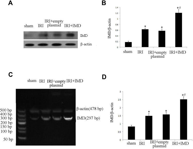
We used both semiquantitative RT-PCR and Western blot analysis to determine the efficiency of ultrasound microbubble-mediated gene transfection in rat kidneys. The results in Figure 1 show that, compared to the sham, IRI and IRI+empty plasmid groups, the IRI+IMD group had a significantly higher level of expression of IMD. The results indicated that the pcDNA3.1-IMD plasmid was successfully transfected into the rat kidneys.
Figure 1.
The transfection efficiency of IMD by ultrasound-mediated gene delivery into the kidneys. (A) The IMD protein expression measured by Western blot in rat kidneys. (B) Quantitative analysis of IMD by Western blots in rats. (C) The IMD mRNA expression measured by RT-PCR in rat kidneys. (D) The densitometric quantifications of band intensities from RT-PCR for IMD/β-actin in rat kidneys. Data in bar graphs are the means ± SD, n = 20. *P < 0.05 vs sham group; #P < 0.05 vs IRI group, respectively.
Histological and Renal Function Assessment
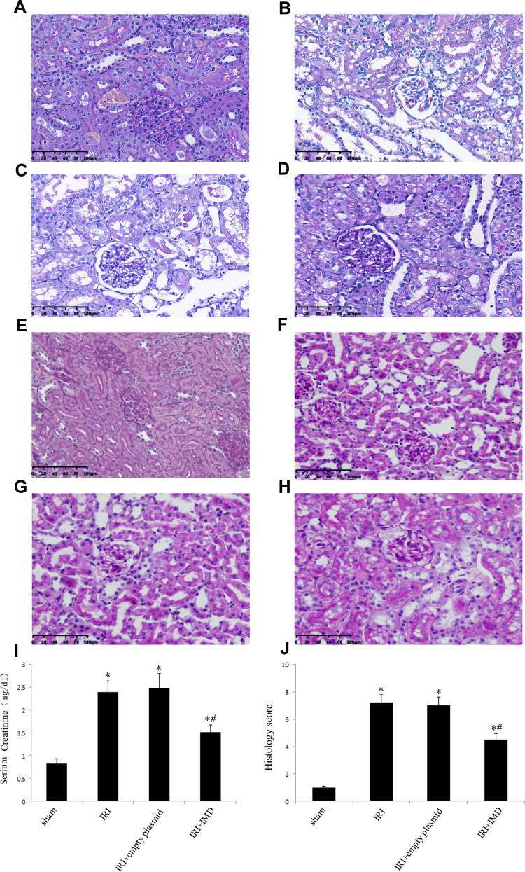
As shown in Figure 2, compared to the Sham group, the IRI (2.39±0.21 mg/dl), IRI+empty plasmid (2.46±0.26 mg/dl), and IRI+IMD (1.48±0.16 mg/dl) groups had significantly higher levels of serum creatinine after renal ischemia reperfusion (P<0.05). The level of serum creatinine in the IRI+IMD group was significantly lower than those of the IRI and IRI+empty plasmid groups (P<0.05). The level of serum creatinine showed no difference between the IRI and IRI+empty plasmid groups (P>0.05). Histological examinations (HE and PAS staining) results indicated that renal ischemia reperfusion-induced significant changes in rat kidneys compared to the Sham group, such as a loss of brush-border membranes, tubular dilatation, flattened tubular epithelia, cast formation, luminal debris, and interstitial infiltration. However, in the group of rats overexpressing IMD, these pathological renal abnormalities were clearly attenuated. Therefore, we may conclude that the overexpression of IMD improves renal function and renal pathological changes in rats.
Figure 2.
Histological and renal function assessments. (A–D) Renal pathomorphological changes after renal ischemia-reperfusion injury (IRI) shown by H&E staining, original magnification, ×400. (A) Sham groups. (B) IRI groups. (C) IRI+empty plasmid groups. (D) IRI+IMD groups. (E–H) Renal pathomorphological changes after renal ischemia-reperfusion injury (IRI) shown by PAS staining, original magnification, ×400. (E) Sham groups. (F) IRI groups. (G) IRI+empty plasmid groups. (H) IRI+IMD groups. (I) Serum creatinine concentration. (J) Semiquantitation of the morphological changes by the histological grading system. Data in bar graphs are the means ± SD, n = 20. *P < 0.05 vs sham group; #P < 0.05 vs IRI group.
IMD Increased the CD31+ MVD Level in Kidney After Ischemia Reperfusion
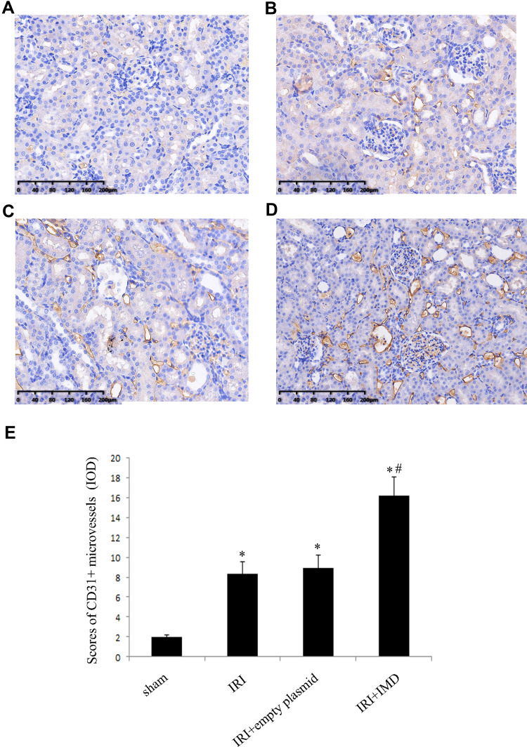
To determine whether IMD could induce early angiogenesis in the kidney after IRI, we used CD31-labeled sections to examine the angiogenic state. As shown in Figure 3, an increased number of CD31+ microvessels, which are suggestive of active angiogenesis, were widespread in renal tissues that underwent ischemia reperfusion compared to the Sham group (P<0.05). Moreover, we found the group with over expression of IMD (IRI+IMD group) had a greater number of microvessels than the IRI and IRI+empty plasmid groups (P<0.05). We may conclude that IMD could increase renal angiogenesis after ischemia reperfusion.
Figure 3.
Evaluation of CD31-labelled microvessel density in kidneys. (A–D) CD31-labelled microvessels in kidney from various groups, original magnification, ×400. (A) Sham groups. (B) IRI groups. (C) IRI+empty plasmid groups. (D) IRI+IMD groups. Each positive area was calculated as one microvessel. (E) Scores of CD31+ MVD levels. The results are expressed as the mean ± SD (n = 20 in each group). *P < 0.05 vs sham group; #P < 0.05 vs IRI group.
IMD Upregulates the Factors Promoting Angiogenesis
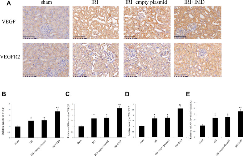
VEGF is generally thought to be the key factor in neovascularization. Therefore, we tested the protein and mRNA expression levels of VEGF and its receptor VEGFR2 among the groups. As shown in Figure 4, compared to the Sham group, the VEGF and VEGFR2 protein and mRNA expression levels were lower than those in all the rats subjected to renal ischemia reperfusion (P<0.05). Compared to the IRI group, the group with IMD intervention (IRI+IMD group) showed higher expression levels of VEGF and VEGFR2 (P<0.05). Therefore, we may conclude that IMD could promote neovascularization by upregulating the expression of VEGF and VEGFR2 in the kidney after ischemia reperfusion.
Figure 4.
The expression of VEGF and VEGFR2 in rat kidneys. (A) Localization of VEGF and VEGFR2 expression in kidneys, immunohistochemistry, Original magnification, ×400. (B) The relative density of VEGF, was quantified as the fold expression change relative to the control rats. (C) The mRNA expression levels of VEGF were determined by real-time PCR. (D) The relative density of VEGFR2, were quantified as the fold expression change relative to the control rats. (E) The mRNA expression levels of VEGFR2 were determined by real time-PCR. Data in bar graphs are the means ± SD, n = 20. *P < 0.05 vs sham group; #P < 0.05 vs IRI group.
IMD Downregulates the Micrangium Injury Factors
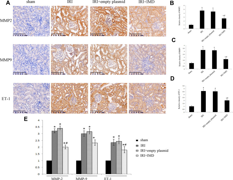
MMP2, MMP9 and ET-1 have been shown to play important roles in renal angiogenesis after ischemia reperfusion. We tested differences in the mRNA and protein expression levels of MMP2, MMP9 and ET-1 among groups. As shown in Figure 5, compared to the Sham group, MMP2, MMP9 and ET-1 had higher protein and mRNA expression levels in all rats subjected to renal ischemia reperfusion (P<0.05). However, compared to the IRI group, the IMD group had lower expression levels (P<0.05). The results indicated that the overexpression of IMD could downregulate MMP2, MMP9 and ET-1 expression.
Figure 5.
Expression of MMP2, MMP9 and ET-1 in rat kidneys. (A) The expression of MMP2, MMP9 and ET-1 in kidneys tested by immunohistochemistry, Original magnification ×400. (B–D) The relative density of MMP2, MMP9 and ET-1 expression levels in kidneys, was quantified as the fold expression change relative to the control rats. (E) The relative mRNA expression levels of MMP2, MMP9 and ET-1, determined by real-time PCR. Data in bar graphs are the means ± SD, n = 20. *P < 0.05 vs sham group; #P < 0.05 vs IRI group.
Discussion
Intermedin, a member of the calcitonin gene-related peptide (CGRP) family, has been shown to participate in the pathophysiological processes of many diseases.19 The nephron, the basic structural and functional unit of the kidney, is composed of the glomerulus and the attached tubule. Our previous research has demonstrated that IMD can clearly alleviate renal IRI in multiple ways, including reducing oxidative stress and endoplasmic reticulum stress, promoting renal tubular cell proliferation.14,15,20
The early-stage damage of capillary endothelial cells after reperfusion has been shown, and this damage may promote inflammation and procoagulant activity, thereby contributing to the reduction in microvasculature density. These results may play critical roles in the progression of chronic kidney disease following the initial recovery from ischemia/reperfusion-induced AKI.21 IMD has been shown to have antiapoptotic activity in human endothelial cells and to exert a clear pro-angiogenic effect both in vitro and in vivo.22,23 IMD has also been shown to improve the function of blood-rich organs, such as the heart, pulmonary system and brain, by promoting angiogenesis.19,24 As the kidney is a blood-rich organ, we conjectured that IMD could show angiogenic auxo-action in the kidney after IRI. In our research, we observed that the overexpression of IMD in rat kidneys could clearly mitigate the degree of renal pathological injury and improve renal function after IRI. These results were consistent with those of our previous reports. Our conjecture confirmed that the level of angiogenesis in the rat kidneys overexpressing IMD was significantly increased after IRI.
The most commonly reported adaptive response to ischemia is the stabilization of the transcription factor Hypoxia-Inducible Factor-1α (HIF-1α), which could regulate hundreds of genes and influence pathophysiological processes such as glucose uptake and metabolism, angiogenesis, inflammatory cell chemotaxis, cell proliferation and survival, and extracellular matrix formation and turnover.25 VEGF is the most well-studied HIF-1α-activated growth factor; it is known to act on microcirculation through various mechanisms and can stimulate endothelial cell proliferation and differentiation, increase vascular permeability, and mediate endothelium-dependent vasodilation. The existing studies suggest that the VEGF-induced endothelial cell physiological or pathological changes are mainly mediated by VEGFR-2 and that VEGF-VEGFR2 is an important signaling pathway in angiogenesis.26 Although the IMD expression level has been shown to be increased under anaerobic conditions, the same results have not been proven in a rat model of renal ischemia reperfusion.27 In this study, our results correspond to those of previous studies, showing that the expression levels of VEGF and VEGFR2 in rats subjected to renal ischemia reperfusion were lower than those in the sham group. However, compared to the IRI group, the IMD group had significantly higher expression levels of VEGF and VEGFR2, although they were still lower than those in the sham group.
The low expression levels of VEGF and VEGFR2 following IR could be the reason for the loss of renal vasculature. However, this result is puzzling because hypoxia following ischemia reperfusion may be expected to stimulate the expression of proangiogenic molecules, particularly VEGF, in a HIF1α-dependent fashion.28 Some scholars thought the overexpression of anti-angiogenesis factors such as endostatin, PAI-I, MMPs, ADAMTS1 and angiostatin may lead to the downregulation of VEGF and the lack of angiogenesis.29,30
MMPs are a family of zinc-dependent proteases responsible for extracellular matrix turnover as well as the degradation of bioactive proteins.31 MMP2 and MMP9 have been shown to play important roles in the pathophysiology of ischemia/reperfusion injury in the kidney, and the overexpression of MMP2 and MMP9 after ischemia/reperfusion could decrease the microvascular density by breaking down the perivascular matrix and increasing microvascular leakage.32 Additionally, ET-1 infusion in the perfused kidney induces the reduction of renal blood flow and glomerular filtration through its vasoconstriction effect.33 The ischemia-induced increase in oxidative stress provokes endothelial dysfunction, which is mediated by ET-1-dependent activation, and the increase in oxidative stress by upregulated ET-1 could directly injure the neovasculature.13,34
Angiogenesis and vessel repair after IRI are complex processes that may last for weeks even months after IRI. However, as in this research, all the samples were collected 24 hours after ischemia/reperfusion, during the acute phase of IRI, and the results of CD31-positive microvascular density between groups were not completely consistent with the trends in MMP2, ET-1 and MMP9 expression levels. In the IMD-overexpressing rat kidneys, the expression levels of MMP2, ET-1 and MMP9 were significantly lower, and the density of CD31-positive microvasculature was higher compared to the IRI group. However, the expression levels of MMP2, ET-1 and MMP9 in rat kidneys after ischemia/reperfusion were increased compared to the levels in the sham group, and the density of CD31-positive microvasculature were higher. These results do not indicate that our results were contrary to those of previous studies, and similar results were presented in Xiangyu Zou’s study.35 These results indicate that the decrease in microvascular density induced by MMP2, ET-1 and MMP9 may be the final result of the entire injury and repair period. IMD could induce angiogenesis by reducing the expression levels of MMP2, ET-1 and MMP9 in the early stage of IRI.
Conclusion
In this study, the overexpression of IMD by ultrasound microbubble-mediated transfection of the pcDNA3.1-IMD plasmid in the rat kidney could protect the kidney after renal ischemia-reperfusion injury by promoting angiogenesis and reducing the destruction of the perivascular matrix. IMD could be a feasible method of reducing renal injury after ischemia reperfusion. However, as our research mainly focused on the early stage of renal IRI, the results still need to be demonstrated in further studies.
Acknowledgments
This work was supported by the National Nature Science Fund Project (No. 81500518, 81500529), the Applied Basic Research Programs of Shanxi Province (No. 201901D111187, 201901D111188), the Key Research and Development (R&D) Projects of Shanxi Province (201803D31163), the Doctoral Startup Research Fund of Shanxi Medical University (No. 03201302, 03201403), the Science and Technology Innovation Fund of Shanxi Medical University (No. 01201403), and the 331 Early Career Researcher Grant fund of Basic Medical College, Shanxi Medical University (No. 201406).
Author Contributions
All authors contributed to data analysis, drafting or revising the article, have agreed on the journal to which the article will be submitted, gave final approval of the version to be published, and agree to be accountable for all aspects of the work.
Disclosure
The authors report no conflicts of interest in this work.
References
- 1.Hoste EA, Schurgers M. Epidemiology of acute kidney injury. Contrib Nephrol. 2010;165:1–8. [DOI] [PubMed] [Google Scholar]
- 2.Bonventre JV, Yang L. Cellular pathophysiology of ischemic acute kidney injury. J Clin Invest. 2011;121(11):21–4210. doi: 10.1172/JCI45161 [DOI] [PMC free article] [PubMed] [Google Scholar]
- 3.Mehta RL, Pascual MT, Soroko S, et al. Spectrum of acute renal failure in the intensive care unit: the PICARD experience. Kidney Int. 2004;66(4):1613–1621. doi: 10.1111/j.1523-1755.2004.00927.x [DOI] [PubMed] [Google Scholar]
- 4.White LE, Hassoun HT. Inflammatory mechanisms of organ crosstalk during ischemic acute kidney injury. Int J Nephrol. 2011;2012:505197. [DOI] [PMC free article] [PubMed] [Google Scholar]
- 5.Roh J, Chang CL, Bhalla A, Klein C, Hsu SY. Intermedin is a calcitonin/calcitonin gene-related peptide family peptide acting through the calcitonin receptor-like receptor/receptor activity-modifying protein receptor complexes. J Bio Chem. 2004;279(8):7264–7274. [DOI] [PubMed] [Google Scholar]
- 6.Takei Y, Hyodo S, Katafuchi T, Minamino N. Novel fish-derived adrenomedullin in mammals: structure and possible function. Peptides. 2004;25(10):1643–1656. doi: 10.1016/j.peptides.2004.06.026 [DOI] [PubMed] [Google Scholar]
- 7.Bosman FT, Stamenkovic I. Functional structure and composition of the extracellular matrix. J Pathol. 2003;200(4):8–423. doi: 10.1002/path.1437 [DOI] [PubMed] [Google Scholar]
- 8.Dejonckheere E, Vandenbroucke RE, Libert C. Matrix metalloproteinases as drug targets in ischemia/reperfusion injury. Drug Discov Today. 2011;16(17):78–762. [DOI] [PubMed] [Google Scholar]
- 9.Cavdar Z, Ozbal S, Celik A, et al. The effects of alpha-lipoic acid on MMP-2 and MMP-9 activities in a rat renal ischemia and re-perfusion model. Biotech Histochem. 2014;89(4):304–314. [DOI] [PubMed] [Google Scholar]
- 10.Kunugi S, Shimizu A, Kuwahara N, et al. Inhibition of matrix metalloproteinases reduces ischemia-reperfusion acute kidney injury. Lab Invest. 2011;91(2):80–170. doi: 10.1038/labinvest.2010.174 [DOI] [PubMed] [Google Scholar]
- 11.Kaneko T, Shimizu A, Mii A, et al. Role of matrix metalloproteinase-2 in recovery after tubular damage in acute kidney injury in mice. S Karger AG. 2012;122(1):23–35. [DOI] [PubMed] [Google Scholar]
- 12.Yanagisawa M, Kurihara H, Kimura S, et al. A novel potent vasoconstrictor peptide produced by vascular endothelial cells. Nature. 1988;332(6163):411–415. doi: 10.1038/332411a0 [DOI] [PubMed] [Google Scholar]
- 13.Arfian N, Emoto N, Vignon-Zellweger N, Nakayama K, Yagi K, Hirata K-I. ET-1 deletion from endothelial cells protects the kidney during the extension phase of ischemia/reperfusion injury. Biochem Biophys Res Commun. 2012;425(2):443–449. doi: 10.1016/j.bbrc.2012.07.121 [DOI] [PubMed] [Google Scholar]
- 14.Qiao X, Li RS, Li H, et al. Intermedin protects against renal ischemia-reperfusion injury by inhibition of oxidative stress. Am J Physiol. 2013;304(1):F112–F119. [DOI] [PubMed] [Google Scholar]
- 15.Wang Y, Tian J, Qiao X, et al. Intermedin protects against renal ischemia-reperfusion injury by inhibiting endoplasmic reticulum stress. BioMed Central. 2015;16(1):169. [DOI] [PMC free article] [PubMed] [Google Scholar]
- 16.Sheashaa H, Lotfy A, Elhusseini F, et al. Protective effect of adipose-derived mesenchymal stem cells against acute kidney injury induced by ischemia-reperfusion in Sprague-Dawley rats. Exp Ther Med. 2016;11(5):1573–1580. [DOI] [PMC free article] [PubMed] [Google Scholar]
- 17.Tian J, Wang Y, Zhou X, et al. Rapamycin slows IgA nephropathy progression in the rat. Am J Nephrol. 2014;39(3):29–218. doi: 10.1159/000358844 [DOI] [PubMed] [Google Scholar]
- 18.Saito K, Matsuo Y, Imafuji H, et al. Xanthohumol inhibits angiogenesis by suppressing nuclear factor-κB activation in pancreatic cancer. Cancer Sci. 2018;109(1):132–140. doi: 10.1111/cas.13441 [DOI] [PMC free article] [PubMed] [Google Scholar]
- 19.Holmes D, Campbell M, Harbinson M, Bell D. Protective effects of intermedin on cardiovascular, pulmonary and renal diseases: comparison with adrenomedullin and CGRP. Curr Protein Pept Sci. 2013;14(4):294–329. doi: 10.2174/13892037113149990049 [DOI] [PubMed] [Google Scholar]
- 20.Wang Y, Li R, Qiao X, et al. Intermedin/adrenomedullin 2 protects against tubular cell hypoxia‐reoxygenation injury in vitro by promoting cell proliferation and upregulating cyclin D 1 expression. Nephrology. 2013;18(9):623–632. doi: 10.1111/nep.12114 [DOI] [PubMed] [Google Scholar]
- 21.Basile D-P. The endothelial cell in ischemic acute kidney injury: implications for acute and chronic function. Kidney Int. 2007;72(2):151–156. doi: 10.1038/sj.ki.5002312 [DOI] [PubMed] [Google Scholar]
- 22.Pearson LJ, Yandle TG, Nicholls MG, Evans JJ. Intermedin (adrenomedullin-2): a potential protective role in human aortic endothelial cells. Cell Physiol Biochem. 2009;23(1):97–108. doi: 10.1159/000204098 [DOI] [PubMed] [Google Scholar]
- 23.Smith RS Jr, Gao L, Bledsoe G, Chao L, Chao J. Intermedin is a new angiogenic growth factor. Am J Physiol Heart Circ Physiol. 2009;297(3):0–7. doi: 10.1152/ajpheart.00404.2009 [DOI] [PMC free article] [PubMed] [Google Scholar]
- 24.Takei Y, Inoue K, Ogoshi M, Kawahara T, Bannai H, Miyano S. Identification of novel adrenomedullin in mammals: a potent cardiovascular and renal regulator. FEBS Lett. 2004;556(1):53–58. doi: 10.1016/S0014-5793(03)01368-1 [DOI] [PubMed] [Google Scholar]
- 25.Kaelin-William G. The von Hippel-Lindau tumour suppressor protein: O2 sensing and cancer. Nat Rev Cancer. 2008;8(11):73–865. [DOI] [PubMed] [Google Scholar]
- 26.Cai Y, Xu MJ, Teng X, et al. Intermedin inhibits vascular calcification by increasing the level of matrix gamma-carboxyglutamic acid protein. Cardiovasc Res. 2009;85(4):73–864. [DOI] [PubMed] [Google Scholar]
- 27.Pallet N, Thervet E, Timsit M-O. Angiogenic response following renal ischemia reperfusion injury: new players. Prog Urol. 2014;24:S20–S25. doi: 10.1016/S1166-7087(14)70059-4 [DOI] [PubMed] [Google Scholar]
- 28.Eltzschig HK, Eckle T. Ischemia and reperfusion–from mechanism to translation. Nat Med. 2011;17(11):401–1391. doi: 10.1038/nm.2507 [DOI] [PMC free article] [PubMed] [Google Scholar]
- 29.Basile DP, Fredrich K, Weihrauch D, Hattan N, Chilian WM. Angiostatin and matrix metalloprotease expression following ischemic acute renal failure. Am J Physiol Renal Physiol. 2004;286(5):0–902. doi: 10.1152/ajprenal.00328.2003 [DOI] [PubMed] [Google Scholar]
- 30.Basile DP, Fredrich K, Chelladurai B, Leonard EC, Parrish AR. Renal ischemia reperfusion inhibits VEGF expression and induces ADAMTS-1, a novel VEGF inhibitor. Am J Physiol Renal Physiol. 2008;294(4):0–36. doi: 10.1152/ajprenal.00596.2007 [DOI] [PubMed] [Google Scholar]
- 31.Nagase H, Visse R, Murphy G. Structure and function of matrix metalloproteinases and TIMPs. Cardiovasc Res. 2006;69(3):562–573. doi: 10.1016/j.cardiores.2005.12.002 [DOI] [PubMed] [Google Scholar]
- 32.Sutton T-A. Minocycline reduces renal microvascular leakage in a rat model of ischemic renal injury. Am J Physiol Renal Physiol. 2004;288(1). [DOI] [PubMed] [Google Scholar]
- 33.Firth JD, Raine AEG, Ratcliffe PJ, Ledingham JGG. Endothelin: an important factor in acute renal failure? Lancet. 1988;332(8621):1179–1182. [DOI] [PubMed] [Google Scholar]
- 34.Maczewski M, Beresewicz A. The role of endothelin, protein Kinase C and free radicals in the mechanism of the post-ischemic endothelial dysfunction in guinea-pig hearts. J Mol Cell Cardiol. 2000;32(2):297–310. doi: 10.1006/jmcc.1999.1073 [DOI] [PubMed] [Google Scholar]
- 35.Zou X, Gu D, Xing X, et al. Human mesenchymal stromal cell-derived extracellular vesicles alleviate renal ischemic reperfusion injury and enhance angiogenesis in rats. Am J Transl Res. 2016;8(10):4289–4299. [PMC free article] [PubMed] [Google Scholar]


