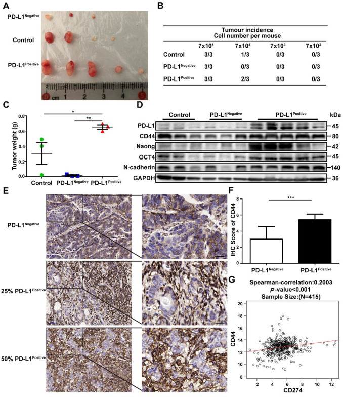
Figure 5.
The PD-L1Positive subpopulation of GC cells has higher tumor-initiation potential. SGC-7901 were treated with GCMSC-CM, and then the PD-L1Negative and PD-L1Positive cells were sorted by flow cytometry, counted, and injected subcutaneously in limiting dilution assays into BALB/c nude mice. After 20 d, the mice were sacrificed and the tumor tissues were collected. (A) Representative images of tumors from the indicated group were shown. (B) The table displays the number of mice that developed tumors. (C) Tumors Weights in mice on day 20 after injection with 7×10⁵ sorted GC cells (n=3). *, P<0.05, **, P<0.01. (D) The levels of PD-L1, CTCF, CD44, Nanog, Oct4, and N-cadherin in tumor tissues were detected by western blot. (E, F) Representative IHC staining and quantification of the CD44 levels in PD-L1Negative and PD-L1Positive tumor tissues from GC patients (scale bar, 50 μm, n=20). ***, P<0.001. (G) Correlations between PD-L1 and CD44 in GC tissues from 415 patients in TCGA data set. ***, P<0.001.

