Abstract
Peripheral ameloblastoma (PA) is a prototype form of extraosseous odontogenic tumor. As knowledge of PA has accumulated on the basis of more than 200 cases reported worldwide over a 60-year timeframe, it is important to comprehend the historical evolution of this entity. In 2018, we summarized the American history of PA, stressing the important early strides made by Bloodgood in 1904 with his many original observations of the “epulis form of ameloblastoma”. During the preparation of our previous report, we were able to find several earlier and interesting descriptions in the literature. This review covers the early history of PA since the nineteenth century, chronologically focusing on meritorious articles published in the United States and Europe.
Keywords: Alveolar border, Basal cell carcinoma, Endothelioma, Epithelial epulis, Gingiva, Peripheral ameloblastoma, Superficial ameloblastoma
Peripheral Ameloblastoma (PA) in the American Literature I (1904–1943)
Proposal and Acceptance of the Previous Terminology
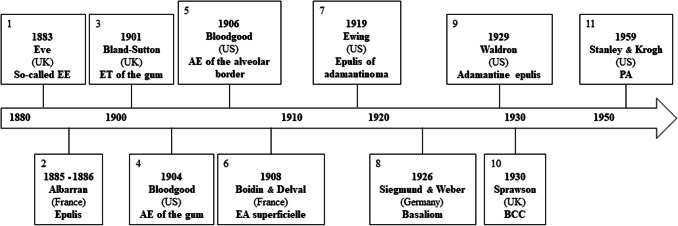
Following the original descriptions of “ameloblastoma of the gingiva” or the “ameloblastoma of the alveolar border” by Bloodgood in 1904 and 1906 [1, 2] (Fig. 1), the concept was perpetuated among clinicians [3–14] and thereafter facilitated effective management of patients with the lesion. In the field of pathology, Ewing [15] introduced the terms “superficial ameloblastoma” and “epulis of ameloblastoma” in 1919 (Fig. 1), and soon after, Carnathan [16] adopted the latter terminology to describe PA of the left mandible in a 64-year-old woman [9, 11].
Fig. 1.
Terminological evolution of PA from 1883 to 1959. So-called “EE” in the British literature includes miscellaneous lesions. Many cases of AE of the alveolar border reported in the American literature were not true PAs. So-called “Basaliom” in the German literature was heterogenous in nature. PA peripheral ameloblastoma, UK United Kingdom, EE epithelial epulis, ET endothelioma, US United States, AE adamantine epithelioma, EA épithélioma adamantin, BCC basal-celled carcinoma
In the field of dentistry, Moorehead and Dewey [17] stated in the chapter of cystoma (synonym: ameloblastoma) of their 1925 book that “occasionally the tumors occur as polypus-like growths on the alveolar border and have the appearance of the so-called epulis”. This view pervaded the dental literature for years [18–20]. From 1929 to 1935, Waldron [21–23] repeatedly referred to superficial ameloblastoma springing from the epithelial border and also coined the term “adamantine epulis” for a fibrous tumor with conspicuous cords of the enamel organ epithelium (Fig. 1). Without giving a specific name, Robinson [24, 25] published a photomicrograph of PA (case 3, a nodule on the buccal gingiva of the right mandibular second molar in a 49-year-old man) in 1937, adding a brief comment that it had a structure suggestive of basal cell carcinoma (BCC).
PA in the American Literature II (1956–1977)
Birth and Popularization of the Current Terminology
Apart from the impact of Bloodgood’s contributions [1, 2, 26–28], a clinical textbook published in 1956 proposed the catchy term “gingival type” for ameloblastoma of alveolar crest origin [29]. It is now 60 years since Stanley and Krogh [30] wrote the best-known English-language case report in 1959 using the term PA for the first time (Fig. 1). They applied Bernier’s classification of giant cell reparative granuloma (central versus peripheral) to ameloblastoma [31]. Thereafter, the terms “alveolar border ameloblastoma” and “epulis-type ameloblastoma” fell out of favor. In 1977, Gardner [32], a leading authority on odontogenic tumor pathology working in Canada at the time, conducted an exhaustive study of PA, addressing nine published and seven unpublished examples and five that had been reported as BCC of the gingiva. PA has since become well known to pathologists worldwide, and there has been a steady stream of reports of either single cases or large series.
PA in the French Literature I (1885–1886)
Albarran’s Original Papers
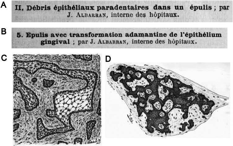
In 1885 and 1886, Albarran [33, 34] wrote two pathology reports on epulis (Fig. 1). His first article described a hazelnut-sized, bilobulated tumor located between the lateral incisor and canine of the left mandible in a 26-year-old man (Fig. 2a) [33], and the second article described a 15-year-old girl who had a hazelnut-sized, lobulated tumor surrounding the left mandibular first premolar (Fig. 2b) [34]. The young Albarran (aged 25 years at the time) consulted Malassez (professor of anatomy at the Collège de France) about the first case [33], and adding another similar example (a chicken egg-sized tumor in the mandibular left first molar area) sent by Hue, Malassez [35] in 1885 coined the term “épulies avec productions épithéliales”. Although no illustrations of either of these lesions were provided [33, 34], it is notable that the former PA was reportedly derived from Malassez’s epithelial rests of the gingival margin (débris épithélial superficiel) [9, 10, 15, 35–39], whereas the latter originated from the surface gingival epithelium. His truly remarkable insight at that time represents the oldest reference to the two different origins of PA [32].
Fig. 2.
a French title of “paradental epithelial debris in an epulis” by Albarran [33] in 1885. b French title of “epulis with ameloblastoma transformation of the gingival epithelium” by Albarran [34] in 1886. c Peripheral ameloblastoma reported by Boidin and Delval [40] in 1908. d Peripheral ameloblastoma depicted by Bland-Sutton [63] in 1901
PA in the French Literature II (1908–1950)
Early Depiction of “Épithélioma Adamantin Superficielle”
In 1908, Boidin and Delval [40] provided a figure of PA, calling it “épithélioma adamantin, variété superficielle ou gingivale” [9] (Fig. 1). They reported two cases of ameloblastoma, one of which was PA on the molar gingiva of the right mandible in a 57-year-old man. This PA is illustrated with an artistic drawing (Fig. 2c) and later discussed in detail in the epulis section of the historic book published by Malassez and Galippe [37]. Boidin and Delval [40] cited the above two reports by Albarran [33, 34] without specific comments. As far as we are aware, one of 8 cases of fibrous epulis (case III, small epulis of ten years’ duration) examined by Delater and Bercher [41] in 1924 was an unequivocal example of PA. Their diagnosis “épithélioma adamantin” was evident in two photomicrographs. Alternative names used in the literature include “épulis épithéliales” and “épulis épithélio-conjonctives” [42–44].
PA in the British Literature I (1851–1871)
What is “Fibrous Epulis with Enlarged Glands”?
In 1851, Paget [45] mentioned in the fibrous tumor chapter of his lecture “Tumours”: “Birkett tells me he has found the glands of the gum much developed in some instances of tumours thus named (later changed into epulis by Heath [46])”. Unfortunately, it is still unclear whether enlargement of “glands of the gum” meant neoplastic hyperplasia of the dental lamina rests of Serres (erroneous and dated synonym: dental or gingival glands of Serres [17, 38, 39, 47–51]) indicative of PA, or simply represented accidental involvement of adjacent glandular tissue in growing epulis. On the basis of the relevant reports [46, 52–54], we consider that such lesions may not represent examples of PA and probably fall within the spectrum of fibrous epulis with entrapped salivary glands.
PA in the British Literature II (1883–1907)
What is “Epithelial Epulis”?
In 1883, Eve [55] introduced the basic concept of PA in Britain for the first time (Fig. 1). He commented “I have met with five epulis from children and adults, composed largely of columns of epithelial cells” in his lecture on multilocular cystic tumor (synonym: ameloblastoma) of the jaw. A microscopic drawing of this type of lesion was presented in his subsequent communication [56]. At that time, the diagnostic terminology “epithelial (epitheliomatous) epulis” was in grate vogue [46, 57–61], and Heath [61] pointed out that “these epitheliomatous growths originated in the rudimentary epithelial structures discovered by Malassez”. It seems likely that several of these cases may have been a form of PA. Indeed, a brief description of PA (an epitheliomatous epulis surrounding the mandibular second molar in a 52-year-old woman) was given by Eve [62] in 1907.
PA in the British Literature III (1901–1923)
True Nature of “Endothelioma of the Gum”
In 1901, Bland-Sutton [63] illustrated PA with a fine drawing in his famous textbook “Tumours innocent and malignant”. It was interpreted as “endothelioma of the gum” (Figs. 1, 2d). At that time, he was of the opinion that many of multilocular cystic (epithelial) tumors are endotheliomas and typical lesions arise on the gingiva. His view was retained in the 6th edition in 1917 [64], but a figure of “endothelioma of the gum” was deleted from the 5th edition six years before [65]. On the other hand, Colyer [66] included this PA in the chapter of connective tissue tumor of the 3rd edition of his well-known book “Dental surgery and pathology” published in 1910. Bland-Sutton’s concept of “endothelioma of the gum” was subsequently discussed in the section of epithelial odontoma (synonym: ameloblastoma) of the 4th and 5th editions [67, 68].
PA in the British Literature IV (1921–1951)
Overlooked Papers
A photomicrographic demonstration of PA was presented in a pathological work “The morbid histology of the teeth” by Mummery [69] in 1921. This case was a large epulis on the incisor gingiva of the mandible and surgically treated by Colyer [68]. Mummery [69] designated it as “fibrous epulis has undergone conversion into an epithelioma”. Because of its unique microscopic appearances, this PA was included in the 5th edition of Colyer’s textbook [68] shortly thereafter. In his opinion, it represents a good example to help support Malassez’s notion that epulis develops in the more superficial paradental epithelial debris [35, 37].
We were also able to retrieve two other reports of PA published in 1933 [70] and 1951 [71], neither of which was covered in the previous studies [30, 32]. The first manuscript briefly mentioned a recurrent PA on the lingual gingiva of the right angle in a 40-year-old man [70], and the second described a series of 20 cases of ameloblastoma, one of which was PA (case 1, a small polyp on the outside of the left maxillary alveolus in a 78-year-old woman) [71]. The former case was summarized by Sprawson, who in 1931 wrote the 6th edition of the above book in collaboration with Colyer [72].
PA in the British Literature V (1930–1952)
Reintroduction of “Superficial Ameloblastoma”
The histologic overlap between ameloblastoma of the jaw and BCC (rodent ulcer) of the skin has been debated in Britain for a long time [70–76]. With the exclusion of Bland-Sutton’s endothelioma from the aforementioned textbook [72, 77], Sprawson [70, 72–74, 77] remarked on several occasions that ameloblastoma should be designated as BCC (Fig. 1). In this context, it is worth mentioning a symposium on the treatment of ameloblastoma held in 1952. On the basis of his 1950 study [78], McWhirter [79] summarized that BCC and ameloblastoma resembled each other in their morphology and biological behavior. He used the dated terminology “superficial ameloblastoma” [10, 15, 40], reflecting a presumptive origin from basal cells of the gingival epithelium. As the number of true cases of intraoral BCC increased [2], there is a consensus among all contemporary pathologists that it should be critically distinguished from PA.
PA in the German Literature (1918–1944)
Inclusion of PA under the Umbrella of “Basaliom”
A somewhat different approach had been employed for PA diagnosis in Germany. The literature traditionally adopted the general term “Basaliom” for all basaloid tumors irrespective of their origin, and thus PA and intraoral BCC were considered to be synonymous [80–82]. For example, the standard 1926 book used the term “adamantinomartiger Basalzellkrebs” for PA [83] (Fig. 1). In 1942, Ritzert [84] published a dissertation, “Über Basaliome in der Mundhöhle”, and two years later republished it as a two-part journal paper in which PA (case 6, a cherry-sized epulis of the left maxillary canine in a 36-year-old patient) was included under the designation “Basaliom mit Adamantinomcharakter”.
PA in the Scandinavian Literature (1935–1954)
Radiotherapy and Recurrence
In 1935, McGregor [85] appears to have been the first to describe PA in Scandinavia. This recurrent PA (case VIII, a small polyp on the alveolar border of the left posterior maxilla in a 69-year-old woman) reportedly disappeared in response to radiation therapy alone [85]. Three years later, a clinical profile of superficial ameloblastoma was provided in a supplement of the Acta Oto-laryngologica [86]. Hertz [87] added three cases of ameloblastoma that showed the clinical characteristics of epulis in 1952. As was common at the time [85, 88, 89], one of Hertz’s PA cases (case 28, an epulis of the mandible in a 30-year-old man) was initially treated by irradiation and recurred a year later [87]. Another example of recurrent PA of the left retromolar area in a 59-year-old woman was reported by Forsberg [90] in 1954.
Conclusion
Current evidence supports the contention that PA is the most common epithelial odontogenic tumor of the gingiva/alveolar mucosa, accounting for up to 10% of all ameloblastomas [91]. Consequently, almost all oral pathologists are experienced in diagnosing PA. Unfortunately, several authors chose to use the terminology “peripheral alveolar type or typical epulis” for ordinary intraosseous ameloblastomas that breached the alveolar bone, grew in the gingiva and exhibited the same clinical appearance as epulis [92, 93]. Although there are many differences in the historical evolution of PA among the old French, British, German and Scandinavian reports, it is evident that two independent French papers on epulis published by Albarran [33, 34] in 1885 and 1886 each documented the earliest cases of PA. Notably, Albarran drew attention to their origins; one appeared to arise from the epithelial rests of Serres [33] and the other from the gingival epithelium [34]. In addition to a pioneering work by Bloodgood [1, 2, 26–28], the American literature contributed to the first proposal of the current term PA [30] and also to the subsequent popularization of this entity [32].
Compliance with Ethical Standards
Conflict of interest
All authors declare that they have no conflict of interest.
Footnotes
Publisher's Note
Springer Nature remains neutral with regard to jurisdictional claims in published maps and institutional affiliations.
References
- 1.Bloodgood JC. Bone cysts. A consideration of the benign and adamantine dentigerous cysts of the jaw and benign cysts of the long pipe bones. Trans Sec Pathol Physiol Am Med Assoc 55th Ann Ses. 1904;249–66. J Am Med Assoc. 1904;43:1124–9.
- 2.Ide F, Kikuchi K, Kusama K, Muramatsu T. Dr. Bloodgood’s contribution to peripheral ameloblastoma. J Oral Maxillofac Surg. 2018;76:1602–1603. doi: 10.1016/j.joms.2018.03.045. [DOI] [PubMed] [Google Scholar]
- 3.Lewis DD. Multilocular cysts of the jaws. Surg Gynec Obstet. 1910;10:28–36. [Google Scholar]
- 4.Scudder CL, editor. Tumors of the jaws. Philadelphia: WB Saunders; 1912. p. 178.
- 5.Kanavel AB. Tumors of the face. Surg Clin Chicago. 1920;4:731–748. [Google Scholar]
- 6.Kirkham HLD. Tumors of the alveolar border of the jaws. Texas State J Med. 1921;17:351–355. [Google Scholar]
- 7.Wheeler WIC. Mouth and face, surgical affections of. Int Med Annu. 1922;40:278–282. [Google Scholar]
- 8.Warthin AS. Adamantinoma. J Cancer Res. 1924;8:157–159. [Google Scholar]
- 9.Murphy JT. Adamantine epithelioma. Radiology. 1924;3:377–387. [Google Scholar]
- 10.Carter BN. Adamantinoma of the lower jaw. Ann Surg. 1931;94:1–6. doi: 10.1097/00000658-193107000-00001. [DOI] [PMC free article] [PubMed] [Google Scholar]
- 11.Crawford RH. Adamantinoma. Trans Med Soc State NC. 1934;81:396–408. [Google Scholar]
- 12.Edwards HGF. Tumors of the mouth and jaws. New Orleans Med Surg J. 1936;88:460–466. [Google Scholar]
- 13.Haam E. Adamantinomas. New Orleans Med Surg J. 1936;88:646–648. [Google Scholar]
- 14.Thrift GN. The inter-relationship of dentistry and otolaryngology. Virginia Med Month. 1943;70:30–35. [Google Scholar]
- 15.Ewing J, editor. Neoplastic diseases. A text-book on tumors. Philadelphia: WB Saunders; 1919. pp. 686–688, 691.
- 16.Carnathan WG. A study of adamantinoma with report of a case. J Tenn State Med Assoc. 1922;14:408–411. [Google Scholar]
- 17.Moorehead FB, Dewey KW, editors. Pathology of the mouth. Philadelphia: WB Saunders; 1925. pp. 432–436, 480.
- 18.New GB. Cystic odontomas. In: Endelman J, Wagner AF, editors. General and dental pathology. With special reference to etiology and pathologic anatomy. A treatise for students and practitioners. St. Louis: CV Mosby; 1920. p. 536. [Google Scholar]
- 19.Simpson BT. Malignancy and its relation to dentistry. Dent Cos. 1927;69:815–825. [Google Scholar]
- 20.Mead SV, editor. Diseases of the mouth. 3rd ed. St. Louis: CV Mosby; 1928. p. 560.
- 21.Waldron CW. Tumors of the oral cavity. In: Bunting RW, editor. A text-book of oral pathology for students and practitioners of dentistry. Philadelphia: Lea & Febiger; 1929. pp. 466–468. [Google Scholar]
- 22.Waldron CW. Cysts of the jaws. A consideration of their etiology, pathology, clinical course, diagnosis and treatment. Ill Dent J. 1933;2:677–695. [Google Scholar]
- 23.Waldron CW. Cysts and neoplasms of the jaws. A consideration of their etiology, pathology, clinical course, diagnosis and treatment. J Can Dent Assoc. 1935;1(103–20):161–170. [Google Scholar]
- 24.Robinson HBG. Histologic study of the ameloblastoma. Arch Pathol. 1937;23:664–673. [Google Scholar]
- 25.Robinson HBG. Tumors, anomalies and cysts of dental origin. Wash Univ Dent J. 1937;4:35–45. [Google Scholar]
- 26.Bloodgood JC. Surgery of the extremities, fractures, dislocations, tumors, surgery of joints, shock, anaesthesia, and infections. Progress Med. 1907;4:135–225. [Google Scholar]
- 27.Bloodgood JC. Conservative operations on bone tumors, based on the clinical and pathologic study of the relative degree of malignancy. J Am Med Assoc. 1908;50:325–7. Am J Surg. 1908;22:91.
- 28.Bloodgood JC. Recent progress in surgical treatment of malignant growths. Trans Sec Pathol Physiol Am Med Assoc 61th Ann Ses. 1910;107–43.
- 29.Sharp GS, Bullock WK, Hazlet JW, editors. Oral cancer and tumors of the jaws. New York: McGraw-Hill; 1956. pp. 277–282. [Google Scholar]
- 30.Stanley HR, Krogh HW. Peripheral ameloblastoma. Report of a case. Oral Surg Oral Med Oral Pathol. 1959;12:760–765. doi: 10.1016/0030-4220(59)90124-0. [DOI] [PubMed] [Google Scholar]
- 31.Bernier JL, editor. The management of oral disease. A treatise on the recognition, identification, and treatment of diseases of the oral regions. St. Louis: CV Mosby; 1955. p. 689.
- 32.Gardner DG. Peripheral ameloblastoma. A study of 21 cases, including 5 reported as basal cell carcinoma of the gingiva. Cancer. 1977;39:1625–1633. doi: 10.1002/1097-0142(197704)39:4<1625::aid-cncr2820390437>3.0.co;2-v. [DOI] [PubMed] [Google Scholar]
- 33.Albarran J. Débris épithéliaux paradentaires dans un épulis. Bull la Soc Anat Paris 4th Ser. 1885;10;307–9.
- 34.Albarran J. Epulis avec transformation adamantine de l’épithélium gingival. Bull la Soc Anat Paris 4th ser. 1886;11:25–9. L’odontol. 1886;6:397–400.
- 35.Malassez L. Sur l’existence d’amas épithéliaux autour de la racine des dents chez l’homme adulte et a l’état normal (débris épithéliaux paradentaires). In: Ranvier L, Malassez L, Vignal W, Darier J, Suchard E, editors. École pratique des hautes études. Laboratoire d’histologie du Collège de France. Paris: Masson; 1885. pp. 1–121. Arch Physiol Norm Pathol 3rd ser. 1885;6:129–48, 309–40, 379–449.
- 36.Malassez L. On the existence of masses of epithelium round the roots of adult teeth in a normal state. J Br Dent Assoc. 1885;6:370–7, 430–3, 484–7.
- 37.Malassez L, Galippe V, editors. Les débris épithéliaux paradentaires. Origine, variétes, rôle physiologique et tumeurs qui en dérivent. Paris: Masson; 1910. pp. 45–112, 172–179.
- 38.Mummery JH. On the structure of the dental follicle and its relations to pathological conditions. Br Dent J new ser. 1920;41:1–60. [Google Scholar]
- 39.Hatton EH. The origin and pathologic significance of the epithelium found about the roots of teeth. Arch Pathol. 1934;17:130–4. Trans Chicago Pathol Soc. 1928–1936;13–14:141–5.
- 40.Boidin L, Delval. L’épithélioma adamantin, ses deux formes. Anatomo-cliniques profonde et superficielle. La Press Méd. 1908;16:793–5.
- 41.Delater, Bercher. L’épulis. Débuté par un granulome superficiel, de la gencive autour d’un épithélium irrité, souvent dysembryoplastique. Rev Stomatol. 1924;26:1–35.
- 42.Delater B. Comment les tumeurs des mâchoires se développent aux dépens d’une dysembryoplasie épithéliale. Contribution à l’étude de l’histogenése du cancer. Bull Assoc Fr Etud Cancer. 1923;12:252–282. [Google Scholar]
- 43.Ruppe C. Classification de l’épulis La Press Méd. 1928;36:1192–1193. [Google Scholar]
- 44.Bercher, Fargin-Fayolle, Fleury, Friez, Lacaisse. Pathologie dentaire. In: 0; 1950. pp. 475–476.
- 45.Paget J. Lectures on tumours. London Med Gaz New Ser. 1851;13:89–95. [Google Scholar]
- 46.Heath C, editor. Injuries and diseases of the jaws. London: John Churchill & Sons; 1867. pp. 195–196. [Google Scholar]
- 47.Serres A, editor. Essai sur l’anatomie et la physiologie des dents, ou nouvelle théorie de la dentition. Paris: Méquignon-Marvis; 1817. pp. 28–33. [Google Scholar]
- 48.Talbot ES, editor. Interstitial gingivitis or so-called pyorrhoea alveolaris. Philadelphia: SS White; 1899. pp. 46–59. [Google Scholar]
- 49.Noyes FB. Epithelial structures in the peridental membrane. Dent Reg. 1899;53:374–393. [PMC free article] [PubMed] [Google Scholar]
- 50.James WW. A preliminary note on the eruption of the teeth. Proc R Soc Med. 1909;2:121–144. doi: 10.1177/003591570900200912. [DOI] [PMC free article] [PubMed] [Google Scholar]
- 51.Mummery JH. Studies in dental histology. V. The epithelial rests (or so-called glands) of Serres. Dent Cos. 1922;64:275–282. [Google Scholar]
- 52.Hutchinson Myeloid epulis of the lower jaw, with enlarged glands. Trans Pathol Soc London. 1857;8:380–382. [Google Scholar]
- 53.The London and provincial practice of medicine and surgery. Report on tumours of the jaws and their operative treatment. Med Times Gaz New Ser. 1859;19:229–33.
- 54.Ashhurst J, editor. The principles and practice of surgery. Philadelphia: Henry C. Lea; 1871. p. 712.
- 55.Eve FS. Lectures on cystic tumours of the jaws, and on the etiology of tumours. Br Med J. 1883;1(1149):1–6. Br J Dent Sci. 1883;26:188–203. [DOI] [PMC free article] [PubMed]
- 56.Eve FS. On cystic and encysted solid tumours of the jaws, with observations on the structure of the enamel organ. Trans Odontol Soc Gr Br New Ser. 1886;18:39–63. [Google Scholar]
- 57.Bryant T, editor. The practice of surgery. A manual. London: J & A Churchill; 1872. pp. 273–274. [Google Scholar]
- 58.Mahomed HAS. On epulis Br J Dent Sci. 1882;25:222–228. [Google Scholar]
- 59.Pepper AJ. Diseases of the jaws. In: Treves F, editor. A manual of surgery. Philadelphia: Lea Brothers; 1886. p. 88. [Google Scholar]
- 60.Burghard FF. On some cases of epulis. Trans Odontol Soc Gr Br New Ser. 1888;20:339–350. [Google Scholar]
- 61.Heath C, editor. Injuries and diseases of the jaws. 4. London: J & A Churchill; 1894. pp. 237–238. [Google Scholar]
- 62.Eve F. A lecture on the pathology and treatment of tumours of the jaws. Br Med J. 1907;1(2426):1525–1532. doi: 10.1136/bmj.1.2426.1525. [DOI] [PMC free article] [PubMed] [Google Scholar]
- 63.Bland-Sutton J, editor. Tumours innocent and malignant. Their clinical features and appropriate treatment, 2nd ed. London: Cassell; 1901. pp. 47–48, 187–189.
- 64.Bland-Sutton J, editor. Tumours innocent and malignant. Their clinical features and appropriate treatment, 6th ed. London: Cassell; 1917. p. 232.
- 65.Bland-Sutton J, editor. Tumours innocent and malignant. Their clinical features and appropriate treatment, 5th ed. New York: Funk & Wagnalls; 1911. p. 405.
- 66.Colyer JF, editor. Dental surgery and pathology, 3rd ed. London: Longmans, Green; 1910. pp. 921–922.
- 67.Colyer JF, editor. Dental surgery and pathology, 4th ed. London: Longmans, Green; 1919. pp. 626, 839–840.
- 68.Colyer JF, editor. Dental surgery and pathology, 5th ed. London: Longmans, Green; 1923. pp. 837–839.
- 69.Mummery JH. The morbid histology of the teeth. Br Dent J New Ser. 1921;42:185–203. [Google Scholar]
- 70.Sprawson E, Keizer WR. On the similarity of rodent ulcers (basal-celled carcinomata), multilocular cysts of the jaws (“adamantinomata”), and epithelial tumours of the anterior (suprasellar) portion of the pituitary body. Dent Rec. 1933;53:369–374. [Google Scholar]
- 71.Lucas RB, Thackray AC. The histology of adamantinoma. Br J Cancer. 1951;5:289–300. doi: 10.1038/bjc.1951.30. [DOI] [PMC free article] [PubMed] [Google Scholar]
- 72.Colyer JF, Sprawson E, editors. Dental surgery and pathology. 6th ed. London: Longmans, Green; 1931. pp. 605, 815–816.
- 73.Harborow G, Turner JG. A complex composite odontoma. Proc R Soc Med. 1930;23:1719–1728. doi: 10.1177/003591573002301221. [DOI] [PMC free article] [PubMed] [Google Scholar]
- 74.Sprawson E. Odontomes. Br Dent J. 1937;62:177–201. [Google Scholar]
- 75.Schulenburg CAR. Adamantinoma Ann R Coll Surg Eng. 1951;8:329–353. [PMC free article] [PubMed] [Google Scholar]
- 76.Hewer TF. Primary epithelial tumours of the jaw. J Clin Pathol. 1952;5:225–233. doi: 10.1136/jcp.5.3.225. [DOI] [PMC free article] [PubMed] [Google Scholar]
- 77.Colyer JF, Sprawson E, editors. Dental surgery and pathology, vol. 2. 7th ed. London: Longmans, Green; 1938. p. 976.
- 78.McWhirter R. Adnexal tumours J Fac Radiol. 1950;2:31–42. [Google Scholar]
- 79.McWhirter R. Symposium on the treatment of adamantinoma. Proc R Soc Med. 1952;45:701–705. doi: 10.1177/003591575204501011. [DOI] [PMC free article] [PubMed] [Google Scholar]
- 80.Krompecher E. Zur Histogenese und Morphologie der Adamantinome und sonstiger Kiefergeschwülste. Beitrag pathol Anat. 1918;64:165–197. [Google Scholar]
- 81.Fuchs P. Zur Kenntnis des Adamantinoms und seiner Ableitung von Basaliom der Schleimhaut der Mundhöhle nebst Ausführungen über die Kiefergeschwülste im allgemeinen (Inaugural-Dissertation) Kiel: Schmidt & Klaunig; 1921. [Google Scholar]
- 82.Häupl K, editor. Grundriß der Histo-pathologie des Zahnes und seines Stützapparates. Leipzig: Verlag; 1940. pp. 172–174. [Google Scholar]
- 83.Siegmund H, Weber R, editors. Pathologische Histologie der Mundhöhle. Leipzig: Verlag; 1926. p. 80.
- 84.Ritzert A. Über Basaliome in der Mundhöhle. Z Stomatol. 1944;42(179–89):223–236. [Google Scholar]
- 85.McGregor L. A report of eleven instances of adamantinoma with a review of the malignant cases in the literature. Acta Radiol. 1935;16:254–274. [Google Scholar]
- 86.Ringertz N. Pathology of malignant tumors arising in the nasal and paranasal cavities and maxilla. Acta Oto-laryngol. 1938;S27:83–94. [Google Scholar]
- 87.Adamantinoma HJ. Histo-pathologic and prognostic studies. Acta Chir Scand. 1952;102:405–432. [PubMed] [Google Scholar]
- 88.Lübschitz K. Adamantinomas of the jaw, with reference especially to their treatment. Acta Radiol. 1945;26:441–455. [Google Scholar]
- 89.Anda L. Adamantinomas of the jaw. Acta Odontol Scand. 1949;8:197–212. doi: 10.3109/00016354909014461. [DOI] [PubMed] [Google Scholar]
- 90.Forsberg A. A contribution to the knowledge of the histology, histogenesis and etiology of adamantinomas. Acta Odontol Scad. 1954;12:39–64. doi: 10.3109/00016355409026076. [DOI] [PubMed] [Google Scholar]
- 91.Vered M, Muller S, Heikinheimo K. Ameloblastoma, extraosseous/peripheral type. In: El-Naggar AK, Chan JKC, Grandis JR, Takata T, Slootweg PJ, editors. WHO classification of head and neck tumours. 4. Lyon: IARC; 2017. p. 218. [Google Scholar]
- 92.Hertzler AE, editor. Surgical pathology of the diseases of the mouth and jaws. Philadelphia: JB Lippincott; 1938. pp. 166–180. [Google Scholar]
- 93.Girdwood W. Jaw tumours. III: adamantinoma. S Afr Med J. 1952;26:340–346. [PubMed] [Google Scholar]


