Abstract
Cues of maternal and paternal origins interact to control seed development, and the underlying molecular mechanisms are still far from clear. Here, we show that TOPOISOMERASE Iα (TOP1α), UP-FRAMESHIFT SUPPRESSOR 1 (UPF1), and TRANSPARENT TESTA GLABRA2 (TTG2) gametophytically, biparentally regulate seed size in Arabidopsis. TOP1α and UPF1 are mainly expressed in antipodal cells, and loss of their function leads to ectopic TTG2 expression in these female gametophytic cells. We further demonstrate that TOP1α and UPF1 directly repress TTG2 expression through affecting its chromatin status and determine its relative expression in antipodal cells versus sperm cells, which controls seed size in a dosage-dependent and parent-of-origin-dependent manner. The molecular interplay among these three genes explains their biparental gametophytic effect during diploidy and interploidy reciprocal crosses. Taken together, our findings reveal a molecular framework of parental interaction for seed size control.
Cues of maternal and paternal origin interact to control seed development, and the underlying molecular mechanisms are still far from clear. This study shows that in Arabidopsis, the relative dosage of the transcription factor TTG2 between antipodal cells and sperm cells at the beginning of seed development determines seed size under the control of TOP1α and UPF1.
Introduction
The seed of angiosperm arises from double fertilization, in which two sperm nuclei (1n) fuse with the egg cell (1n) and the central cell (2n), respectively. The fertilization product from the sperm-egg fusion, the zygote (2n), develops into the embryo that could potentially generate the new plant, whereas the product from sperm–central cell fusion gives rise to the endosperm (3n), which provides nutrition to the embryo or seedling development. The female gametophyte of Arabidopsis thaliana contains other accessory cells in addition to the gametes, including two synergid cells and three antipodal cells. Synergid cells function in pollen tube guidance before and during fertilization, whereas the role of antipodal cells is largely elusive.
The endosperm of Arabidopsis undergoes karyokinesis repeatedly at early stages. Subsequently, the cell wall appears to separate individual nuclei, a process called the endosperm cellularization. The timing of this process determines seed size, although embryo enlargement replaces the space of endosperm to fill the seed cavity afterward. The small-seed mutants of the IKU-pathway genes, such as haiku1 (iku1), iku2, and miniseed3 (mini3), exhibit early endosperm cellularization [1–3], whereas the gain-of-function mutation of SHORT HYPOCOTYL UNDER BLUE1 (SHB1) delays endosperm cellularization, resulting in large seeds [2, 4, 5]. In addition, endosperm development is also affected by neighboring tissues. For example, maternal sporophytic mutation of TRANSPARENT TESTA GLABRA2 (TTG2) restricts integument elongation and causes precocious endosperm cellularization. TTG2 genetically interacts with the IKU-pathway as ttg2 iku2 double mutants exhibit stronger seed phenotypes than either of the single mutants [6]. However, how TTG2 is regulated to control seed development is unclear so far. Besides, seed coat–derived small RNAs can also control endosperm development by regulating the gene expression in the endosperm [7, 8].
In Arabidopsis, smaller seeds result from pollinating tetraploids by diploids, which is due to early endosperm cellularization. In contrast, the seeds from pollinating diploids by tetraploids are highly abortive because of late or failed endosperm cellularization [9, 10]. The reciprocal phenotypes of interploidy crosses could be due to altered dosage of maternally expressed genes (MEGs) and paternally expressed genes (PEGs), which are imprinted genes exhibiting uniparental expression in the endosperm.
However, endosperm development could also be affected by dosage-dependent factors in addition to imprinting. For example, sole duplication of the endosperm ploidy by nitrous oxide treatment at the beginning of seed development in maize, from 3n (2 maternal versus 1 paternal) to 6n (4 maternal versus 2 paternal), is sufficient to cause defective endosperms. Although the maternal/paternal ratio within this sort of endosperms per se is not changed by nuclear duplication, the dosage balance is altered between the components in the nascent endosperm nucleus and those inherited from female gametophyte [11, 12]. Therefore, the dosage effect could be expanded outside the endosperm nuclei, which is similar to the functional mode of TTG2 in affecting endosperm cellularization [6]. Furthermore, female sporophytic mutation of TTG2 suppresses the interploidy abortion without affecting the parental dosage within endosperm [13]. These observations suggest that parental-inherited factors, no matter whether they function in the endosperm or not, may exert dosage-dependent effects to regulate seed size. Nevertheless, the potential non-imprinting parent-of-origin effects are largely elusive in Arabidopsis.
Here, we report that TOPOISOMERASE Iα (TOP1α; a DNA topoisomerase) and UP-FRAMESHIFT SUPPRESSOR 1 (UPF1; an ATP-dependent RNA helicase) regulate seed size though TTG2 (a WRKY transcription factor). These non-imprinted genes exert parent-of-origin roles, including a major maternal gametophytic effect and a minor paternal effect. Loss of TOP1α or UPF1 leads to ectopic expression of TTG2 in antipodal cells. TOP1α and UPF1 directly repress TTG2 by affecting its chromatin status. Genetic analysis consistently shows that TOP1α and UPF1 function upstream of TTG2. Our findings suggest that the relative TTG2 dosage in antipodal cells to sperms determines seed size, thus revealing a novel parental dosage–sensitive molecular framework that mediates the interplay of maternal and paternal cues in seed development.
Results
TOP1α interacts with UPF1 to regulate seed size
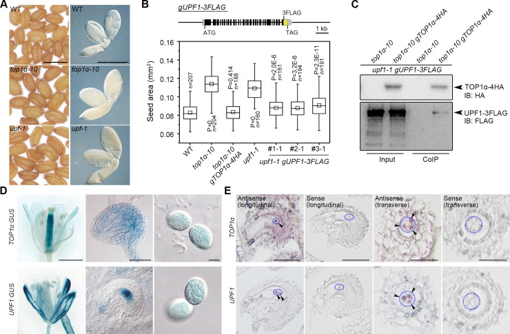
Topoisomerases add or remove DNA supercoils accumulated during replication and transcription [14]. In Arabidopsis, TOP1α (AT5G55300) is the major type I topoisomerase [15]. Interestingly, TOP1α not only affects flowering time [16] but also influences seed size (Fig 1A and 1B and S1A Fig). To identify interacting partners of TOP1α, we performed co-immunoprecipitation (CoIP) coupled with liquid chromatography coupled with tandem mass spectrometry (LC-MS/MS) using nuclear extracts of young siliques from the established top1α-10 gTOP1α-4HA line [16]. A peptide corresponding to UPF1 was identified, and the corresponding loss-of-function mutant upf1-1 also produced large seeds (Fig 1A and 1B and S1A–S1C Fig) as reported previously [17]. Notably, the large-seed phenotype of top1α-10 and upf1-1 was not due to low fertilization in siliques (S1D Fig). The genomic fragment of TOP1α or UPF1 restored the seed size in the respective mutants, indicating that they are essential for regulating seed size (Fig 1B). Their interaction in vivo was subsequently confirmed by CoIP using the extracts from the homozygous progenies created from top1α-10 gTOP1α-4HA crossed with upf1-1 gUPF1-3FLAG (Fig 1C).
Fig 1. TOP1α and UPF1 regulate seed size and interact in the nucleus.
(A) top1α-10 and upf1-1 produce larger seeds than WT. Left panels, mature seeds; right panels, mature embryos. Scale bars, 0.5 mm. (B) Seed sizes of top1α-10 and upf1-1 are rescued by correspondent genomic fragments. Upper panel: schematic diagram of the gUPF1-3FLAG construct. Lower panel: seed sizes of different genetic backgrounds. P values were determined by two-tailed Mann-Whitney U-test compared to WT. (C) In vivo interaction between TOP1α and UPF1 shown by CoIP. Nuclear protein extracts from pistils of the specified genotypes were immunoprecipitated by anti-HA antibody. The input and CoIP proteins were detected by anti-HA (upper panel) and anti-FLAG antibody (lower panel), respectively. (D) Representative GUS staining of TOP1α:GUS and UPF1:GUS in opening flowers (left panels), ovules (middle panels), and pollen grains (right panels). Scale bars, 1 mm, 50 μm, 10 μm from left to right. (E) In situ localization of TOP1α and UPF1 mRNA expression in ovules. Blue dotted ellipses indicate the regions where antipodal cells are located, and arrowheads indicate antipodal cells with signals. Scale bars, 50 μm, 50 μm, 25 μm, and 25 μm from left to right. The data underlying this figure are included in S1 Data and S1 Raw Images. CoIP, co-immunoprecipitation; GUS, β-glucuronidase; HA, hemagglutinin; n, number of seeds examined; IB, immunoblotting; WT, wild type.
TOP1α and UPF1 share an overlapping expression pattern
Both TOP1α and UPF1 were highly expressed in flowers and nascent siliques, but their expression decreased during seed development (S1E and S1F Fig). We generated β-glucuronidase (GUS) reporter lines to visualize their expression in detail. TOP1α:GUS staining signal appeared in the pistil and the whole ovule with a relatively strong signal at the chalazal end of embryo sac, whereas UPF1:GUS signal was observed as dots in the pistil and at the chalazal end (Fig 1D). Both TOP1α:GUS and UPF1:GUS were weakly expressed in pollen grains (Fig 1D), which is consistent with pollen transcriptome data [18]. To confirm the expression of TOP1α and UPF1 at the chalazal end, we performed in situ hybridization and found that UPF1 was specifically localized in the three antipodal cells, whereas TOP1α was expressed in antipodal cells and their surrounding region (Fig 1E).
Although cytosolic UPF1 is involved in non-sense mRNA decay (NMD) together with UPF2 and UPF3 [19], loss-of-function of UPF2 and UPF3 did not produce large seeds as upf1-1 (S1G Fig), implying a non-NMD function of UPF1. Although UPF1 was mostly localized in the cytosol, but also present in the nucleus, its interaction with TOP1α was only detected in the nucleus in bimolecular fluorescence complementation (BiFC) assay (S1H–S1J Fig). Moreover, UPF1-3FLAG was co-immunoprecipitated with histone H3, supporting its chromatin association in vivo (S1K Fig). We also inserted a nuclear localization signal (NLS) at the proximal C-terminal of UPF1 to generate gUPF1-NLS-3FLAG. The derived fusion protein, which was only localized in the nucleus in planta (S1L Fig), fully rescued the seed phenotype of upf1-1 (S1M Fig), demonstrating that nuclear-localized UPF1 regulates seed size.
top1α-10, upf1-1, and ttg2-6 exhibit a parent-of-origin effect on diploidy crosses
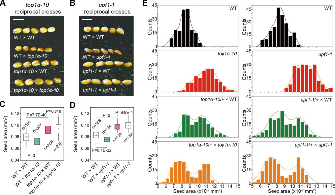
We performed reciprocal crosses between top1α-10 or upf1-1 and wild-type plants to test the potential parent-of-origin effect. For top1α-10 and upf1-1, their maternal and paternal mutations generated larger and smaller seeds than wild-type plants, respectively (Fig 2A–2D). On the one hand, seeds of hand-pollinated top1α-10 and upf1-1 were of similar size to the seeds of top1α-10 and upf1-1 pollinated with wild-type plants, indicating their maternal effects (Fig 2A–2D). There were bimodal distributions of seed size of heterozygotes (top1α-10/+ or upf1-1/+) pollinated with homozygous mutants (top1α-10 or upf1-1) or wild-type plants, which supports the maternal gametophytic effect (Fig 2E). This is in line with their overlapping localization in female gametophyte (antipodal cells; Fig 1E). On the other hand, wild-type plants pollinated with top1α-10 or upf1-1 produced smaller seeds than self-crossed wild-type plants, indicating their paternal effect. However, such a paternal effect could be overridden by the dominant effect of a maternal mutation of either TOP1α or UPF1 (Fig 2C and 2D). These data suggest that the seed size is determined by the dosage-dependent maternal and paternal TOP1α or UPF1.
Fig 2. top1α-10 and upf1-1 exhibit a parent-of-origin effect on diploidy crosses.
(A) Representative seeds from reciprocal crosses between WT and top1α-10. Scale bars, 0.5 mm. (B) Representative seeds from reciprocal crosses between WT and upf1-1. Scale bars, 0.5 mm. (C, D) Parent-of-origin effect of top1α-10 (C) and upf1-1 (D) pertaining to seed size. (E) Histograms of the size distributions of seeds produced by top1α-10-related testcrosses (left panels) and upf1-1-related testcrosses (left panels). The distribution curves were determined by kernel-density estimation. The data underlying this figure are included in S1 Data. WT, wild-type plants.
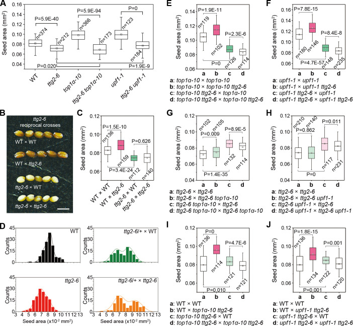
The TTG2 (AtWRKY44) has been reported to regulate interploidy barrier as maternal TTG2 mutations confer tolerance to paternal excess (diploids pollinated with tetraploids) abortion [13], implying a maternal influence on paternal dosage. We then carried out a detailed investigation on ttg2-6 (SALK_206852) in the Col background (S2A Fig), which produced small seeds like other ttg2 alleles [20] (Fig 3A). ttg2-6 was epistatic to both top1α-10 and upf1-1 (Fig 3A) and had an opposite gametophytic parent-of-origin effect compared to top1α-10 and upf1-1 (Fig 3B–3D). Moreover, the parent-of-origin effect of ttg2-6 was observed in the top1α-10 or upf1-1 background (Fig 3E and 3F), but the same parent-of-origin effect of top1α-10 or upf1-1 was attenuated in ttg2-6 (Fig 3G and 3H). In addition, ttg2-6 top1α-10 or ttg2-6 upf1-1 double mutants caused a similar effect to ttg2-6 in reciprocal crosses (Fig 3I and 3J). Collectively, our results indicate that TTG2 is one of the genetic downstream targets of TOP1α and UPF1 in the control of seed size.
Fig 3. TTG2 acts downstream of TOP1α and UPF1.
(A) ttg2-6 is epistatic to top1α-10 and upf1-1 pertaining to seed size. (B) Representative seeds from reciprocal crosses between WT and ttg2-6. Scale bars, 0.5 mm. (C) Parent-of-origin effect of ttg2-6 pertaining to seed size. (D) Histograms of the size distributions of seeds produced by ttg2-6-related testcrosses. The distribution curves were determined by kernel-density estimation. (E, F) Parent-of-origin effect of ttg2-6 in top1α-10 (E) and upf1-1 backgrounds (F). (G, H) Parent-of-origin effect of top1α-10 (G) and upf1-1 (H) in ttg2-6 background. (I, J) Parent-of-origin effect of ttg2-6 top1α-10 (I) and ttg2-6 upf1-1 (J) pertaining to seed size. (A, C, and E-J) P values were determined by two-tailed Mann-Whitney U-test. The data underlying this figure are included in S1 Data. n, number of seeds examined; WT, wild-type plants.
Since it has been reported that TTG2 functions as a sporophytic regulator in the integument in Ler [6] and is also a female gametophytic expressed gene (DD91) [21] before fertilization, it is possible that TTG2 may combine sporophytic and gametophytic effects. Since Ler encodes a weak TTG2 [13], the gametophytic role of TTG2 could be concealed during crosses. Even Ler × ttg2-1 resulted in larger seeds than Ler × Ler, which cannot be explained by sporophytic maternal effect or integument cell elongation [6]. As top1α-10 and upf1-1 did not exhibit seed coat phenotypes like those of ttg2-6 (S2B Fig), the seed size phenotype of top1α-10 and upf1-1 is likely related to the gametophytic rather than the sporophytic TTG2.
TOP1α and UPF1 directly regulate TTG2
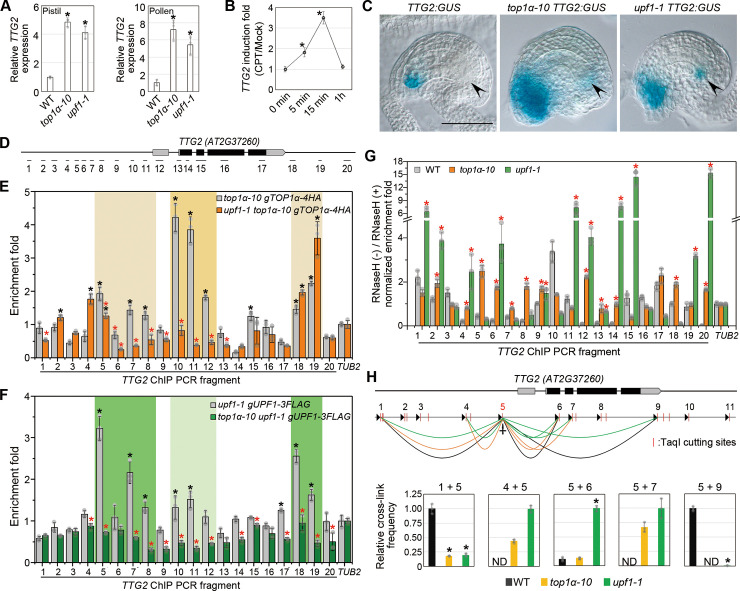
To understand the molecular link among TOP1α, UPF1, and TTG2, we proceeded to investigate their expression profiles. Quantitative real-time PCR revealed that TTG2 expression was up-regulated in both pistils and pollens of top1α-10 and upf1-1 (Fig 4A). Moreover, TTG2 expression increased during early seed development till 4 days after pollination (DAP) (S2C Fig), whereas the expression of TOP1α and UPF1 decreased (S1F Fig). Camptothecin (CPT; a topoisomerase I–specific inhibitor) treatment also quickly induced TTG2 expression in pistils (Fig 4B). We generated a transcriptionally fused TTG2:GUS reporter line, which exhibited the typical trichome signal as previously reported [20] (S2D Fig), suggesting that the reporter line could be used to examine TTG2 expression. Using this reporter, we observed ectopic signals in antipodal cells and higher signals around the micropyle in top1α-10 or upf1-1 compared to wild-type plants (Fig 4C). Ectopic TTG2 expression in antipodal cells was in line with the localization of TOP1α and UPF1 and persisted at least until 1 DAP (S2E Fig). Despite the known sporophytic TTG2 effect on seed size [6], our results also revealed a gametophytic effect of ttg2-6 (Fig 3D), which is consistent with the gametophytic effect exhibited by top1α-10 and upf1-1 (Fig 2E). Although TTG2 expression was also elevated in the whole micropyle region (including the sporophytic tissues) of top1α-10 and upf1-1, these two mutants did not show any seed coat phenotypes related to ttg2-6 (S2B Fig). These observations indicate that ectopic expression of TTG2 in antipodal cells rather than in micropyle contributes to the gametophytic effect of ttg2-6 associated with the seed phenotypes exhibited by top1α-10 and upf1-1.
Fig 4. TOP1α and UPF1 directly repress TTG2.
(A) TTG2 expression in pistils (left panel) or pollens (right panel) in different genetic backgrounds. Expression values normalized against U-BOX are shown relative to the expression level in WT. Values are mean ± s.d. of three biological replicates. *P < 0.05 as compared to WT, two-tailed Student’s t test. (B) CPT treatment induces TTG2 expression. Time-course experiments were conducted by treating the pistils with 10 μM CPT or mock solution. Fold change of CPT-treated versus mock-treated at each time point is presented as mean ± s.d. of three biological replicates. *P < 0.05 as compared to “0 min,” two-tailed Student’s t test. (C) Representative GUS staining of TTG2:GUS in WT (left), top1α-10 (middle), and upf1-1 (right) backgrounds. Scale bar, 50 μm. Arrowheads indicate antipodal cells. (D) Schematic diagram of the fragments amplified in ChIP and DRIP analysis spanning the TTG2 genomic region. The coding and untranslated regions are indicated by black and gray boxes, respectively, and introns and other genomic regions are indicated by black lines. (E, F) ChIP analysis of TOP1α-4HA (E) and UPF1-3FLAG (F) binding to the TTG2 genomic region. Pistil samples were harvested for ChIP analysis using anti-HA (E) or anti-FLAG (F). Values are mean ± s.d. of three biological replicates. The black asterisks indicate significant enrichment compared to that of TUB2. The red asterisks indicate significantly decreased enrichment when UPF1 is absent (E) or TOPα is absent (F). *P < 0.05, two-tailed Student’s t test. (G) DRIP analysis at TTG2 genomic region. Pistil samples were harvested for DRIP analysis using S9.6 antibody. Values are mean ± s.d. of three biological replicates. The red asterisks indicate significantly higher enrichment in comparison with WT. *P < 0.05, two-tailed Student’s t test. (H) 3C analysis of chromatin looping at the TTG2 locus. Upper panel: Schematic diagram of the spatial interactions at TTG2 genomic locus. Detectable spatial interactions are linked by black (in WT), orange (in top1α-10), and green arcs (in upf1-1) between the anchor point (site 5) and surrounding loci. Arrowheads indicate the primers for qPCR. Lower panel: the cross-link frequencies are shown relative to the strongest interaction at each site. Values are mean ± s.d. of three biological replicates. *P < 0.05 as compared to WT, two-tailed Student’s t test. The data underlying this figure are included in S1 Data. 3C, chromatin conformation capture; ChIP, chromatin immunoprecipitation; CPT, camptothecin; DRIP, DNA:RNA hybrid immunoprecipitation; GUS, β-glucuronidase; HA, hemagglutinin; ND, not detectable; qPCR, quantitative PCR; WT, wild-type plants.
We further detected the binding of TOP1α and UPF1 to the TTG2 genomic locus by chromatin immunoprecipitation (ChIP) assay (Fig 4D–4F). The UPF1-NLS-3FLAG displayed stronger binding than UPF1-3FLAG to similar regions (S3A Fig), further supporting the role of nuclear-localized UPF1. Interestingly, there was an overlap in the regions bound by TOP1α and UPF1, although their binding preference differed (Fig 4E and 4F). TOP1α binding to the proximal promoter region was strikingly weakened in upf1-1, whereas UPF1’s binding to the whole region was also weakened in top1α-10 (Fig 4E and 4F), indicating that their protein interaction may strengthen their respective binding to the TTG2 locus.
Loss of TOP1 could accumulate R-loops (DNA:RNA hybrid) as previously reported [22, 23]. DNA:RNA hybrid immunoprecipitation (DRIP) revealed that R-loops were accumulated in the 5′ TTG2 promoter region in top1α-10 and in the entire TTG2 gnomic region in upf1-1 (Fig 4G). Higher expression of TTG2 in top1α-10 and upf1-1 suggests a promotional role of the R-loops, as they can keep chromatin open and protect hypomethylated promoters from being silenced [24, 25]. Through chromatin conformation capture (3C) assays of either TaqI- or CviQI-digested chromatin, we found that the proximal promoter region spatially interacted with the distal 5′ and 3′ regions, indicating a folded chromatin in wild-type plants (Fig 4H and S3B Fig). In top1α-10 and upf1-1, the distal chromatin interactions were significantly weakened, whereas the short-range interactions were strengthened (Fig 4H and S3B Fig). Moreover, we conducted formaldehyde-assisted isolation of regulatory elements coupled with quantitative PCR (FAIRE-qPCR) to check the chromatin accessibility. The open chromatin was predominantly located at the coding and distal promoter regions, which became more accessible in top1α-10 and upf1-1 (S3C Fig). These data substantiate that loss of TOP1α and UPF1 decondenses the TTG2 locus.
We also tested if R-loops could protect hypomethylated promoters from being silenced. The met1 (mainly CpG hypomethylation), but not cmt3 (mainly CpHpG hypomethylation), showed a parent-of-origin effect on seed size [26]. ago4 ago6, nrpd2a-2 nrpd2b-1, and cmt3 drm1 drm2 had greatly hypomethylated CpHpH or CpHpG sites [27, 28] but did not display parent-of-origin effects on seed size (S4A–S4C Fig). Thus, we only quantified CpG methylation at the TTG2 locus. Hypomethylated TTG2 promoters in top1α-10 and upf1-1 compared to wild type were revealed by CpG-sensitive restriction enzymes digestion coupled qPCR (S4D and S4E Fig). The hypomethylation was verified by methyl-cytosine immunoprecipitation (mCIP) at the TOP1α-occupied promoter region (m10 and m11) (S4D and S4F Fig). Taken together, our results suggest that TOP1α and UPF1 bind to the TTG2 locus interdependently and keep this locus folded and silenced. Loss of TOP1α and UPF1 generates ectopic R-loops in this region, resulting in decondensed and hypomethylated chromatin and thus permitting a basal TTG2 transcription.
TOP1α, UPF1, and TTG2 are non-imprinted genes and provide feedback regulation of ploidy increase
We are wondering how TTG2, TOP1α, and UPF1 could contribute to the parent-of-origin effect. Imprinting is the best-studied cause for parent-of-origin effects. However, reciprocal crosses carried out in this study revealed that these genes did not act like either MEG or PEG (S5A and S5B Fig). We found that TTG2 was biparentally expressed (S5C Fig), and TTG2, TOP1α, and UPF1 were also reported as non-imprinted genes [29]. Imprinting happens in the endosperm [30, 31], whereas TOP1α and UPF1 are mainly expressed in antipodal cells rather than central cells (Fig 1D and 1E).
The parent-of-origin effects of TOP1α, UPF1, and TTG2, which are independent of imprinting, imply that they may be involved in a hitherto unknown module that senses and regulates parental dosage balance. Pollinating 2x Col with top1α-10 produced small seeds (Fig 2A and 2C), which is similar to tetraploids pollinated with diploids. Thus, top1α-10 likely simulates a decreased genome dosage compared to 2x Col. Interestingly, tetraploids have larger seeds than diploids, whereas top1α-10 produced large seeds even when it simulates a decreased genomic dosage. Thus, it is highly possible that TOP1α, UPF1, and TTG2 act in a feedback loop that compensates for the effects of ploidy increase (S6B Fig). To test this assumption, we generated tetraploid mutants (S7 Fig). TOP1α and UPF1 expression was reduced in the tetraploids versus diploidy wild-type plants, whereas TTG2 expression was elevated (S6C Fig), indicating that they respond to ploidy increase. Moreover, the seeds of 4x Col were 34.0% larger than those of diploidy wild type. Such an increase weakened in top1α-10 and upf1-1 backgrounds (25.9% and 19.1% larger than diploids, respectively) but enhanced in the ttg2-6 background (48.3% larger than diploids) (S6D Fig). Seeds of 8x ttg2-6 were larger than those of 4x ttg2-6, whereas 8x top1α-10 produced seeds with similar size to those of 4x top1α-10 (S6D Fig). These data support that loss of TTG2 or TOP1α enhances or weakens the effect of ploidy increase, respectively. In this scenario, changes in the expression of TOP1α, UPF1, and TTG2 may mimic altered parental dosage, resulting in parent-of-origin effect in reciprocal crosses.
top1α-10, upf1-1, and ttg2-6 affect the reciprocal phenotypes of interploidy crosses
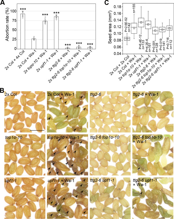
Interploidy crosses are an ideal platform to test the parent-of-origin effect and dosage effect because of the distinct phenotype of paternal excess (diploids pollinated with tetraploids) and maternal excess (tetraploids pollinated with diploids). Moreover, maternal mutation of TTG2 suppresses the interploidy barrier [13]. Thus, we investigated the effects of top1α-10, upf1-1, and ttg2-6 on interploidy crosses as well. Firstly, we examined various mutants in paternal excess. Warschau (Wa-1) is a natural autotetraploid ecotype. Since the abortion rate of 2x Col pollinated with 4x Col or Wa-1 was 92.38% or 27.45%, respectively, Wa-1 was used as a male parent because the interploidy barrier in Col background was too strong to show the potential variances (Fig 5A). Seeds of 2x top1α-10 or 2x upf1-1 pollinated with Wa-1 had higher abortion rates than the seeds of 2x Col pollinated with Wa-1, whereas the seeds of 2x ttg2-6, 2x ttg2-6 top1α-10, or 2x ttg2-6 upf1-1 pollinated with Wa-1 had lower abortion rates than seeds of 2x Col pollinated with Wa-1 (Fig 5A and 5B). Similarly, maternal TOP1α and UPF1 mutations also strengthened the large-seed phenotype of paternal excess (Fig 5C). In contrast, the seeds of 2x Col pollinated with 4x top1α-10 or 4x upf1-1 had lower abortion rates than the seeds of 2x Col pollinated with 4x Col or 4x ttg2-6 (S8A Fig). Since most of the seeds resulting from these paternal excesses in the Col background were aborted or in irregular shapes (S8B Fig), we did not measure their seed size.
Fig 5. TOP1α, UPF1, and TTG2 affect the phenotypes of paternal-excess interploidy crosses.
(A) Maternal mutations affect the abortion rate of paternal excess. Values are mean ± s.d. ***P < 0.001 as compared to 2x Col × Wa-1, two-tailed Student’s t test. (B) Seed morphology of paternal excess using Wa-1 and diploidy mutants. Arrowheads indicate the aborted seeds. Scale bar, 0.5 mm. (C) Maternal mutations affect the seed size of paternal excess. Aborted seeds are excluded before measurements. P values were determined by two-tailed Mann-Whitney U-test as compared to 2x Col × Wa-1. The data underlying this figure are included in S1 Data. n, number of seeds examined; Wa-1, Warschau.
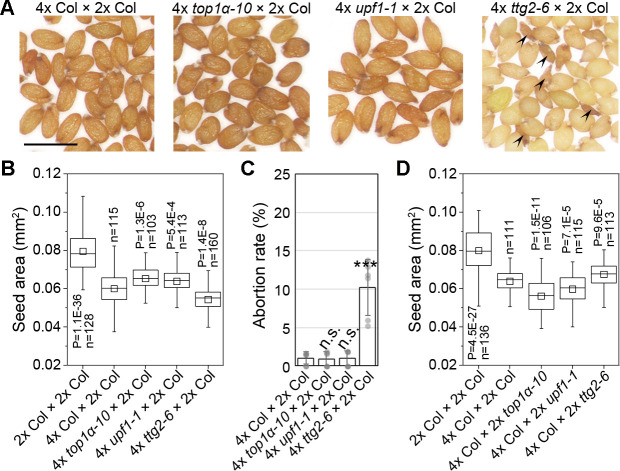
We further investigated seeds produced from maternal excess. Maternal excess produces viable seeds so we can test seed size in the Col background. Pollinating 4x top1α-10 or 4x upf1-1 with 2x Col produced larger seeds than pollinating 4x Col with 2x Col, whereas 4x ttg2-6 pollinated with 2x Col produced smaller seeds than 4x Col pollinated with 2x Col (Fig 6A and 6B). In addition, only 4x ttg2-6 pollinated with 2x Col produced some small aborted seeds, implying an extreme phenotype of maternal excess as shown in hexaploid pollinated with diploid [10] (Fig 6A and 6C). When pollinating 4x Col with different diploidy males, paternal TTG2 mutations produced larger seeds, whereas paternal TOP1α and UPF1 mutations produced smaller seeds, compared to the seeds of 4x Col pollinated with 2x Col (Fig 6D). Regarding that top1α-10 and upf1-1 simulate a decreased genomic dosage, they caused enlarged dosage differences compared to tetraploids. In contrast, ttg2-6 simulates an increased genomic dosage, thus narrowing the dosage difference between ttg2-6 and tetraploids. These results support that the mutations of these genes can skew the parental balance to affect the phenotypes of interploidy crosses.
Fig 6. TOP1α, UPF1, and TTG2 affect the phenotypes of maternal-excess interploidy crosses.
(A) Morphology of the seeds produced by 4x mutants pollinated with diploidy wild type. Arrowheads indicate the aborted seeds. Scale bar, 0.5 mm. (B) Maternal mutations affect the seed size of maternal excess. P values were determined by two-tailed Mann-Whitney U-test as compared to 4x Col × 2x Col. (C) Maternal mutations affect the abortion rate of maternal excess. Values are mean ± s.d. ***P < 0.001 as compared to 4x Col × 2x Col, two-tailed Student’s t test. (D) Paternal mutations affect the seed size of maternal excess. P values were determined by two-tailed Mann-Whitney U-test as compared to 4x Col × 2x Col. The data underlying this figure are included in S1 Data. n, number of seeds examined.
Discussion
TOP1α and UPF1 are fundamental regulators of DNA and RNA structures, respectively, and have been reported to affect plant development, including the floral transition [16, 32]. In this study, we have revealed that TOP1α and UPF1 regulate seed size through TTG2 in a parent-of-origin manner. TOP1α interacts with UPF1, and both of them repress TTG2 biparentally by maintaining the condensed chromatin at the TTG2 locus correlated with the level of R-loops and CG-methylation, which is consistent with the observations on the epistatic analysis. This mechanism also brings insight into non-NMD functions of UPF1 as a transcription regulator in Arabidopsis, which is implied by other studies in Drosophila and human cells [33, 34].
In the developmental scope, seed size is controlled in multiple dimensions, such as the capacity restriction by the integument, the growth of the embryo, and the timing of endosperm cellularization. In addition, parental cues from gametophytes may also contribute to seed development at the very beginning of seed development. For example, pollen-derived SHORT SUSPENSOR (SSP) mRNA is translated in the zygote to control the asymmetric zygotic cell division [35]. It is noteworthy that TOP1α and UPF1 function gametophytically and are enriched in antipodal cells rather than female gametes, whereas the function of antipodal cells is by now unclear. One hypothesis is that antipodal cells are backup gametes as they can be transformed into gametes as if the normal gamete development failed [36, 37]. Although TTG2 has been reported to act sporophytically [6], our reciprocal crosses and test crosses reveal a surprising gametophytic role of TTG2 in seed development. This role is associated with TTG2 expression in antipodal cells, which is specifically suppressed by TOP1α and UPF1, and derepression of TTG2 contributes to the parent-of-origin effect of top1α-10, upf1-1, and ttg2-6. As antipodal cells are not transmitted to the filial generation as gametes, they exist only for a short period of time after fertilization [38], implying that the related regulators may only work in a narrow time window at the beginning of seed development. Besides, as the paternal-inherited mutations of TOP1α, UPF1, and TTG2 also plays roles in determining seed size, we believe that the parental interplay related to these genes happens between antipodal cells and sperms.
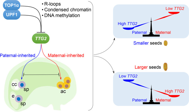
Taken together, we propose a TTG2 dosage–dependent molecular framework wherein the parental TTG2 dosage between antipodal and sperm cells regulated by TOP1α and UPF1 determines seed size (Fig 7A). Elevated maternal to paternal TTG2 ratios result in large seeds, whereas the declined ratios cause an opposite phenotype that partially mimics parental imbalance (Fig 7B and S1 Table). This model is consistently valid as evidenced by interploidy crosses. Both top1α-10 and upf1-1 simulate a decreased genomic dosage, whereas ttg2-6 simulates an increased genomic dosage. Thus, they influence the phenotype strength of interploidy crosses. On the other hand, paternal-inherited top1α-10 or upf1-1 causes smaller seeds, whereas paternal-inherited ttg2-6 causes larger seeds in interploidy crosses. In contrast, paternal-inherited top1α-10, upf1-1, or ttg2-6 acted opposite to maternal ones. These features are also observed in interploidy crosses, indicating that the parent-of-origin effects of TOP1α, UPF1, and TTG2 mutations are not altered by the ploidy level. Therefore, the model described here provides a new layer of regulation on the phenotypes of interploidy crosses. This is reasonable because the molecular framework consisting of TOP1α, UPF1, and TTG2 is independent of imprinting, which also affects interploidy phenotypes [39–44].
Fig 7. A model depicting the regulation of seed size by parental TTG2 dosage.
TOP1α interacts with UPF1, which facilitates their binding to the TTG2 locus. This binding compromises R-loops that hamper the chromatin condensation. Loss of TOP1α and UPF1 makes the TTG2 locus more accessible with less GC-methylation, permitting a higher TTG2 expression. The repressive effects of TOP1α and UPF1 on TTG2 exist in sperms from the pollen and in antipodal cells from the female gametophyte, whereas paternal-inherited and maternal-inherited TTG2 affect seed size in an opposite manner. The relative parental dosage of TTG2 determines the seed size so that an increase in maternal or paternal TTG2 results in larger or smaller seeds, respectively. ac, antipodal cells; cc, central cell; e, egg cell; sp, sperm.
Our results suggest that a parental dosage–sensitive regulatory module may be controlled by signals existing outside the endosperm in Arabidopsis. Although this module acts biparentally, the maternal effect is stronger than the paternal one. For example, the seed phenotypes of homozygous mutants of TOP1α, UPF1, and TTG2 are determined by their respective maternal genotype. In addition, maternal mutations of TOP1α, UPF1, and TTG2 have a greater influence on the phenotypes of interploidy crosses than their corresponding paternal mutations, respectively. Thus, a key question that remains unanswered is why the maternal and paternal TTG2 display opposite effects. It is possible that TTG2 protein may have different interacting partners or target genes in antipodal and sperm cells. The antipodal cell fate is controlled by the adjacent gamete, the central cell, so that intensive cell-cell communication should exist between antipodal cells and the central cell [45]. The communication may continuously exist between antipodal cells and the nascent endosperm, which is produced from the fusion between sperm and central cells. Further addressing whether this sort of communication is relevant to different effects of the maternal and paternal TTG2 may help to understand their downstream regulatory events for seed size control.
Materials and methods
Plant materials and growth conditions
A. thaliana plants were grown under long-day conditions (16 h light/8 h dark) at 22°C. The top1α-10 (SALK_013164), upf1-1 (CS6940), upf2-1 (SAIL_512_G03), upf3-1 (CS9900), ttg2-6 (SALK_026852), cmt3 drm1 drm2 (CS16384), and nrpd2a-2 nrpd2b-1 (CS66155) are in Col-0 background. The ago4 ago6 (CS66095) is in Ler background. The mutants, the natural tetraploid Wa-1 (CS6885) and Bur-0 (CS22679) ecotype, were obtained from ABRC. The 4x Col, 4x top1α-10, 4x upf1-1, 4x ttg2-6, 8x ttg2-6, and 8x top1α-10 were generated in this research by colchicine treatment. Before being used in genetic analysis, the newly generated tetraploids and octoploids were self-pollinated for two generations by single seed descent and checked by flow cytometry in every generation. All transgenic plants were generated by Agrobacterium tumefaciens–mediated transformation.
Ploidy analysis by flow cytometry
One stage 12 flower bud was collected for each plant and was ground to powder in liquid nitrogen in a 1.5-mL tube. Five hundred microliters of TMT buffer (200 mM Tris-HCl [pH 7.5], 4 mM MgCl2, 0.5% [v/v] Triton X-100) was added to each sample and vortexed immediately. Fifty microliters of propidium iodide (PI) stain solution (1 mg/mL) was added in each tube and keep at 4°C for 15 min. Filter the slurry by 60-μm membrane before mounting flow cytometry analysis. As the PI staining is sample-amount sensitive, the plant should be grown in the same condition and the flower bud harvested should keep in the same size. Moreover, in each bath of analysis, 2x Col and Wa-1 were involved as controls to calibrate the signals.
Plasmid construction for plant transformation
The top1α-10 gTOP1α-4HA was validated in the previous study [16]. upf1-1 top1α-10 gTOP1α-4HA was generated by genetic crossing. To construct upf1-1 gUPF1-3FLAG, the genomic region of UPF1 was amplified into two fragments (−1,636 bp to +8,845 bp, and +8,850 bp to +12,658 bp, numbered relative to start code) and the two fragments were inserted before and after the 3xFLAG tag in pC1305, respectively. To construct upf1-1 gUPF1-NLS-3FLAG, an artificial nuclear localization sequence (NLS) was incorporated into gUPF1-3FLAG fragment at the beginning of exon 27, between Q1129 and A1130. Both pC1305-gUPF1-3FLAG and pC1305-gUPF1-NLS-3FLAG were transformed into upf1-1 background. top1α-10 upf1-1 gUPF1-3FLAG was generated by genetic crossing. To construct UPF1:GUS, 1,636-bp 5′ upstream sequence (from −1,636 to −1) and 3,809-bp 3′ downstream sequence (from +8,850 to +12,658) of UPF1 were assembled before and after the GUS coding sequence. The entire fragment was cloned to pCAMBIA1300. To construct TOP1α:GUS, a 2,593-bp 5′ upstream sequence was cloned to pBI101-GUS. Both UPF1:GUS and TOP1α:GUS were transformed into wild-type background. To construct TTG2:GUS, a 3,459-bp 5′ upstream sequence was cloned to pBI101-GUS. TTG2:GUS was transformed into upf1-1 background, and then the typical line was crossed with top1α-10. The TTG2:GUS in wild-type and top1α-10 backgrounds were segregated from the F2 population. The primers used are listed in S2 Table.
Seed size measurement
Only the seeds growing at the same time and the same condition were used in the comparison. All the seeds of reciprocal crosses were produced by hand-pollination including the homozygous controls. Mature seeds were spread on a plain white paper. Photos were taken by stereomicroscope (Nikon). The seed area and length/width ratio were analyzed by ImageJ. Box plots were visualized by Origin. Boxes indicated upper quartile to lower quartile, whiskers indicated 1.5 interquartile range (IQR), the means were shown by open squares, and the medians were shown by transverse lines. Two-tailed Mann-Whitney U-tests were applied between samples by Origin, and the P values were indicated with the number of the seeds examined.
Microscopy and histochemical analysis
For the clearing of ovules and developing seeds, the pistils or young siliques were fixed with 10% acetic acid in ethanol for 1 h, washed for 0.5 h in 90% ethanol, 0.5 h in 70% ethanol, and then cleared overnight in chloralhydrate solution (8 g chloralhydrate, 2 mL glycerol, and 4 mL H2O). The samples were observed under differential interference contrast (DIC) microscopy (Leica). GUS staining was conducted by normal procedure and the samples were cleared in chloralhydrate solution before being mounted to microscopy.
In situ hybridization
Unfertilized pistils were cut longitudinally before being fixed in 4% paraformaldehyde at 4°C overnight. They were then dehydrated through an ethanol series and Histoclear, embedded into paraffin, sectioned to 8 μm, and mounted on poly-d-lysine-coated slides (Fisher Scientific). For the synthesis of RNA probes, a gene-specific region of TOP1α or UPF1 was amplified by a pair of primers (S2 Table), cloned into the pGEM-T Easy vector (Promega), and in vitro transcribed using the DIG RNA Labelling Kit (Roche). In situ hybridization was performed as previously reported [46].
Cell-fraction assay
The cell-fractionation analysis was carried out as described before with modifications [47]. Pistils were ground in liquid nitrogen and were lysed with nuclear fractionation buffer (20 mM Tris-HCl [pH 7.0], 250 mM sucrose, 25% glycerol, 20 mM KCl, 2 mM EDTA, 2.5 mM MgCl2, 30 mM β-mercaptoethanol, protease inhibitor cocktail, and 0.7% Triton X-100). The obtained slurry was filtrated (BD Falcon, 100 μM cell strainer) to remove tissue debris, and then the total filtrate was centrifuged at 1,000g for 5 min. The supernatant was the cytosolic fraction and record the volume (Vcyt). The supernatant was filtrated with membrane filters (0.22-μm pore size) to avoid nuclear contamination. The pellet was further washed with resuspension buffer (20 mM Tris-HCl [pH 7.0], 20 mM KCl, 2 mM EDTA, 2.5 mM MgCl2, 30 mM β-mercaptoethanol, protease inhibitor cocktail) three to four times until the pellet was no longer green, and the white pellet was resuspended as nuclear fraction in nuclear lysis buffer (50 mM HEPES [pH 7.5], 150 mM NaCl, 1 mM EDTA, 1% SDS, 1% Triton X-100, 30 mM β-mercaptoethanol, protease inhibitor cocktail) and record the volume (Vnuc). Loadings of fractions were normalized by Vcyt and Vnuc ratio. Samples were loaded to SDS-PAGE and detected by anti-FLAG antibody (F3165, sigma, 1:5,000 dilution).
CoIP assay
Pistils in various genetic backgrounds were collected, and the cell fractionation was carried out as described above. Nuclear protein extracts were 4-fold diluted by extraction buffer (50 mM Tris-HCl [pH 7.5], 150 mM NaCl, 1 mM EDTA, 5% glycerol, 0.5% Triton X-100, 1 mM PMSF, proteinase inhibitor cocktail, and 25 μM MG132) and incubated with anti-HA agarose beads (Sigma) or anti-FLAG magnetic beads (Sigma) at 4°C for 2 h and then washed four to six times by extraction buffer. Immunoprecipitated proteins and nuclear protein extracts as inputs were resolved by SDS–polyacrylamide gel electrophoresis and detected by the corresponding antibody (anti-FLAG: F3165, sigma, 1:5,000 dilution; anti-HA: sc-7392 HRP, Santa Cruz, 1:2,000 dilution; anti-H3: ab1791, abcam, 1:3,000 dilution). For IP-MS, immunoprecipitated proteins were eluted and analyzed by TripleTOF 5600 System (AB Sciex).
Transient expression assays in tobacco
For subcellular localization analysis, coding sequences of TOP1α was fused with EGFP at C-terminus in pC1302E, whereas the coding sequence of UPF1 was fused with mCherry at C-terminus in pC1300mCherry. For BiFC, the coding sequence of TOP1α was fused to cYPF at C-terminus in pXY104, whereas the coding sequence of UPF1 was fused to nYFP at N-terminus in pXY106. The plasmids were transformed into A. tumefaciens, and then the transformed A. tumefaciens were infiltrated into Nicotiana benthamiana leaves. Leaves were observed 2 d after infiltration under a confocal microscope (Zeiss LSM710).
ChIP assay
ChIP assays were carried out in various genetic backgrounds using pistils, following the previous protocol with minor modifications [48]. The extracted chromatin was sonicated to produce DNA fragments between 200 and 500 bp. The solubilized chromatin was incubated with anti-HA agarose beads (Sigma) and anti-FLAG magnetic beads (Sigma) for 2 h at 4°C. A genomic fragment of TUBULIN2 (TUB2) was amplified as a control. ChIP assays were repeated with three biological replicates. The primers used are listed in S2 Table.
DRIP assay
Pistils were ground to fine powder in liquid nitrogen, and DNA was extracted by extraction buffer (10 mM HEPES [pH 7.5], 400 mM sucrose, 25 mM EDTA, 1 mM MgCl2, 0.5% SDS, 1 mM PMSF, RNase inhibitor). DNA was recovered by phenol/chloroform extraction and then fragmented into length around 500 bp by pulsing sonication on ice. Normalize the DNA concentration of different samples by nanodrop before subjecting to S9.6 antibody (Kerafast) immunoprecipitation. To generate negative controls, RNaseH (NEB) were added during immunoprecipitation. The immunoprecipitation and following qPCR procedure were the same as ChIP assay. The primers used are listed in S2 Table.
3C
The 3C procedure was modified from the previous protocol [49]. Pistils were ground to fine powder in liquid nitrogen and were incubated with cross-link buffer (10 mM HEPES [pH 7.5], 400 mM sucrose, 5 mM MgCl2, 1 mM EDTA, 1 mM PMSF, proteinase inhibitor cocktail, 1% [v/v] formaldehyde). Vacuum the mixture for 15 min on ice and stop the cross-link reaction by adding glycine to 125 mM, and then vacuum for an additional 2 min. The mixture was diluted four times by nucleus isolation buffer (15 mM PIPES [pH 6.8], 150 mM sucrose, 5 mM MgCl2, 60 mM KCl, 15 mM NaCl, 1 mM CaCl2, 1 mM PMSF, proteinase inhibitor cocktail, 0.9% Triton X-100) and was incubated for 15 min at 4°C using rotating wheel. The obtained slurry was filtrated (BD Falcon, 100 mM cell strainer) to remove tissue debris, and then the total filtrate was centrifuged at 1,000g for 5 min. Wash the pellet once by resuspension buffer (20 mM Tris-HCl [pH 7.0], 20 mM KCl, 2 mM EDTA, 2.5 mM MgCl2, 30 mM β-mercaptoethanol, protease inhibitor cocktail). Then the nuclei were resuspended in the 1X NEB buffer 3.1 containing 0.1% SDS and incubated for 10 min at 65°C. TritonX-100 was added to 1% (v/v) and samples were then digested with CviQI (37°C) and TaqI (55°C) overnight. SDS was added to 1.6% (v/v) and heat 10 min at 65°C in order to inactivate the restriction enzyme. Triton X-100 was added to the final concentration 1% (v/v) and then samples were incubated at 37°C for 0.5 h, mixing occasionally. Then, scale up the total volume to 15 mL and ligate DNA by T4 DNA ligase overnight at room temperature. Subsequently, 200 μg of protease K was added and incubated at 65°C overnight to reverse cross-link and digest the protein. Then DNA was purified by phenol-chloroform extraction and ethanol precipitation. The purified DNA was used as qPCR templates. An 8-kb whole genome sequence of TTG2 was amplified, digested by CviQI and TaqI, and randomly ligated, as a control template. An amplicon of a TTG2 genome fragment without CviQI and TaqI cutting site (region 17 in ChIP) was used as the internal control in qPCR. The primers used are listed in S2 Table.
FAIRE
FAIRE procedure was modified from the previous protocol [50]. Pistils in various genetic backgrounds were used to prepare cross-linked chromatin (for “FAIRE” samples) by the same procedure as ChIP assays. “Un-FAIRE” samples were prepared as the same procedure, just without formaldehyde-mediated cross-linking. The suspended chromatin was extracted by phenol:Chloroform:Isoamyl alcohol (25:24:1) three times and then was ethanol-precipitated before qPCR. Quantification of FAIRE/un-FAIRE ratio was presented to measure chromatin accessibility. The primers used are listed in S2 Table.
mCIP
Pistils were ground to fine powder in liquid nitrogen. Extract genomic DNA by standard CTAB method. Genomic DNA (250 μg) of each material was sonicated to about 500 bp and subjected to immunoprecipitation by anti-methyl-cytosine. The immunoprecipitation and following qPCR procedure were the same as ChIP assay. The primers used are listed in S2 Table.
Expression analysis
Total RNA from various tissues was extracted using FavorPrep Plant Total RNA Mini Kit (Favorgen) and reverse-transcribed using the M-MLV Reverse Transcriptase (Promega) according to the manufacturers’ instructions. Quantitative real-time PCR was performed on three biological replicates using the CFX384 real-time PCR detection system with iQ SYBR Green Supermix (Bio-Rad). The expression of U-BOX was used as an internal control. Relative expression levels of genes were calculated by the ΔCt method or ΔΔCt method. The primers used are listed in S2 Table.
Supporting information
(A) Comparison of 100-seed weight of top1α-10, upf1-1, and WT. Values are mean ± s.d. Asterisks indicate significant differences in comparison to wild type. *P < 0.05, two-tailed Student’s t test. (B) top1α-10 and upf1-1 produce slender seeds as indicated by the length/width ratio. P values were determined by two-tailed Mann-Whitney U-test compared to WT. (C) The peptide of UPF1 identified by IP-MS/MS. (D) Dissected siliques from plants with various genetic backgrounds. Scale bar, 1 mm. (E) qRT-PCR analysis of the expression profiles of TOP1α and UPF1 in adult plants. Relative expression of GOI was normalized against U-BOX expression. Values are mean ± s.d. of three biological replicates. (F) qRT-PCR analysis of the expression profiles of TOP1α and UPF1 during seed development at different days after pollination. Relative expression of GOI was normalized against U-BOX expression. Values are mean ± s.d. of three biological replicates. (G) Seed size of NMD-related mutants. The seed size of upf2-1 is N.A. because of embryonic lethality. P values were determined by two-tailed Mann-Whitney U-test in comparison to wild type. (H) Subcellular localization of TOP1α-GFP (upper panels) and UPF1-mCherry (lower panels) in tobacco leaf epidermal cells. Scale bar, 20 μm. (I) BiFC analysis of the interaction between TOP1α and UPF1 in tobacco leaf epidermal cells. Scale bar, 20 μm. (J) UPF1 subcellular localization shown by cell-fractionation assay. UPF1 protein in nuclear (“N”) or cytoplasmic (“C”) fractions extracted from pistils were detected by anti-FLAG. The nuclear fraction was loaded 10-fold in excess compared to the cytosol fraction. The RUBISCO large subunit (RbcL) stained with Ponceau S and immunoblot analysis using anti-H3 are used as the indicators for cytosol and nuclear fractions, respectively. (K) UPF1 is associated with H3 in vivo as revealed by CoIP. Nuclear protein extracts from the pistils were immunoprecipitated by anti-FLAG. The input and co-immunoprecipitated proteins were detected by anti-FLAG and anti-H3. (L) UPF1-NLS subcellular localization shown by cell-fractionation assay. Upper panel, schematic diagram of the gUPF1-NLS-3FLAG construct. UPF1-NLS protein in nuclear (“N”) or cytoplasmic (“C”) fractions extracted from pistils were detected by anti-FLAG. The nuclear fractions and cytosolic fractions were loaded in an equivalent dose. (M) The seed size of upf1-1 is rescued by gUPF1-NLS-3FLAG. P values were determined by two-tailed Mann-Whitney U-test compared to wild type. The data underlying this figure are included in S2 Data and S1 Raw Images. BiFC, bimolecular fluorescence complementation; CoIP, co-immunoprecipitation; FB, flower bud; GFP, green fluorescent protein; GOI, genes-of-interest; H3, histone 3; IP-MS/MS, liquid chromatography coupled with tandem mass spectrometry; L, leaf; n, number of seeds examined; N.A., not applicable; NLS, nuclear localization signal; NMD, non-sense mRNA decay; OS, old silique (4–7 days after pollination); qRT-PCR, quantitative real-time PCR; R, Root; St, stem; WT, wild-type plants; YS, young silique (0–4 days after pollination).
(TIF)
(A) Characterization of ttg2-6. Left panel: Schematic diagram of ttg2-6 insertion site. The coding and untranslated regions are indicated by black and gray boxes, respectively, and introns and other genomic regions are indicated by black lines. Right panel: Relative TTG2 expression in WT and ttg2-6. Values are means ± s.d of three biological replicates. Asterisks indicate significant differences in comparison to wild type. *P < 0.001, two-tailed Student’s t test. (B) Seed coat phenotypes of WT, ttg2-6, top1α-10, and upf1-1. Upper panels: SEM of the seed coat. Scale bar, 50 μm. “C” indicates columella, and “PCC” indicates partially collapsed columella. Middle panels: Seed coat mucilage staining with ruthenium red. Scale bar, 200 μm. Bottom panels: Seed color. Scale bar, 0.5 mm. The data underlying this figure are included in S2 Data. (C) qRT-PCR analysis of the expression profiles of TTG2 during seed development at different DAP. Relative expression of TTG2 was normalized against U-BOX expression. Values are mean ± s.d. of three biological replicates. (D) Typical trichome expression of TTG2:GUS. (E) Representative GUS staining of TTG2:GUS in WT (upper row), top1α-10 (middle row), and upf1-1 (bottom row) backgrounds, and at 0 DAP (left column), 1 DAP (middle column), and 2 DAP (right column). Scale bars, 50 μm (left column), 50 μm (left column), and 200 μm (right column). Arrowheads indicate antipodal cells. DAP, days after pollination; GUS, β-glucuronidase; qRT-PCR, quantitative real-time PCR; SEM, scanning electron microscopy; WT, wild-type plants.
(TIF)
(A) ChIP analysis of UPF1-NLS-3FLAG binding to the TTG2 genomic region. ChIP was performed by anti-FLAG. A TUB2 fragment was amplified as a negative control. Values are mean ± s.d. of three biological replicates. Asterisks indicate significantly high enrichment in comparison to the TUB2 fragment. *P < 0.05, two-tailed Student’s t test. (B) 3C analysis of chromatin looping status at the TTG2 locus. Upper panel: Schematic diagram of the spatial interactions at TTG2 genomic locus. Detectable spatial interactions are linked by black (in wild type), orange (in top1α-10), and green arcs (in upf1-1) between the anchor point (site 6) and surrounding loci. Arrowheads indicate the primers for qPCR. Lower panel: The cross-link frequencies are shown relative to the strongest interaction at each site. Values are mean ± s.d. of three biological replicates. *P < 0.05 as compared to WT, two-tailed Student’s t test. (C) FAIRE analysis of chromatin accessibility of TTG2 locus. Amplicons of cross-linked samples (FAIRE) versus un-cross-linked samples (un-FAIRE) at each site are presented as mean ± s.d. Asterisks indicate significant differences in comparison to wild type. *P < 0.05, two-tailed Student’s t test. The data underlying this figure are included in S2 Data. 3C, chromatin conformation capture; ChIP, chromatin immunoprecipitation; FAIRE, formaldehyde-assisted isolation of regulatory elements; ND, not detectable; qPCR, quantitative PCR; WT, wild-type plants.
(TIF)
(A-C) Reciprocal crosses of ago4 ago6 (ago4/6) (A), cmt3 drm1 drm2 (cdd) (B), and nrpd2a-2 nrpd2b-1 (nrpd2a/2b) (C) with WT plants. P values were determined by two-tailed Mann-Whitney U-test. (D) The recognition map of selected CpG-sensitive restriction enzymes at the TTG2 locus. The cut sites were indicated in blue (HpyCH4IV), orange (BstUI), red (HhaI), and green (HpaII) strings. The regions to be tested in quantitative real-time PCR are marked as m1 to m23 with the color code of restriction enzymes that digest the corresponding region. PCR fragments of ChIP analysis are aligned above the map. (E) Relative methylation levels at the TTG2 genomic locus. Genomic DNA from pistil was digested by CpG-sensitive restriction enzymes. Undigested genomic DNA was used as an input. The CpG methylation levels were measured by comparing the digested DNA with the input. The relative methylation levels in top1α-10 and upf1-1 backgrounds were normalized against those of WT in each region. Values are mean ± s.d. of three biological replicates. Asterisks indicate significantly low methylation in comparison to WT. *P < 0.05, two-tailed paired Student’s t test. (F) mCIP analysis of the selected regions of TTG2. ChIP PCR fragment 3 and 12 are selected as controls because of no methylation site (fragment 3) or no difference in methylation levels (fragment 12), as indicated in (D and E). ChIP PCR fragment 10 and 11 overlap with region m9–13 as indicated in (D and E). Values are mean ± s.d. of three biological replicates. The asterisks indicate significantly low enrichment compared to WT. *P < 0.05, two-tailed Student’s t test. No statistical difference (n.s), P > 0.05. The data underlying this figure are included in S2 Data. ChIP, chromatin immunoprecipitation; mCIP, methyl-cytosine immunoprecipitation; n, number of seeds examined; ND, not detected; WT, wild-type plants.
(TIF)
(A and B) The parent-of-origin effect of top1α-10 and ttg2-6 is distinct from that of the mutants of imprinted genes. Schematic diagrams represent the patterns of reciprocal crosses that are related to top1α-10 (A) and ttg2-6 (B). Left panel: Actual observation in this study. Middle panel: Patterns under PEG assumption. Right panel: Patterns under MEG assumption. upf1-1 displays a similar behavior to top1α-10. As the maternal PEG and the paternal MEG are not expressed, the corresponding genotypes are indicated as “/” in the PEG and MEG assumption, respectively. Seed size is marked as WT-like (“w”), mutant-like (“m”), or additional type (“a”). (C) TTG2 is not an imprinted gene. Left panel: The SNP in the coding region of TTG2Bur-0 was used to develop the CAPS marker. Restriction enzyme PsiI digests Bur-0 amplicon, but not Col amplicon. Right panel: CAPS test on cDNA derived from the mRNA of F1 siliques at 2 DAP. CAPS, cleaved amplified polymorphic sequences; DAP, days after pollination; MEG, maternally expressed imprinted gene; PEG, paternally expressed imprinted gene; WT, wild-type plants.
(TIF)
(A) Schematic diagrams that summarize the behaviors in reciprocal crosses. top1α-10 (left panel) and ttg2-6 (middle panel) displayed distinctive phenotypes compared to tetraploids (right panel). upf1-1 displays a similar phenotype to top1α-10. Seed size of reciprocal crosses is marked as “w” (WT-like), “m” (mutant-like) or “a” (additional type). Seed size of interploidy crosses is marked as “Pex” (paternal excess), “4” (tetraploid), “2” (diploid), or “Mex” (maternal excess). (B) TOP1α, UPF1, and TTG2 may compose a feedback console in ploidy increase response. Loss of TOP1α or UPF1 mimics a genome-dosage decrease, whereas loss of TTG2 mimics a genome-dosage increase. (C) Relative gene expression in pistils in tetraploids compared to diploid WT. The relative expression of TOP1α, UPF1, TTG2, and commonly used control genes are presented. Expression values normalized against U-BOX are shown relative to the expression levels in diploid WT. Values are mean ± s.d. of three biological replicates. Asterisks indicate significant differences in comparison to diploid WT. *P < 0.05, two-tailed Student’s t test. TOP1α expression in 4x top1α-10 and UPF1 expression in 4x upf1-1 were not tested (N.A.). (D) Comparison of seed size among diploids, tetraploids, and octoploids in different genetic backgrounds. The percentages of size increase are based on the means of the seed area. P values were determined by two-tailed Mann-Whitney U-test in comparison to corresponding diploids. The data underlying this figure are included in S2 Data. n, number of seeds examined; WT, wild-type plants.
(TIF)
Histograms of 5,000 events are shown with the nuclei in different ploidy indicated as 2C, 4C, 8C, and 16C according to DNA contents. Wild-type plants (Col-0) and Wa-1 (an autotetraploidy accession) are used as controls to calibrate the peaks of 2C, 4C, 8C, and 16C nuclei. Pollinating 8x ttg2-6 with 4x ttg2-6 generated 6x ttg2-6. This is used to show that the peaks are accurate and sensitive enough for determining the ploidy levels. Stage 12 flower buds were used for flow cytometry analysis. Wa-1, Warschau.
(TIF)
(A) Paternal mutations affect the abortion rate of paternal excess. Values are mean ± s.d. *P < 0.05 as compared to 2x Col × 4x Col, two-tailed Student’s t test. (B) Morphology of the seeds produced by paternal excess with 4x mutants as male parents. Asterisks indicate viable seeds. Scale bar, 0.5 mm. The data underlying this figure are included in S2 Data.
(TIF)
(PDF)
(PDF)
From Figs 1B, 2C–2E, 3A, 3C–3J, 4A, 4B, 4E–4H, 5A, 5C, and 6B–6D.
(XLSX)
From S1A, S1B, S1E–S1G, S1M, S2A, S2C, S3A–S3C, S4A–S4C, S4E, S4F, S6C–S6D and S8A Figs.
(XLSX)
Acknowledgments
We thank the Arabidopsis Biological Resource Centre for providing seeds. We thank the Protein and Proteomics Centre (PPC) in the Department of Biological Sciences, National University of Singapore, for mass spectrometry service, and members of the Dr. Yu’s lab for discussion and comments on the manuscript.
Abbreviations
- 3C
chromatin conformation capture
- BiFC
bimolecular fluorescence complementation
- ChIP
chromatin immunoprecipitation
- CoIP
co-immunoprecipitation
- CPT
camptothecin
- DAP
days after pollination
- DRIP
DNA:RNA hybrid immunoprecipitation
- FAIRE
formaldehyde-assisted isolation of regulatory elements
- GUS
β-glucuronidase
- iku1
haiku1
- LC-MS/MS
liquid chromatography coupled with tandem mass spectrometry
- mCIP
methyl-cytosine immunoprecipitation
- MEG
maternally expressed gene
- mini3
miniseed3
- NLS
nuclear localization signal
- NMD
non-sense mRNA decay
- PEG
paternally expressed gene
- qPCR
quantitative PCR
- SHB1
SHORT HYPOCOTYL UNDER BLUE1
- SSP
SHORT SUSPENSOR
- TOP1α
TOPOISOMERASE Iα
- TTG2
TRANSPARENT TESTA GLABRA2
- UPF1
UP-FRAMESHIFT SUPPRESSOR 1
- Wa-1
Warschau
Data Availability
All relevant data are within the paper and its Supporting Information files.
Funding Statement
This work was supported by the Singapore National Research Foundation Investigatorship Programme (NRF-NRFI2016-02) granted to H.Y. The funders had no role in study design, data collection and analysis, decision to publish, or preparation of the manuscript.
References
- 1.Wang A, Garcia D, Zhang H, Feng K, Chaudhury A, Berger F, et al. The VQ motif protein IKU1 regulates endosperm growth and seed size in Arabidopsis. Plant J. 2010;63(4):670–9. 10.1111/j.1365-313X.2010.04271.x [DOI] [PubMed] [Google Scholar]
- 2.Luo M, Dennis ES, Berger F, Peacock WJ, Chaudhury A. MINISEED3 (MINI3), a WRKY family gene, and HAIKU2 (IKU2), a leucine-rich repeat (LRR) kinase gene, are regulators of seed size in Arabidopsis. Proc. Natl. Acad. Sci. USA. 2005;102(48):17531–6. 10.1073/pnas.0508418102 . [DOI] [PMC free article] [PubMed] [Google Scholar]
- 3.Garcia D, Saingery V, Chambrier P, Mayer U, Jurgens G, Berger F. Arabidopsis haiku mutants reveal new controls of seed size by endosperm. Plant Physiol. 2003;131(4):1661–70. 10.1104/pp.102.018762 . [DOI] [PMC free article] [PubMed] [Google Scholar]
- 4.Kang X, Li W, Zhou Y, Ni M. A WRKY transcription factor recruits the SYG1-like protein SHB1 to activate gene expression and seed cavity enlargement. PLoS Genet. 2013;9(3):e1003347 10.1371/journal.pgen.1003347 . [DOI] [PMC free article] [PubMed] [Google Scholar]
- 5.Zhou Y, Zhang X, Kang X, Zhao X, Zhang X, Ni M. SHORT HYPOCOTYL UNDER BLUE1 associates with MINISEED3 and HAIKU2 promoters in vivo to regulate Arabidopsis seed development. Plant Cell. 2009;21(1):106–17. 10.1105/tpc.108.064972 . [DOI] [PMC free article] [PubMed] [Google Scholar]
- 6.Garcia D, Fitz Gerald JN, Berger F. Maternal control of integument cell elongation and zygotic control of endosperm growth are coordinated to determine seed size in Arabidopsis. Plant Cell. 2005;17(1):52–60. 10.1105/tpc.104.027136 . [DOI] [PMC free article] [PubMed] [Google Scholar]
- 7.Kirkbride RC, Lu J, Zhang C, Mosher RA, Baulcombe DC, Chen ZJ. Maternal small RNAs mediate spatial-temporal regulation of gene expression, imprinting, and seed development in Arabidopsis. Proc. Natl. Acad. Sci. USA. 2019;116(7):2761–6. 10.1073/pnas.1807621116 [DOI] [PMC free article] [PubMed] [Google Scholar]
- 8.Lu J, Zhang C, Baulcombe DC, Chen ZJ. Maternal siRNAs as regulators of parental genome imbalance and gene expression in endosperm of Arabidopsis seeds. Proc. Natl. Acad. Sci. USA. 2012;109(14):5529–34. 10.1073/pnas.1203094109 . [DOI] [PMC free article] [PubMed] [Google Scholar]
- 9.Adams S, Vinkenoog R, Spielman M, Dickinson HG, Scott RJ. Parent-of-origin effects on seed development in Arabidopsis thaliana require DNA methylation. Development. 2000;127(11):2493–502. . [DOI] [PubMed] [Google Scholar]
- 10.Scott RJ, Spielman M, Bailey J, Dickinson HG. Parent-of-origin effects on seed development in Arabidopsis thaliana. Development. 1998;125(17):3329–41. . [DOI] [PubMed] [Google Scholar]
- 11.Kato A, Birchler JA. Induction of tetraploid derivatives of maize inbred lines by nitrous oxide gas treatment. J. Hered. 2006;97(1):39–44. 10.1093/jhered/esj007 . [DOI] [PubMed] [Google Scholar]
- 12.Birchler JA. Interploidy hybridization barrier of endosperm as a dosage interaction. Front. Plant Sci. 2014;5:281 10.3389/fpls.2014.00281 . [DOI] [PMC free article] [PubMed] [Google Scholar]
- 13.Dilkes BP, Spielman M, Weizbauer R, Watson B, Burkart-Waco D, Scott RJ, et al. The maternally expressed WRKY transcription factor TTG2 controls lethality in interploidy crosses of Arabidopsis. PLoS Biol. 2008;6(12):2707–20. 10.1371/journal.pbio.0060308 . [DOI] [PMC free article] [PubMed] [Google Scholar]
- 14.Vos SM, Tretter EM, Schmidt BH, Berger JM. All tangled up: how cells direct, manage and exploit topoisomerase function. Nat. Rev. Mol. Cell Biol. 2011;12(12):827 10.1038/nrm3228 . [DOI] [PMC free article] [PubMed] [Google Scholar]
- 15.Zhang Y, Zheng L, Hong JH, Gong X, Zhou C, Perez-Perez JM, et al. TOPOISOMERASE1α acts through two distinct mechanisms to regulate stele and columella stem cell maintenance in the Arabidopsis root. Plant Physiol. 2016;171:483–93. 10.1104/pp.15.01754 . [DOI] [PMC free article] [PubMed] [Google Scholar]
- 16.Gong X, Shen L, Peng YZ, Gan Y, Yu H. DNA topoisomerase I α affects the floral transition. Plant Physiol. 2017;173(1):642–54. 10.1104/pp.16.01603 . [DOI] [PMC free article] [PubMed] [Google Scholar]
- 17.Yoine M, Nishii T, Nakamura K. Arabidopsis UPF1 RNA helicase for nonsense-mediated mRNA decay is involved in seed size control and is essential for growth. Plant Cell Physiol. 2006;47(5):572–80. 10.1093/pcp/pcj035 . [DOI] [PubMed] [Google Scholar]
- 18.Loraine AE, McCormick S, Estrada A, Patel K, Qin P. RNA-seq of Arabidopsis pollen uncovers novel transcription and alternative splicing. Plant Physiol. 2013;162(2):1092–109. 10.1104/pp.112.211441 . [DOI] [PMC free article] [PubMed] [Google Scholar]
- 19.Hug N, Longman D, Caceres JF. Mechanism and regulation of the nonsense-mediated decay pathway. Nucleic Acids Res. 2016;44(4):1483–95. 10.1093/nar/gkw010 . [DOI] [PMC free article] [PubMed] [Google Scholar]
- 20.Johnson CS. TRANSPARENT TESTA GLABRA2, a trichome and seed coat development gene of Arabidopsis, encodes a WRKY transcription factor. Plant Cell. 2002;14(6):1359–75. 10.1105/tpc.001404 . [DOI] [PMC free article] [PubMed] [Google Scholar]
- 21.Steffen JG, Kang IH, Macfarlane J, Drews GN. Identification of genes expressed in the Arabidopsis female gametophyte. Plant J. 2007;51(2):281–92. 10.1111/j.1365-313X.2007.03137.x . [DOI] [PubMed] [Google Scholar]
- 22.Shafiq S, Chen C, Yang J, Cheng L, Ma F, Widemann E, et al. DNA Topoisomerase 1 prevents R-loop accumulation to modulate auxin-regulated root development in rice. Mol. plant. 2017;10(6):821–33. 10.1016/j.molp.2017.04.001 . [DOI] [PubMed] [Google Scholar]
- 23.El Hage A, French SL, Beyer AL, Tollervey D. Loss of Topoisomerase I leads to R-loop-mediated transcriptional blocks during ribosomal RNA synthesis. Genes Dev. 2010;24(14):1546–58. 10.1101/gad.573310 . [DOI] [PMC free article] [PubMed] [Google Scholar]
- 24.Powell WT, Coulson RL, Gonzales ML, Crary FK, Wong SS, Adams S, et al. R-loop formation at Snord116 mediates topotecan inhibition of Ube3a-antisense and allele-specific chromatin decondensation. Proc. Natl. Acad. Sci. USA. 2013;110(34):13938–43. 10.1073/pnas.1305426110 . [DOI] [PMC free article] [PubMed] [Google Scholar]
- 25.Ginno PA, Lott PL, Christensen HC, Korf I, Chédin F. R-loop formation is a distinctive characteristic of unmethylated human CpG island promoters. Mol. Cell. 2012;45(6):814–25. 10.1016/j.molcel.2012.01.017 . [DOI] [PMC free article] [PubMed] [Google Scholar]
- 26.Xiao W, Brown RC, Lemmon BE, Harada JJ, Goldberg RB, Fischer RL. Regulation of seed size by hypomethylation of maternal and paternal genomes. Plant Physiol. 2006;142(3):1160–8. 10.1104/pp.106.088849 . [DOI] [PMC free article] [PubMed] [Google Scholar]
- 27.Duan CG, Zhang H, Tang K, Zhu X, Qian W, Hou YJ, et al. Specific but interdependent functions for Arabidopsis AGO4 and AGO6 in RNA‐directed DNA methylation. EMBO J. 2015;34(5):581–92. 10.15252/embj.201489453 . [DOI] [PMC free article] [PubMed] [Google Scholar]
- 28.Henderson IR, Jacobsen SE. Tandem repeats upstream of the Arabidopsis endogene SDC recruit non-CG DNA methylation and initiate siRNA spreading. Genes Dev. 2008;22(12):1597–606. 10.1101/gad.1667808 . [DOI] [PMC free article] [PubMed] [Google Scholar]
- 29.Schon MA, Nodine MD. Widespread contamination of Arabidopsis embryo and endosperm transcriptome data sets. Plant Cell. 2017;29(4):608–17. 10.1105/tpc.16.00845 . [DOI] [PMC free article] [PubMed] [Google Scholar]
- 30.Luo M, Taylor JM, Spriggs A, Zhang H, Wu X, Russell S, et al. A genome-wide survey of imprinted genes in rice seeds reveals imprinting primarily occurs in the endosperm. PLoS Genet. 2011;7(6):e1002125 10.1371/journal.pgen.1002125 . [DOI] [PMC free article] [PubMed] [Google Scholar]
- 31.Hsieh T-F, Shin J, Uzawa R, Silva P, Cohen S, Bauer MJ, et al. Regulation of imprinted gene expression in Arabidopsis endosperm. Proc. Natl. Acad. Sci. USA. 2011;108(5):1755–62. 10.1073/pnas.1019273108 . [DOI] [PMC free article] [PubMed] [Google Scholar]
- 32.Yoine M, Ohto Ma, Onai K, Mita S, Nakamura K. The lba1 mutation of UPF1 RNA helicase involved in nonsense‐mediated mRNA decay causes pleiotropic phenotypic changes and altered sugar signalling in Arabidopsis. Plant J. 2006;47(1):49–62. 10.1111/j.1365-313X.2006.02771.x. [DOI] [PubMed] [Google Scholar]
- 33.Singh AK, Choudhury SR, De S, Zhang J, Kissane S, Dwivedi V, et al. The RNA helicase UPF1 associates with mRNAs co-transcriptionally and is required for the release of mRNAs from gene loci. Elife. 2019;8 10.7554/eLife.41444 . [DOI] [PMC free article] [PubMed] [Google Scholar]
- 34.Hong D, Park T, Jeong S. Nuclear UPF1 Is Associated with Chromatin for Transcription-Coupled RNA Surveillance. Mol. and cells. 2019;42(7):523–9. 10.14348/molcells.2019.0116 . [DOI] [PMC free article] [PubMed] [Google Scholar]
- 35.Bayer M, Nawy T, Giglione C, Galli M, Meinnel T, Lukowitz W. Paternal control of embryonic patterning in Arabidopsis thaliana. Science. 2009;323(5920):1485–8. 10.1126/science.1167784 . [DOI] [PubMed] [Google Scholar]
- 36.Gross-Hardt R, Kagi C, Baumann N, Moore JM, Baskar R, Gagliano WB, et al. LACHESIS restricts gametic cell fate in the female gametophyte of Arabidopsis. PLoS Biol. 2007;5(3):e47 10.1371/journal.pbio.0050047 . [DOI] [PMC free article] [PubMed] [Google Scholar]
- 37.Moll C, von Lyncker L, Zimmermann S, Kagi C, Baumann N, Twell D, et al. CLO/GFA1 and ATO are novel regulators of gametic cell fate in plants. Plant J. 2008;56(6):913–21. 10.1111/j.1365-313X.2008.03650.x . [DOI] [PubMed] [Google Scholar]
- 38.Song X, Yuan L, Sundaresan V. Antipodal cells persist through fertilization in the female gametophyte of Arabidopsis. Plant Reprod. 2014;27(4):197–203. 10.1007/s00497-014-0251-1 . [DOI] [PubMed] [Google Scholar]
- 39.Wang G, Jiang H, Del Toro de Leon G, Martinez G, Kohler C. Sequestration of a transposon-derived siRNA by a target mimic imprinted gene induces postzygotic reproductive isolation in Arabidopsis. Dev. Cell. 2018;46(6):696–705 e4. 10.1016/j.devcel.2018.07.014 . [DOI] [PubMed] [Google Scholar]
- 40.Martinez G, Wolff P, Wang Z, Moreno-Romero J, Santos-Gonzalez J, Conze LL, et al. Paternal easiRNAs regulate parental genome dosage in Arabidopsis. Nat. Genet. 2018;50(2):193–8. 10.1038/s41588-017-0033-4 . [DOI] [PubMed] [Google Scholar]
- 41.Borges F, Parent JS, van Ex F, Wolff P, Martinez G, Kohler C, et al. Transposon-derived small RNAs triggered by miR845 mediate genome dosage response in Arabidopsis. Nat Genet. 2018;50(2):186–92. 10.1038/s41588-017-0032-5 . [DOI] [PMC free article] [PubMed] [Google Scholar]
- 42.Huang F, Zhu QH, Zhu A, Wu X, Xie L, Wu X, et al. Mutants in the imprinted PICKLE RELATED 2 gene suppress seed abortion of fertilization independent seed class mutants and paternal excess interploidy crosses in Arabidopsis. Plant J. 2017;90(2):383–95. 10.1111/tpj.13500 . [DOI] [PubMed] [Google Scholar]
- 43.Wolff P, Jiang H, Wang G, Santos-Gonzalez J, Köhler C. Paternally expressed imprinted genes establish postzygotic hybridization barriers in Arabidopsis thaliana. Elife. 2015;4:e10074 10.7554/eLife.10074 [DOI] [PMC free article] [PubMed] [Google Scholar]
- 44.Kradolfer D, Wolff P, Jiang H, Siretskiy A, Kohler C. An imprinted gene underlies postzygotic reproductive isolation in Arabidopsis thaliana. Dev. Cell. 2013;26(5):525–35. 10.1016/j.devcel.2013.08.006 [DOI] [PubMed] [Google Scholar]
- 45.Kagi C, Baumann N, Nielsen N, Stierhof YD, Gross-Hardt R. The gametic central cell of Arabidopsis determines the lifespan of adjacent accessory cells. Proc. Natl. Acad. Sci. USA. 2010;107(51):22350–5. 10.1073/pnas.1012795108 . [DOI] [PMC free article] [PubMed] [Google Scholar]
- 46.Yu H, Yang SH, Goh CJ. DOH1, a class 1 knox gene, is required for maintenance of the basic plant architecture and floral transition in orchid. Plant Cell. 2000;12(11):2143–59. 10.1105/tpc.12.11.2143 . [DOI] [PMC free article] [PubMed] [Google Scholar]
- 47.Li C, Shen H, Wang T, Wang X. ABA regulates subcellular redistribution of OsABI-LIKE2, a negative regulator in ABA signaling, to control root architecture and drought resistance in Oryza sativa. Plant Cell Physiol. 2015;56(12):2396–408. 10.1093/pcp/pcv154 . [DOI] [PubMed] [Google Scholar]
- 48.Li C, Zhang B, Chen B, Ji L, Yu H. Site-specific phosphorylation of TRANSPARENT TESTA GLABRA1 mediates carbon partitioning in Arabidopsis seeds. Nat. Commun. 2018;9(1):571 10.1038/s41467-018-03013-5 . [DOI] [PMC free article] [PubMed] [Google Scholar]
- 49.Louwers M, Splinter E, Van Driel R, De Laat W, Stam M. Studying physical chromatin interactions in plants using Chromosome Conformation Capture (3C). Nat. protoc. 2009;4(8):1216 10.1038/nprot.2009.113 . [DOI] [PubMed] [Google Scholar]
- 50.Omidbakhshfard MA, Winck FV, Arvidsson S, Riaño‐Pachón DM, Mueller‐Roeber B. A step‐by‐step protocol for formaldehyde‐assisted isolation of regulatory elements from Arabidopsis thaliana. J. Integr. Plant Biol. 2014;56(6):527–38. 10.1111/jipb.12151 . [DOI] [PubMed] [Google Scholar]
Associated Data
This section collects any data citations, data availability statements, or supplementary materials included in this article.
Supplementary Materials
(A) Comparison of 100-seed weight of top1α-10, upf1-1, and WT. Values are mean ± s.d. Asterisks indicate significant differences in comparison to wild type. *P < 0.05, two-tailed Student’s t test. (B) top1α-10 and upf1-1 produce slender seeds as indicated by the length/width ratio. P values were determined by two-tailed Mann-Whitney U-test compared to WT. (C) The peptide of UPF1 identified by IP-MS/MS. (D) Dissected siliques from plants with various genetic backgrounds. Scale bar, 1 mm. (E) qRT-PCR analysis of the expression profiles of TOP1α and UPF1 in adult plants. Relative expression of GOI was normalized against U-BOX expression. Values are mean ± s.d. of three biological replicates. (F) qRT-PCR analysis of the expression profiles of TOP1α and UPF1 during seed development at different days after pollination. Relative expression of GOI was normalized against U-BOX expression. Values are mean ± s.d. of three biological replicates. (G) Seed size of NMD-related mutants. The seed size of upf2-1 is N.A. because of embryonic lethality. P values were determined by two-tailed Mann-Whitney U-test in comparison to wild type. (H) Subcellular localization of TOP1α-GFP (upper panels) and UPF1-mCherry (lower panels) in tobacco leaf epidermal cells. Scale bar, 20 μm. (I) BiFC analysis of the interaction between TOP1α and UPF1 in tobacco leaf epidermal cells. Scale bar, 20 μm. (J) UPF1 subcellular localization shown by cell-fractionation assay. UPF1 protein in nuclear (“N”) or cytoplasmic (“C”) fractions extracted from pistils were detected by anti-FLAG. The nuclear fraction was loaded 10-fold in excess compared to the cytosol fraction. The RUBISCO large subunit (RbcL) stained with Ponceau S and immunoblot analysis using anti-H3 are used as the indicators for cytosol and nuclear fractions, respectively. (K) UPF1 is associated with H3 in vivo as revealed by CoIP. Nuclear protein extracts from the pistils were immunoprecipitated by anti-FLAG. The input and co-immunoprecipitated proteins were detected by anti-FLAG and anti-H3. (L) UPF1-NLS subcellular localization shown by cell-fractionation assay. Upper panel, schematic diagram of the gUPF1-NLS-3FLAG construct. UPF1-NLS protein in nuclear (“N”) or cytoplasmic (“C”) fractions extracted from pistils were detected by anti-FLAG. The nuclear fractions and cytosolic fractions were loaded in an equivalent dose. (M) The seed size of upf1-1 is rescued by gUPF1-NLS-3FLAG. P values were determined by two-tailed Mann-Whitney U-test compared to wild type. The data underlying this figure are included in S2 Data and S1 Raw Images. BiFC, bimolecular fluorescence complementation; CoIP, co-immunoprecipitation; FB, flower bud; GFP, green fluorescent protein; GOI, genes-of-interest; H3, histone 3; IP-MS/MS, liquid chromatography coupled with tandem mass spectrometry; L, leaf; n, number of seeds examined; N.A., not applicable; NLS, nuclear localization signal; NMD, non-sense mRNA decay; OS, old silique (4–7 days after pollination); qRT-PCR, quantitative real-time PCR; R, Root; St, stem; WT, wild-type plants; YS, young silique (0–4 days after pollination).
(TIF)
(A) Characterization of ttg2-6. Left panel: Schematic diagram of ttg2-6 insertion site. The coding and untranslated regions are indicated by black and gray boxes, respectively, and introns and other genomic regions are indicated by black lines. Right panel: Relative TTG2 expression in WT and ttg2-6. Values are means ± s.d of three biological replicates. Asterisks indicate significant differences in comparison to wild type. *P < 0.001, two-tailed Student’s t test. (B) Seed coat phenotypes of WT, ttg2-6, top1α-10, and upf1-1. Upper panels: SEM of the seed coat. Scale bar, 50 μm. “C” indicates columella, and “PCC” indicates partially collapsed columella. Middle panels: Seed coat mucilage staining with ruthenium red. Scale bar, 200 μm. Bottom panels: Seed color. Scale bar, 0.5 mm. The data underlying this figure are included in S2 Data. (C) qRT-PCR analysis of the expression profiles of TTG2 during seed development at different DAP. Relative expression of TTG2 was normalized against U-BOX expression. Values are mean ± s.d. of three biological replicates. (D) Typical trichome expression of TTG2:GUS. (E) Representative GUS staining of TTG2:GUS in WT (upper row), top1α-10 (middle row), and upf1-1 (bottom row) backgrounds, and at 0 DAP (left column), 1 DAP (middle column), and 2 DAP (right column). Scale bars, 50 μm (left column), 50 μm (left column), and 200 μm (right column). Arrowheads indicate antipodal cells. DAP, days after pollination; GUS, β-glucuronidase; qRT-PCR, quantitative real-time PCR; SEM, scanning electron microscopy; WT, wild-type plants.
(TIF)
(A) ChIP analysis of UPF1-NLS-3FLAG binding to the TTG2 genomic region. ChIP was performed by anti-FLAG. A TUB2 fragment was amplified as a negative control. Values are mean ± s.d. of three biological replicates. Asterisks indicate significantly high enrichment in comparison to the TUB2 fragment. *P < 0.05, two-tailed Student’s t test. (B) 3C analysis of chromatin looping status at the TTG2 locus. Upper panel: Schematic diagram of the spatial interactions at TTG2 genomic locus. Detectable spatial interactions are linked by black (in wild type), orange (in top1α-10), and green arcs (in upf1-1) between the anchor point (site 6) and surrounding loci. Arrowheads indicate the primers for qPCR. Lower panel: The cross-link frequencies are shown relative to the strongest interaction at each site. Values are mean ± s.d. of three biological replicates. *P < 0.05 as compared to WT, two-tailed Student’s t test. (C) FAIRE analysis of chromatin accessibility of TTG2 locus. Amplicons of cross-linked samples (FAIRE) versus un-cross-linked samples (un-FAIRE) at each site are presented as mean ± s.d. Asterisks indicate significant differences in comparison to wild type. *P < 0.05, two-tailed Student’s t test. The data underlying this figure are included in S2 Data. 3C, chromatin conformation capture; ChIP, chromatin immunoprecipitation; FAIRE, formaldehyde-assisted isolation of regulatory elements; ND, not detectable; qPCR, quantitative PCR; WT, wild-type plants.
(TIF)
(A-C) Reciprocal crosses of ago4 ago6 (ago4/6) (A), cmt3 drm1 drm2 (cdd) (B), and nrpd2a-2 nrpd2b-1 (nrpd2a/2b) (C) with WT plants. P values were determined by two-tailed Mann-Whitney U-test. (D) The recognition map of selected CpG-sensitive restriction enzymes at the TTG2 locus. The cut sites were indicated in blue (HpyCH4IV), orange (BstUI), red (HhaI), and green (HpaII) strings. The regions to be tested in quantitative real-time PCR are marked as m1 to m23 with the color code of restriction enzymes that digest the corresponding region. PCR fragments of ChIP analysis are aligned above the map. (E) Relative methylation levels at the TTG2 genomic locus. Genomic DNA from pistil was digested by CpG-sensitive restriction enzymes. Undigested genomic DNA was used as an input. The CpG methylation levels were measured by comparing the digested DNA with the input. The relative methylation levels in top1α-10 and upf1-1 backgrounds were normalized against those of WT in each region. Values are mean ± s.d. of three biological replicates. Asterisks indicate significantly low methylation in comparison to WT. *P < 0.05, two-tailed paired Student’s t test. (F) mCIP analysis of the selected regions of TTG2. ChIP PCR fragment 3 and 12 are selected as controls because of no methylation site (fragment 3) or no difference in methylation levels (fragment 12), as indicated in (D and E). ChIP PCR fragment 10 and 11 overlap with region m9–13 as indicated in (D and E). Values are mean ± s.d. of three biological replicates. The asterisks indicate significantly low enrichment compared to WT. *P < 0.05, two-tailed Student’s t test. No statistical difference (n.s), P > 0.05. The data underlying this figure are included in S2 Data. ChIP, chromatin immunoprecipitation; mCIP, methyl-cytosine immunoprecipitation; n, number of seeds examined; ND, not detected; WT, wild-type plants.
(TIF)
(A and B) The parent-of-origin effect of top1α-10 and ttg2-6 is distinct from that of the mutants of imprinted genes. Schematic diagrams represent the patterns of reciprocal crosses that are related to top1α-10 (A) and ttg2-6 (B). Left panel: Actual observation in this study. Middle panel: Patterns under PEG assumption. Right panel: Patterns under MEG assumption. upf1-1 displays a similar behavior to top1α-10. As the maternal PEG and the paternal MEG are not expressed, the corresponding genotypes are indicated as “/” in the PEG and MEG assumption, respectively. Seed size is marked as WT-like (“w”), mutant-like (“m”), or additional type (“a”). (C) TTG2 is not an imprinted gene. Left panel: The SNP in the coding region of TTG2Bur-0 was used to develop the CAPS marker. Restriction enzyme PsiI digests Bur-0 amplicon, but not Col amplicon. Right panel: CAPS test on cDNA derived from the mRNA of F1 siliques at 2 DAP. CAPS, cleaved amplified polymorphic sequences; DAP, days after pollination; MEG, maternally expressed imprinted gene; PEG, paternally expressed imprinted gene; WT, wild-type plants.
(TIF)
(A) Schematic diagrams that summarize the behaviors in reciprocal crosses. top1α-10 (left panel) and ttg2-6 (middle panel) displayed distinctive phenotypes compared to tetraploids (right panel). upf1-1 displays a similar phenotype to top1α-10. Seed size of reciprocal crosses is marked as “w” (WT-like), “m” (mutant-like) or “a” (additional type). Seed size of interploidy crosses is marked as “Pex” (paternal excess), “4” (tetraploid), “2” (diploid), or “Mex” (maternal excess). (B) TOP1α, UPF1, and TTG2 may compose a feedback console in ploidy increase response. Loss of TOP1α or UPF1 mimics a genome-dosage decrease, whereas loss of TTG2 mimics a genome-dosage increase. (C) Relative gene expression in pistils in tetraploids compared to diploid WT. The relative expression of TOP1α, UPF1, TTG2, and commonly used control genes are presented. Expression values normalized against U-BOX are shown relative to the expression levels in diploid WT. Values are mean ± s.d. of three biological replicates. Asterisks indicate significant differences in comparison to diploid WT. *P < 0.05, two-tailed Student’s t test. TOP1α expression in 4x top1α-10 and UPF1 expression in 4x upf1-1 were not tested (N.A.). (D) Comparison of seed size among diploids, tetraploids, and octoploids in different genetic backgrounds. The percentages of size increase are based on the means of the seed area. P values were determined by two-tailed Mann-Whitney U-test in comparison to corresponding diploids. The data underlying this figure are included in S2 Data. n, number of seeds examined; WT, wild-type plants.
(TIF)
Histograms of 5,000 events are shown with the nuclei in different ploidy indicated as 2C, 4C, 8C, and 16C according to DNA contents. Wild-type plants (Col-0) and Wa-1 (an autotetraploidy accession) are used as controls to calibrate the peaks of 2C, 4C, 8C, and 16C nuclei. Pollinating 8x ttg2-6 with 4x ttg2-6 generated 6x ttg2-6. This is used to show that the peaks are accurate and sensitive enough for determining the ploidy levels. Stage 12 flower buds were used for flow cytometry analysis. Wa-1, Warschau.
(TIF)
(A) Paternal mutations affect the abortion rate of paternal excess. Values are mean ± s.d. *P < 0.05 as compared to 2x Col × 4x Col, two-tailed Student’s t test. (B) Morphology of the seeds produced by paternal excess with 4x mutants as male parents. Asterisks indicate viable seeds. Scale bar, 0.5 mm. The data underlying this figure are included in S2 Data.
(TIF)
(PDF)
(PDF)
From Figs 1B, 2C–2E, 3A, 3C–3J, 4A, 4B, 4E–4H, 5A, 5C, and 6B–6D.
(XLSX)
From S1A, S1B, S1E–S1G, S1M, S2A, S2C, S3A–S3C, S4A–S4C, S4E, S4F, S6C–S6D and S8A Figs.
(XLSX)
Data Availability Statement
All relevant data are within the paper and its Supporting Information files.


