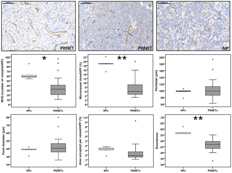
Fig. 1.
Angiogenesis in human pituitary neuroendocrine tumours and in normal pituitary. Microvessel density (MVD) and vasculature architecture parameters differences between human pituitary neuroendocrine tumours (PitNETs) and normal pituitaries (NPs) are shown. PitNETs (n = 24) and NPs (n = 5) tissue sections were stained for CD31. CD31 positive vessels were counted in three different high-power fields (HPF) to obtain MVD (number of vessels/HPF). CD31-stained ×20 magnification fields were analysed with ImageJ and vessel contour was manually traced in ImageJ in order to obtain the vasculature architecture parameters: total microvessel area, area occupied per vessel, vessel perimeter, vessel Feret’s diameter and roundness (vessel roundness correspond to a value comprised between 0 and 1, with 1 = perfect circle). Representative images of vessels from two PitNETs and one NP are shown (×20). Scale bar 100 µm. *p < 0.05, **p < 0.01, ***p < 0.001 (Mann–Whitney U test)

