

Abstract
Background & objectives: Hypertension can be induced by inhibiting nitric oxide synthesis with L-NAME, which also has a role in oxidative stress. Curcumin has strong antioxidant property. Our aim was to examine the possible preventive role of curcumin on renal dysfunction secondary to hypertension. Material & Methods: Twenty-four adult male Albino rats were divided in four groups: normal (N); curcumin (C; received curcumin 100 mg/kg/day by oral gavage for 10 weeks); hypertensive (H; received L-NAME 40 mg/kg/day in their drinking water for 4 weeks); and hypertensive-curcumin (HC; received L-NAME and curcumin). Arterial blood pressure was evaluated non-invasively for 4 weeks. Rats were then sacrificed for assessment of oxidative stress (catalase, lipid peroxidase, reduced glutathione and superoxide dismutase), renal function and structure, and biomarkers of apoptosis (Bcl-2 and caspase-3). AT1R expression and renal mtDNA integrity were also assessed. Results: Curcumin attenuated the effects of L-NAME on blood pressure and renal function. The renal histopathological changes observed in the L-NAME group were improved by curcumin administration. The expression of Bcl2 and caspase-3 was improved associated with downregulation of AT1R in curcumin treated groups. The antioxidant markers and mtDNA fragmentation show marked increase in hypertensive group which significantly decreased after curcumin treatment. Conclusion: Curcumin improved blood pressure elevation renal dysfunction. These improvements mediated through anti-oxidant capabilities and downregulation of AT1R favoring reduced apoptosis and preserved mitochondrial DNA.
Keywords: L-NAME, renal function, curcumin, oxidative stress, AT1R, mtDNA
Introduction
Blockade of NO formation with L-NAME results in impaired vascular function, exemplified by defective endothelial-dependent vasorelaxation, augmented angiotensin II-induced vasoconstriction, and an impaired antithrombotic effect. Moreover, increased oxidative stress has been reported in L-NAME hypertensive rats [1,2]. Administration of L-NAME increases vascular superoxide production and malondialdehyde (MDA) concentrations, with reduced plasma superoxide dismutase (SOD) activity [3]. In cardiovascular diseases, oxidative stress is linked to mitochondrial dysfunction. Furthermore, the anti-remodeling effects of some compounds have been incompletely associated with improved mitochondrial function; therefore, a major challenge to protect against tissue damage is to develop effective antioxidant therapeutics targeting mitochondria [4].
Hypertension is closely associated with progressive kidney dysfunction, manifested as glomerulosclerosis, interstitial fibrosis, proteinuria, and ultimately deteriorating glomerular filtration. It appears probable that tissue hypoxia in the hypertensive kidney increases oxidative stress as does ANG II and elevated pressure. Also, inhibition of ANG II activity, by either blocking ANG II type 1 receptors or angiotensin-converting enzyme, or by preventing oxidative stress by administration of antioxidants may results in improved blood pressure control and its consequences of kidney dysfunction [5,6]. Curcumin (diferuloylmethane) is a natural polyphenol compound isolated from Curcuma longa Linn. (Zingiberaceae), which is tree widely cultivated in the tropical regions of Asia. Curcumin is well recognized as a dietary spice for centuries and its pharmacological activities have been studied in various animal models and clinical studies including anti-inflammatory, anticancer, anti-diabetic, anti-dementia, and antioxidant properties [7,8]. Curcumin derives its effective antioxidant activity through its ability to both scavenge oxygen superoxide and hydroxyl radicals and initiation of antioxidant and cytoprotective enzymes [9]. Curcumin is able to scavenge reactive oxygen species (ROS) of both cytosolic and mitochondrial origin [4].
In this study, we explored the mechanisms by which curcumin prevents renal dysfunction secondary to hypertension, in order to assess its potential as a novel therapeutic intervention to maintain renal function in hypertensive patients.
Material and methods
Animals
Twenty-four adult male Albino rats (200-220 g) were used in this study. Animals were from the Ophthalmology Research Institute in Giza. Rats were housed in polyethylene cages (6/cage) in a 12-h light-dark cycle at controlled temperature and humidity ranges of 20-27°C and 40%-60%, respectively and provided with a typical chow diet and tap water. After delivery, rats were allowed to acclimatize for one week before the start of the study. The procedures and experimental protocols were reviewed and approved by the Suez Canal University Faculty of Medicine ethics committee.
Study groups
Rats were randomly assigned to 4 groups of 6 rats each. The normal group (N) received tap water. The curcumin group (C) received curcumin (100 mg/kg/day) by oral gavage for 10 weeks [10]. The hypertensive group (H) received L-NAME (40 mg/kg/day) in their drinking water for 4 weeks [11]. Finally, the hypertensive-curcumin group (HC) received curcumin (100 mg/kg/day) by gavage for 10 weeks. Hypertension was induced in 6th week by adding L-NAME (40 mg/kg/day) to their drinking water for 4 weeks. After 10 weeks, rats were sacrificed by overdose of the anesthetic drug. Blood samples were drawn from abdominal aorta to determine kidney function. The aorta was rapidly excised from the animal and used for quantitative assessment of AT1R. The right kidney was removed from each rat and fixed in 10% paraformaldehyde for histological analysis. The left kidney was removed, frozen and then homogenized for assessment of oxidative stress and antioxidant markers.
Measurement of arterial blood pressure
Blood Pressure (BP) was measured in conscious rats by tail-cuff plethysmography data acquisition analysis software from BIOPAC Systems. Rats were allowed to habituate for one week. Weekly measurement of blood pressure began with the induction of hypertension and continued until the end of the experiment.
Assay of nitrate/nitrite
Nitrate and nitrite, the end products of NO metabolism, were used as indices of nitric oxide synthase (NOS) activity. Accumulation of nitrate and nitrite was measured in plasma as described previously [12].
Assessment of renal function
Serum creatinine and blood urea nitrogen levels were measured at the end of the 10 weeks in all the study groups. These metabolites were measured using commercially available kits (Fortress diagnostics, UK). In addition, rats were placed in metabolic cages to collect urine, and urine creatinine and was using an autoanalyzer (Mindray BS-120 Chemistry Analyzer, China).
Renal histological & immunohistological assessment
Rats were sacrificed under general anesthesia. The right kidney was removed from each rat and then fixed in 10% formaldehyde and processed through paraffin embedding and prepared for histopathological & immunohistological analysis. Hematoxylin & Eosin stain (H&E) was used to visualize the general ultrastructure of the kidneys and reveal any histopathological changes in the renal cortices. Periodic Acid Schiff (PAS) stain was used to reveal interruption or thickening of the basal laminae with hyaline deposits. Masson’s trichrome stain was used to identify changes in the amount and location of the collagen fibers around blood vessels and in the renal interstitium. Five high power fields (×400) for five serial sections from each animal were examined for assessment of the normal architecture of the renal cortical glomeruli, tubules and the histopathological changes in the form of necrotic damage, glomerular collapse or loss, blood vessels dilatation and congestion, red blood cells (RBCs) extravasation, lymphocytic infiltration and hyaline deposition.
Apoptotic activity in the renal cortical tubules and glomeruli was assessed by immunohisochemical staining of bcl-2 and caspase-3 using a streptavidin biotin peroxidase method. Four-micrometer thick paraffin sections were incubated with either a monoclonal antibody to bcl-2 (DAKO, CA, USA, at a dilution of 1:40) or a polyclonal antibody to caspase-3 (Santa Cruz, USA, at a dilution 1:150). All incubations were achieved for 30 min at room temperture. Diaminobenzidine (Sigma Fast 3,3’-diaminobenzidine tablets, D-4293; Sigma, St. Louis, MO, USA) was used as the chromogen. Brown coloration of the cytoplasm was measured as positive reaction. Fiji image analyzer was used to measure the color area percentage of the greenish collagen, magenta stained basal laminae and the brown reaction in immunostaining.
Quantitative assessment of AT1R by ELISA
Freshly isolated blood vessels were homogenized in 1 mL sterile phosphate buffered saline (PBS; Sigma-Aldrich Ltd, cat. No. P7059) pH 7.4. [13] and the abundance of AT1R determined by ELISA (Rat Angiotensin II receptor 1 ELIZA Kit; Biospes Co. Ltd, cat No. BYEK1426) according to the manufacturer instruction.
Measurement of markers of oxidative in renal tissue
Catalase, lipid peroxidation (malondialdehyde, MDA), reduced glutathione and superoxide dismutase (SOD) were measured in renal tissues as markers of oxidative stress. Tissue samples were homogenized in four volumes of ice-cold Tris-HCl buffer (50 mM, pH 7.4) using an Ultra Turrax homogenizer (Ultra Turrax IKA T18 Basic, USA) for 2 min at 5,000 g at 4°C. Then the level of lipid peroxidase was measured. The homogenate was centrifuged at 5,000 g at 4°C for 60 min and aliquot of the supernatant reserved for assay of catalase activity. An equal volume of an ethanol/chloroform mixture (5:3, v/v) was added to the homogenate and the supernatant solution was extracted. This was elucidated by centrifugation at 5,000 g at 4°C for 30 min. The clear upper layer was collected and assessed for superoxide dismutase (SOD) [14].
Lipid peroxidation was measured using the method of [15] Draper and Hadley. The principle of the method is the spectrophotometric measurement of the color produced during the reaction with thiobarbituric acid (TBA) with lipid peroxides. For this purpose, 2.5 ml of 100 g/l trichloroacetic acid solution was added to 0.5 ml of homogenate in a centrifuge tube and placed in a boiling water bath for 15 min. After refrigeration in tap water, the mixture was centrifuged at 1,000× g for 10 min, and 2 ml of the supernatant was added to 1 ml of 6.7 g/l TBA solution in a test tube and placed in a boiling water bath for 15 min. The solution was then cooled with tap water, and its absorbance was measured using a spectrophotometer (UV-1601, Shimadzu, Japan) at 532 nm. The results were expressed as Nano moles lipid peroxides per gram protein in the renal tissue.
The method for the measurement of SOD was based on the principle that xanthine reacts with xanthine oxidase to generate superoxide radicals that react with 2-(4-iodophenyl)-3-(4-nitrophenol)-5-phenyltetrazolium chloride to form a red formazan dye. The SOD activity is then measured by the degree of inhibition of this reaction. Superoxide dismutase activity was expressed as units per gram protein [16]. The CAT activity was quantified in renal tissue by the method of Aebi [17]. The method is based on the determination of the rate constant (s-1, k) for H2O2 decomposition at 240 nm. Results were expressed as k (rate constant) per gram protein. Reduced glutathione (GSH) levels were measured in renal tissue, according to the method described by Beutler [18]. The amount of total GSH was determined from a standard curve obtained with known amounts of GSH standards. GSH levels were expressed as units per gram protein.
Assessment of renal mtDNA integrity
Isolation of mitochondria
Mitochondria were extracted by differential centrifugations following the method of Chappel and Hansford [19]. Tissue (100 mg) was homogenized in 0.25 M sucrose in 0.7 M Tris-HCl Buffer (pH 7.4) at 1 g tissue 9 ml of Tris-sucrose. EDTA was added to aid disruption of cells. Tissue homogenate was spun at 2500× g for 10 min to remove nuclei and unbroken cells. Supernatant fluid was decanted into centrifuge tubes and spun at 10,000× g for 10 min to form primary mitochondrial pellet. Supernatant fluid was poured, and the pellet was gently resuspended in 10 ml Tris-sucrose for washing. The pellet was recentrifuged and supernatant fluid was decanted. This washing cycle was repeated several times to improve the degree of mitochondrial purity. The final mitochondrial pellet was resuspended in 1 ml Tris-sucrose per gram of original sample.
Isolation of mitochondrial DNA (mtDNA)
Mitochondrial DNA (mtDNA) was isolated using the alkaline denaturation method performed as outlined by Birnboim and Doly [20]. mtDNA samples were subjected to 1% agarose gel electrophoresis at 4 V/cm using TAE solution (40 mM Tris-acetate, pH 8.0, and 1 mM EDTA) as a running buffer. The gel was visualized by ultraviolet trans-illumination and photographed after staining with 0.5 μg g/ml ethidium bromide. A 1 Kb DNA marker was loaded into the gel.
Statistical methods
Data were expressed as mean ± SD and analyzed using the Statistical Package of Social Sciences (SPSS program, version 20, SPSS Inc., Chicago, IL, USA). The difference of mean values among groups was assessed by using one-way analysis of variance (ANOVA) followed by Bonferroni’s multiple comparison test. All p values reported are two-tailed, and P < 0.05 was considered significant. All possible comparisons were made between groups.
Results
Mean arterial blood pressure (MABP)
The arterial blood pressure was measured serially in each treatment group. There was a significant increase in MABP in the hypertensive group in comparison to normal and curcumin groups at all time points. The curcumin- and L-NAME-treated group show a significant decrease in MABP beginning from the second week in comparison to the group receiving L-NAME alone (Figure 1).
Figure 1.
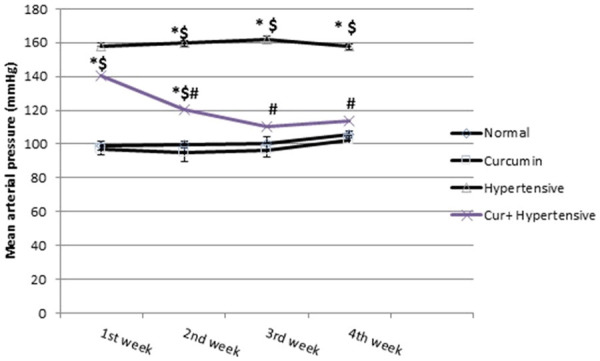
Mean Arterial blood pressure (MABP) in all studied groups. All data are expressed as mean ± SEM and analyzed using one-way ANOVA. *Significant in comparison to normal group. $Significant in comparison to Curcumin group. #Significant in comparison to Hypertensive group.
Plasma nitrate/nitrite concentrations
Plasma nitrate/nitrite levels from the different experimental groups are shown in Figure 2. In L-NAME-treated (hypertensive) rats, plasma nitrate/nitrite concentrations were significantly reduced when compared to those of normal and curcumin groups. Treatment of L-NAME rats with curcumin significantly restored the plasma nitrate/nitrite levels.
Figure 2.
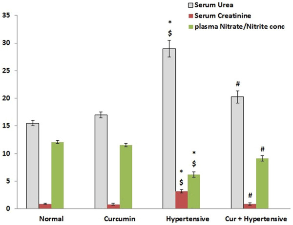
Kidney function tests (serum urea and Creatinine level) and Plasma nitrate/nitrite levels in all studied groups. All data are expressed as mean ± SEM and analyzed using one-way ANOVA. *Significant in comparison to normal group. $Significant in comparison to Curcumin group. #Significant in comparison to Hypertensive group.
Renal functions (serum creatinine and BUN)
Figure 2 shows the serum creatinine and urea in the study groups. Compared to the normal and curcumin groups, serum creatinine and BUN were significantly higher in the hypertensive group (P < 0.05), while the curcumin-treated group (cur + hypertensive) was lower than the hypertensive group (P < 0.05). The creatinine clearance was significantly decreased associated with significant increase in urinary albumin/creatinine ratio compared to the normal and curcumin groups. Treatment with curcumin significantly improve both creatinine clearance and urinary albumin levels (Table 1).
Table 1.
Creatinine clearance and urinary albumin/creatinine between the studied groups
| Normal Group | Curcumin Group | Hypertensive Group | Cur + Hypertensive Group | |
|---|---|---|---|---|
| Creatinine clearance [mL/min] | 2.4±0.2 | 2.9±0.1 | 0.5±0.1*,$ | 1.0±0.1*,$,# |
| Urinary albumin/creatinine [ng/mg] | 1.1±0.1 | 1.3±0.1 | 16.3±1.0*,$ | 9.6±0.7*,$,# |
All data are expressed as mean ± SEM and analyzed using one-way ANOVA.
Significant in comparison to normal group.
Significant in comparison to Curcumin group.
Significant in comparison to Hypertensive group.
Histopathological examination and immunostaining
Table 2 shows the percentage of frequency distribution of the histopathological changes between all study groups as revealed by H&E staining. There was significant tissue damage, congested capillaries associated with RBC extravasation and lymphocytic infiltration in the hypertensive group in comparison to the normal group. All these changes showed significant improvement in the treated group (cur + hypertensive). There were no significant differences between the normal and curcumin groups (Figure 3).
Table 2.
The percentage of frequency distribution of the histopathological changes between the studied groups
| Normal Group % | Curcumin Group % | Hypertensive Group % | Cur + Hypertensive Group % | |
|---|---|---|---|---|
| Congested/Dilated capillaries | 12 | 8 | 44* | 40* |
| RBCs Extravasation | 0 | 0 | 52* | 36* |
| Lymphocytic infiltration | 0 | 0 | 44* | 16*,$ |
| Thickened capillary walls | 0 | 0 | 52* | 8$ |
| PCT damage | 4 | 4 | 68* | 36*,$ |
| DCT damage | 4 | 4 | 68* | 40*,$ |
| Glomerular damage | 0 | 0 | 32* | 20* |
| Hyalinization | 0 | 0 | 56* | 20*,$ |
P < 0.05 (Significant) using chi square test.
Significant in comparison to normal group.
Significant in comparison to Hypertensive group.
Figure 3.

Photomicrographs of renal cortex of all study groups: (A) normal group, (B) curcumin group, (C) hypertensive group and (D) cur + hypertensive group. A &B show renal corpuscles with normal glomerular (G) blood capillaries covered by nuclei of podocytes and mesangial cells and surrounded with urinary space. Proximal convoluted (PT) and the distal convoluted tubules (DT) show normal appearance. It also shows normal blood arterioles (arrows). (C1) Shows collapsed glomeruli (G) or complete loss (LG). The PT and DT show shrinkage of their lining cells with pyknosis of their nuclei. In (C1-I) there is extravasation of RBCs in the renal interstitium (arrows), lymphocytic infiltration (green arrow) and dilated congested capillaries (*). Thickened wall of arterioles (black arrow), are shown in (C1-II). (C2) Shows hyaline material deposits in the renal interstitium (arrows), hydropic degeneration of the cells lining the PCTs (PT) and edematous arteriolar wall (green arrow). (D) Shows few corpuscles have lost glomeruli (★). It shows normal arteriolar wall (green arrow) but surrounded with dilated capillaries (*). No lymphocytic infiltration, RBCs extravasation or hyalinosis is present. H&E ×400.
PAS staining of the renal cortex of the hypertensive group revealed statistically significant increase in the magenta colored area associated with marked thickened basal laminae of The Bowman capsules and the glomeruli. There was significant decrease in the magenta colored area in the curcumin-treated hypertensive group in comparison to the untreated hypertensive group (Figure 4).
Figure 4.
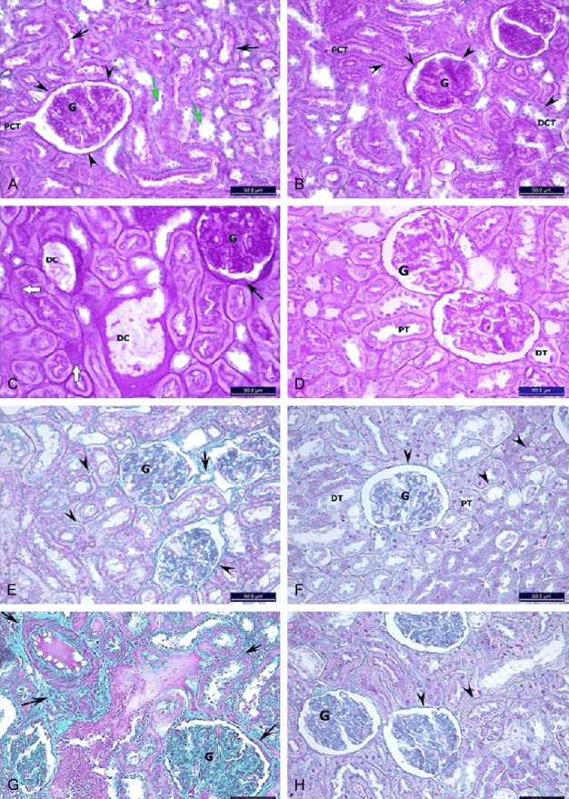
(A) Photomicrograph for the renal cortex of all study groups: (A-D) PAS ×400. (E-H) Masson’s trichrome ×400. (A, E) normal group, (B, F) curcumin group, (C, G) hypertensive group and (D, H) cur + hypertensive group. In (A, B) there are normal regular basal lamina of glomerular capillaries (G), urinary spaces (arrow heads), continuous with that of PCT whose cells have apical brush border (black arrows). It also shows normal basal lamina of the DCT whose cells has no brush border (green arrows). (C) Shows increase in the PAS stained material. The Bowman capsules show increased thickness of their basal laminae (arrow) and so the glomeruli (G). Thickening in the capillary walls (DC) with hyaline material depositions in the interstitium (white arrows). (D) Shows normal thickness of basal laminae around the renal tubules (PT&DT), renal corpuscles and glomerular capillaries (G). No hyaline deposits are present. (E, F) Have little amount of green material in the interstitium, around tubules and corpuscles (arrow heads), surrounding the arterioles (arrow) and in the mesangium (G). (G) Shows increase in the greenish collagen content around the arterioles, renal tubules and corpuscles (arrows) and in the glomerular mesangium (G). (H) Shows normal distribution and amount of greenish collagen. (B) Mean PAS and Masson stain intensity percentage was quantified by Fiji image analyzer software. All data are expressed as mean ± SEM and analyzed using one-way ANOVA. *Significant in comparison to normal group. $Significant in comparison to Curcumin group. #Significant in comparison to Hypertensive group.
Masson’s trichrome stain revealed the presence of only small amounts of collagen in the renal interstitial, around tubules, corpuscles, arterioles and mesangium in normal and curcumin groups. There was a significant increase in collagen staining in the untreated hypertensive group, which was reversed in the curcumin-treated hypertensive group (Figure 4).
The expression levels of Bcl-2 (anti-apoptotic gene) and caspase-3, which is an apoptosis coordination enzyme in renal tissue was analyzed by immunohistochemistry. The expression of Bcl2 in the hypertensive group was significantly decreased, while the expression of caspase-3 in the hypertensive group was significantly increased when compared to the normal, curcumin, and curcumin-treated hypertensive groups. This means that the apoptosis increased in the hypertensive group and was improved by curcumin treatment (Figure 5).
Figure 5.
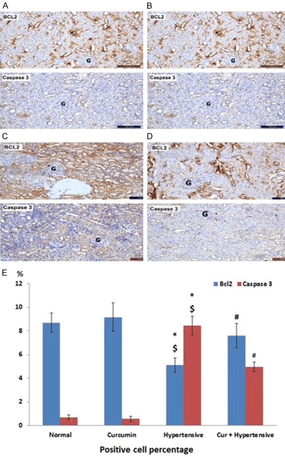
Phptomicrograph of the renal cortex of all study groups: (A) normal group, (B) curcumin group, (C) hypertensive group, (D) cur + hypertensive group. Brown cytoplasmic reaction in the cells linining the tubules. The glomeruli (G) show negative reaction in both stains in all groups. BCL2 & Caspase 3 immunostaining ×200. (E) Bcl-2 and caspase-3 immunohistochemical intensity was quantified by Fiji image analyzer software. All data are expressed as mean ± SEM and analyzed using one-way ANOVA. *Significant in comparison to normal group. $Significant in comparison to Curcumin group. #Significant in comparison to Hypertensive group.
Quantitative assessment of AT1R by ELISA
Compared to the normal and curcumin groups, AT1R immunoreactivity was significantly more abundant in the untreated hypertensive group (P < 0.05), while the curcumin-treated hypertensive group (cur + hypertensive) showed no increase in AT1R (Figure 6).
Figure 6.
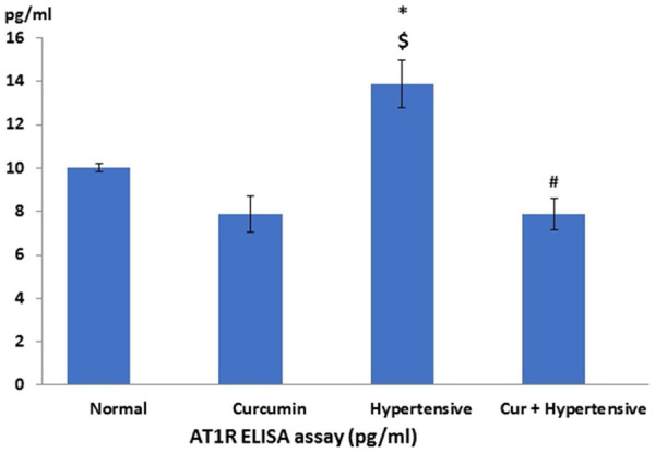
AT1R expression in blood vessels of all studied groups. All data are expressed as mean ± SEM and analyzed using one-way ANOVA. *Significant in comparison to normal group. $Significant in comparison to Curcumin group. #Significant in comparison to Hypertensive group.
Oxidative stress markers in renal tissues
The results of the SOD, catalase, reduced glutathione and MDA analyses are shown in Figure 7. Compared to the normal and curcumin groups, the levels of SOD and reduced glutathione were significantly lower in the renal tissue of the untreated hypertensive group (Figure 7). The curcumin-treated hypertensive group showed a significant increase compared to the untreated hypertensive group. The amount of MDA, reflecting lipid peroxidation, in renal tissues was significantly higher in the untreated hypertensive group when compared to the normal and curcumin groups. No increase in MDA level was observed in the curcumin-treated hypertensive rats relative to the non-hypertensive groups.
Figure 7.
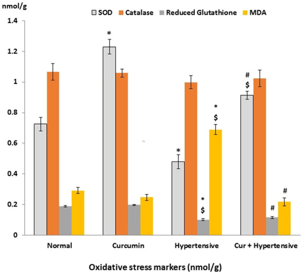
Oxidative stress level in renal tissue of all studied groups. All data are expressed as mean ± SEM and analyzed using one-way ANOVA and Bonferroni post-hoc test. *Significant in comparison to normal group. $Significant in comparison to Curcumin group. #Significant in comparison to Hypertensive group.
Renal mitochondrial DNA integrity
In the normal and curcumin groups, the intact form of mtDNA migrated as a major band of approximately 16.5 kb when electrophoresed (Figure 8, lanes N, C). On the other hand, hypertension markedly reduced the amounts of intact mtDNA in the renal tissue (lane H). However, mtDNA from curcumin-treated hypertensive group (lane HC) migrated as its intact form (Figure 8).
Figure 8.
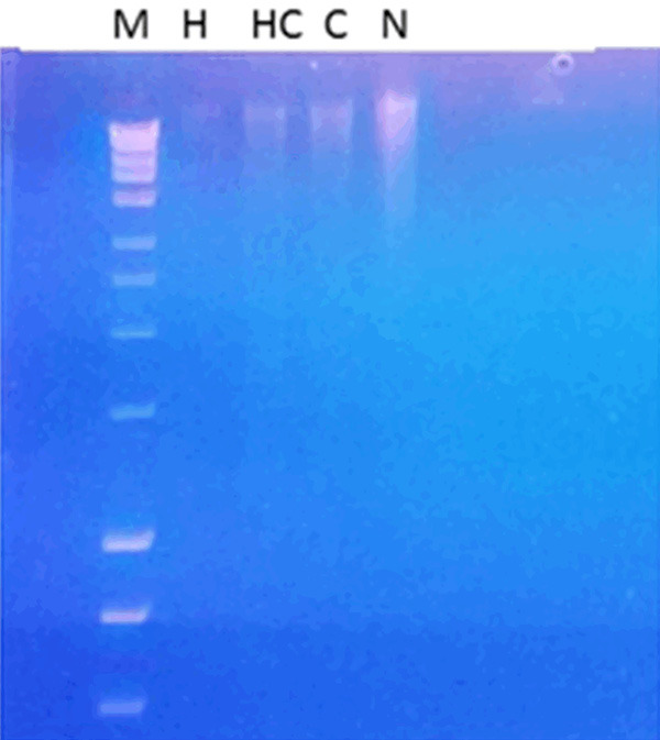
Mitochondrial DNA (mtDNA) in the experimental groups, Lane M, 1 kb DNA ladder; lane N: intact mtDNA isolated from normal group; lane C: mtDNA samples isolated from curcumin group; lane H: mtDNA samples isolated from hypertensive group and lanes HC: mtDNA sample isolated from treated group.
Discussion
The use of natural products such as curcumin to protect renal function may present an efficient and affordable method to protect against harmful effects of hypertension [21]. Curcumin shows therapeutic potential against cancer, lung, renal, neurological, liver, metabolic and cardiovascular diseases [22,23] through its effects differ [21,24]. It is a well-studied natural product with a documented safety profile [25]. In hypertension, the progressive renal impairment, where either dialysis or transplantation become necessary, is inevitable. Both options may be expensive or unavailable in many developing countries [26,27]. Nitric oxide (NO) production is one of the most important defense mechanisms against hypertension [28]. The primary protective mechanisms of curcumin against the renal damage casue by (NO) deficiency are unknown. Hence, we postulated that co-administration of curcumin in an L-NAME-induced model of hypertension would ameliorate most of the renal adverse effects associated with this model. The L-NAME model pf hypertension mimics hypertension in humans, so it is suitable to study the renal effects of curcumin in this model [29]. The active metabolite of L-NAME binds competitively to eNOS [30]. NOS inhibition attenuates both the synthesis and metabolism of NO, which normally mediates vascular relaxation. NO deficiency leads to systemic vasoconstriction and hypertension [31]. In this context, serial measurements of blood pressure showed a progressive increase starting from the end of the first week reaching the peak by the end of the 4th week of L-NAME administration (Figure 1). These findings matched those of Abdelrahman [32]. Curcumin treatment significantly decreased blood pressure in the hypertensive group starting from the 2nd week of L-NAME administration: blood pressure in this group had returned to normal values by the end of the experiment. These findings suggest curcumin may have increased serum nitrite/nitrate level in L-NAME treated rats (Figure 2), resulting in a decrease in systemic oxidative stress markers (Figure 7) and higher bioavailability of NO. Previous studies also demonstrated that curcumin lowers blood pressure in hypertensive rat models by comparable NO dependent mechanisms [28,33]. In these studies, curcumin may have increased NO bioavailability induced by reducing the abundance of ROS [34,35]. We found that L-NAME administration for four weeks in rats significantly increased MDA levels which were accompanied by a decrease in serum nitrite/nitrate and by down regulation of renal SOD and reduced glutathione activity, suggestive of high oxidative stress and impairment of antioxidant defenses. Earlier studies [36,37] established that NO deficiency causes excessive production of ROS and persistent oxidative stress with impaired renal antioxidant defense through inactivation of antioxidant enzymes. Our findings further demonstrate that curcumin administration increased SOD activity in kidneys of L-NAME-treated rats, and therefore counteracted renal oxidative injury. Restored GS reductase contents in renal tissues and normalized MDA content was found to be a potent antioxidant and very effective in reducing ROS production [38].
Collectively, these results support the findings by Ali and his colleges [39], who demonstrated that the antioxidant properties of curcumin may result from its ability to scavenge ROS, the increase in NO bioavailability, and the enhancement of the antioxidant defense system. Because curcumin increases Nrf2 expression in the kidney [40], the activation of this transcription factor is involved in the regulation of cytoprotective genes encoding antioxidant proteins which can antagonize oxidative damage [41]. Subsequently, oxidative stress fortifies renal dysfunction [42] as found in our study. Interestingly, we reported that the beneficial action of curcumin encompassed functional, biochemical and histological markers of hypertensive renal damage in L-NAME treated rats. The tubular necrosis and the occurrence of apoptosis (markers for early events in kidney damage) [43], as well as, the chronic markers including interstitial fibrosis and the sclerotic glomeruli seen in the histopathological staining, albuminuria and uremic toxins [44] were significantly improved by Curcumin administration. We found renal insult in rats with L-NAME characterized by structural and functional alterations in the form of glomerulosclerosis, tubulointerstitial injury, fibrosis and declining glomerular filtration. The present albuminuria and glomerulosclerosis reflect elevations in the mean glomerular transcapillary pressure and flows [45,46]; barotrauma caused by increased capillary pressure due to progressive hypertension parallel to L-NAME administration. Oxidant stress [47], in addition to, AT1R up-regulation (Figure 6) enhancing ANGII activity with altered oxygen availability in the hypertensive kidney [48] may induce the renal tissue lymphocytic infiltration, fibrosis and tubular atrophy (Figures 5, 6 and 7). The L-NAME-treated hypertensive group showed significant decrease in expression of Bcl-2, while the expression of caspase-3 significantly increased. Oxidative stress triggers caspase-3 activation leading to apoptosis. Curcumin restored the balance of pro-apoptotic (caspasce-3) and anti-apoptotic (Bcl-2) proteins which acts as an antioxidant in cells scavanging ROS [49]. In this work, L-NAME induced hypertension was associated with significant increase in the abundance of AT1R, resulting in vasoconstriction and reduction of vascular compliance with subsequent elevated blood pressure. The inhibition of NO production by L-NAME may exaggerate the effect of ROS resulting in more endothelial dysfunction [50]. Moreover, oxidative stress increases AT1R expression in the kidney [51] so, it was not surprising that treatment with curcumin was accompanied by significant down regulation of AT1R, which augments the amelioration of oxidant/anti-oxidant status and NO bioavailability as shown earlier. These data are consistent with many studies showing the anti-oxidative effect of curcumin in hypertensive rats [11,52-54].
Based on the previous findings, increased vascular tone induced by ANGII limited oxygen delivery (renal tissue hypoxia) with further increases in oxidative stress, resulting generally in increased glomerular injury [55], specifically in mitochondrial injury [34]. Besides, the ultimate preservation of mitochondrial content is a consequence of increasing antioxidant system activity. These data are supported by findings of [56]. Moreover, NO inhibition can directly cause renal tissue hypoxia. It seems that L-NAME induced kidney damage contributes to many factors. In this sense, we demonstrated that the preservation of intact mitochondrial DNA plays a key role in the protective effect of curcumin co-treatment against L-NAME induced renal oxidant damage. Curcumin treatment in the L-NAME hypertension model reduced the amount of mtDNA degradation observed in the untreated hypertensive group.
DNA base oxidation by ROS attack and/or damage of repair enzymes can be claimed [57]. These data were in line with antigenotoxic property of herbs extract against mitochondrial DNA damage [24,58].
Based on kidney functional, biochemical and pathological findings, it seems fair to conclude that the ameliorative effect of Curcumin on L-NAME induced hypertensive renal damage is mediated through anti-oxidant capabilities and downregulation of AT1R favoring reduced apoptosis and preserved mitochondrial DNA.
Acknowledgements
The authors would like to thank Bruce G. Allen, PhD-Professor, Department of Medicine, University of Montreal, Canada for providing meticulous revision of the manuscript.
Disclosure of conflict of interest
None.
References
- 1.Kitamoto S, Egashira K, Kataoka C, Usui M, Koyanagi M, Takemoto M, Takeshita A. Chronic inhibition of nitric oxide synthesis in rats increases aortic superoxide anion production via the action of angiotensin II. J Hypertens. 2000;18:1795–1800. doi: 10.1097/00004872-200018120-00013. [DOI] [PubMed] [Google Scholar]
- 2.Tsukahara H, Hiraoka M, Kobata R, Hata I, Ohshima Y, Jiang MZ, Noiri E, Mayumi M. Increased oxidative stress in rats with chronic nitric oxide depletion: measurement of urinary 8-hydroxy-2’-deoxyguanosine excretion. Redox Rep. 2000;5:23–28. doi: 10.1179/rer.2000.5.1.23. [DOI] [PubMed] [Google Scholar]
- 3.Priviero F, Teixeira C, Claudino M, De Nucci G, Zanesco A, Antunes E. Vascular effects of long-term propranolol administration after chronic nitric oxide blockade. Eur J Pharmacol. 2007;571:189–196. doi: 10.1016/j.ejphar.2007.05.060. [DOI] [PubMed] [Google Scholar]
- 4.Correa F, Buelna-Chontal M, Hernández-Reséndiz S, García-Niño WR, Roldán FJ, Soto V, Silva-Palacios A, Amador A, Pedraza-Chaverrí J, Tapia E, Zazueta C. Curcumin maintains cardiac and mitochondrial function in chronic kidney disease. Free Radic Biol Med. 2013;61:119–129. doi: 10.1016/j.freeradbiomed.2013.03.017. [DOI] [PubMed] [Google Scholar]
- 5.Fredrik P, Lina N. Renal oxidative stress, oxygenation, and hypertension. Am J Physiol Regul Integr Comp Physiol. 2011;301:R1229–R1241. doi: 10.1152/ajpregu.00720.2010. [DOI] [PMC free article] [PubMed] [Google Scholar]
- 6.Leong C, Anderson W, O’Connor P, Evans R. Evidence that renal arterial-venous oxygen shunting contributes to dynamic regulation of renal oxygenation. Am J Physiol Renal Physiol. 2007;292:F1726–F1733. doi: 10.1152/ajprenal.00436.2006. [DOI] [PubMed] [Google Scholar]
- 7.Aggarwal BB, Sung B. Pharmacological basis for the role of curcumin in chronic diseases: an age-old spice with modern targets. Trends Pharmacol Sci. 2009;30:85–94. doi: 10.1016/j.tips.2008.11.002. [DOI] [PubMed] [Google Scholar]
- 8.Rinwa P, Kaur B, Jaggi AS, Singh N. Involvement of PPARgamma in curcuminmediated beneficial effects in experimental dementia. Naunyn Schmiedebergs Arch Pharmacol. 2010;381:529–539. doi: 10.1007/s00210-010-0511-z. [DOI] [PubMed] [Google Scholar]
- 9.Nakmareong S, Kukongviriyapan U, Pakdeechote P, Donpunha W, Kukongviriyapan V, Kongyingyoes B, Sompamit K, Phisalaphong C. Antioxidant and vascular protective effects of curcumin and tetrahydrocurcumin in rats with L-NAME-induced hypertension. Naunyn Schmiedebergs Arch Pharmacol. 2011;383:519–529. doi: 10.1007/s00210-011-0624-z. [DOI] [PubMed] [Google Scholar]
- 10.Sompamit K, Kukongviriyapan U, Nakmareong S, Pannangpetch P, Kukongviriyapan V. Curcumin improves vascular function and alleviates oxidative stress in non-lethal lipopolysaccharideinduced endotoxaemia in mice. Eur J Pharmacol. 2009;616:192–199. doi: 10.1016/j.ejphar.2009.06.014. [DOI] [PubMed] [Google Scholar]
- 11.Nakmareong S, Kukongviriyapan U, Pakdeechote P, Kukongviriyapan V, Kongyingyoes B, Donpunha W, Prachaney P, Phisalaphong C. Tetrahydrocurcumin alleviates hypertension, aortic stiffening and oxidative stress in rats with nitric oxide deficiency. Hypertens Res. 2012;35:418–425. doi: 10.1038/hr.2011.180. [DOI] [PubMed] [Google Scholar]
- 12.Kukongviriyapan V, Somparn N, Senggunprai L, Prawan A, Kukongviriyapan U, Jetsrisuparb A. Endothelial dysfunction and oxidant status in pediatric patients with hemoglobin E-beta thalassemia. Pediatr Cardiol. 2008;29:130–135. doi: 10.1007/s00246-007-9107-x. [DOI] [PubMed] [Google Scholar]
- 13.Uehara Y, Takada S, Hnrwa N, Kawabata Y, Ohshnna N, Numabe A, Ishnmtsu T, Goco A, Yagl S, Omata M. Vasoconstrictors and renal protection Induced by Bl-selective adreno-rceptor antagonist bisoprolol. J Cardiovasc Pharmacol. 1994;23:897–90. doi: 10.1097/00005344-199406000-00007. [DOI] [PubMed] [Google Scholar]
- 14.Abogresha N, Greish S, Zakareya E, Khalil W. Remote effect of kidney ischemia-reperfusion injury on pancreas: role of oxidative stress, mitochondrial apoptosis. Arch Med Sci. 2016;12:2. doi: 10.5114/aoms.2015.48130. [DOI] [PMC free article] [PubMed] [Google Scholar]
- 15.Draper HH, Hadley M. Malondialdehyde determination as index of lipid peroxidation. Methods Enzymol. 1990;186:421–31. doi: 10.1016/0076-6879(90)86135-i. [DOI] [PubMed] [Google Scholar]
- 16.Woolliams JA, Wiener G, Anderson PH, McMurray CH. Variation in the activities of glutathione peroxidase and superoxide dismutase and in the concentration of copper in the blood in various breed crosses of sheep. Res Vet Sci. 1983;34:253–6. [PubMed] [Google Scholar]
- 17.Aebi H. Catalase in vitro. Methods Enzymol. 1984;105:121–6. doi: 10.1016/s0076-6879(84)05016-3. [DOI] [PubMed] [Google Scholar]
- 18.Beutler E, Duron O, Kelly BM. Improved method for the determination of blood glutathione. J Lab Clin Med. 1963;61:882–8. [PubMed] [Google Scholar]
- 19.Chappel JB, Hansford RG. Subcellular components. London: Butterworths; 1969. [Google Scholar]
- 20.Birnboim HC, Doly J. A rapid alkaline extraction procedure for screening recombinant plasmid DNA. Nucleic Acids Res. 1979;7:1513–23. doi: 10.1093/nar/7.6.1513. [DOI] [PMC free article] [PubMed] [Google Scholar]
- 21.Trujillo J, Chirino YI, Molina-Jijon E, Anderica-Romero AC, Tapia E, Pedraza-Chaverri J. Renoprotective effect of the antioxidant curcumin: recent findings. Redox Biol. 2013;1:448–56. doi: 10.1016/j.redox.2013.09.003. [DOI] [PMC free article] [PubMed] [Google Scholar]
- 22.Gupta S, Prasad S, Kim J, Patchva S, Webb L, Priyadarsini I, Aggarwal B. Multitargeting by curcumin as revealed by molecular interaction studies. Nat Prod Rep. 2011;28:1937–1955. doi: 10.1039/c1np00051a. [DOI] [PMC free article] [PubMed] [Google Scholar]
- 23.Kannappan R, Gupta S, Kim J, Reuter S, Aggarwal B. Neuroprotection by spice-derived nutraceuticals: you are what you eat! Mol Neurobiol. 2011;44:142–159. doi: 10.1007/s12035-011-8168-2. [DOI] [PMC free article] [PubMed] [Google Scholar]
- 24.Molina-Jijón E, Tapia E, Zazueta C, ElHafidi M, Zatarain-Barrón Z, Hernández-Pando R, Medina-Campos O, Zarco-Márquez G, Torres I, Pedraza-Chaverri J. Curcumin prevents Cr (VI)-induced renal oxidant damage by a mitochondrial pathway. Free Radic Biol Med. 2011;51:1543–1557. doi: 10.1016/j.freeradbiomed.2011.07.018. [DOI] [PubMed] [Google Scholar]
- 25.Gupta SC, Patchva S, Koh W, Aggarwal BB. Discovery of curcumin, a component of golden spice, and its miraculous biological activities. Clin Exp Pharmacol Physiol. 2012;39:283–99. doi: 10.1111/j.1440-1681.2011.05648.x. [DOI] [PMC free article] [PubMed] [Google Scholar]
- 26.Ahmad QZ, Jahan N, Ahmad G, Tajuddin An appraisal of nephroprotection and the scope of natural products in combating renal disorders. J Nephrol Ther. 2014;23:773–81. [Google Scholar]
- 27.Muralidharan A, White S. The need for kidney transplantation in low- and middle-income countries in 2012: an epidemiological perspective. Transplantation. 2015;99:476–81. doi: 10.1097/TP.0000000000000657. [DOI] [PubMed] [Google Scholar]
- 28.Husian K. Interaction of exercise training and chronic NOS inhibition on blood pressure, heart rate, NO and antioxidants in plasma of rats. Pathophysiology. 2003;10:47–56. doi: 10.1016/j.pathophys.2003.06.001. [DOI] [PubMed] [Google Scholar]
- 29.Ramanathan V, Thekkumalai M. Role of chrysin on hepatic and renal activities of Nω-nitro-l-arginine-methylester induced hypertensive rats. Int J Nutr Pharmacol Neurol Dis. 2014;4:58–63. [Google Scholar]
- 30.Ramaswami G, Chai H, Yao Q, Lin PH, Lumsden AB, Chen C. Curcumin blocks homocysteine-induced endothelial dysfunction in porcine coronary arteries. J Vasc Surg. 2004;40:1216–1222. doi: 10.1016/j.jvs.2004.09.021. [DOI] [PubMed] [Google Scholar]
- 31.Hopkins A, Lamm M, Funk J, Ritenbaugh C. Hibiscus sabdariffa L. in the treatment of hypertension and hyperlipidemia: a comprehensive review of animal and human studies. Fitoterapia. 2013;85:84–94. doi: 10.1016/j.fitote.2013.01.003. [DOI] [PMC free article] [PubMed] [Google Scholar]
- 32.Abdel-Rahman RF, Hessin AF, Abdelbaset M, Ogaly HA, Abd-Elsalam RM, Hassan SM. Antihypertensive effects of roselle-olive combination in L-NAME-induced hypertensive rats. Oxid Med Cell Longev. 2017;2017:9460653. doi: 10.1155/2017/9460653. [DOI] [PMC free article] [PubMed] [Google Scholar]
- 33.Kum O, Senturk UK, Kocer G, Ozdem S, Baskurt OK, Cetin A, Yesilkaya A, Gunduz F. Effect of exercise training on resistance arteries in rats with chronic NOS inhibition. J Appl Physiol. 2009;107:896–902. doi: 10.1152/japplphysiol.91180.2008. [DOI] [PubMed] [Google Scholar]
- 34.Correa F, Buelna-Chontal M, Hernández-Reséndiz S, García-Niño WR, Roldán FJ, Soto V, Silva-Palacios A, Amador A, Pedraza-Chaverrí J, Tapia E, Zazueta C. Curcumin maintains cardiac and mitochondrial function in chronic kidney disease. Free Radic Biol Med. 2013;61:119–29. doi: 10.1016/j.freeradbiomed.2013.03.017. [DOI] [PubMed] [Google Scholar]
- 35.Shi X, Guan Y, Jiang S, Li T, Sun B, Cheng H. Renin-angiotensin system inhibitor attenuates oxidative stress induced human coronary artery endothelial cell dysfunction via the PI3K/AKT/mTOR pathway. Arch Med Sci. 2018;15:152–164. doi: 10.5114/aoms.2018.74026. [DOI] [PMC free article] [PubMed] [Google Scholar]
- 36.Quiroz Y, Pons H, Gordon KL, Rincón J, Chavez M, Parra G, Herrera-Acosta J, Gomez-Garrea D, Largo R, Egido J, Johnson RJ, Rodriguez-Iturbe B. Mycophenolate mofetil prevents salt-sensitive hypertension resulting from nitric oxide synthesis inhibition. Am J Physiol Renal Physiol. 2001;281:F38–47. doi: 10.1152/ajprenal.2001.281.1.F38. [DOI] [PubMed] [Google Scholar]
- 37.Vaziri ND. Causal link between oxidative stress, inflammation, and hypertension. Iran J Kidney Dis. 2008;2:1–10. [PubMed] [Google Scholar]
- 38.Porkert M, Sher S, Reddy U, Cheema F, Niessner C, Kolm P, Jones DP, Hooper C, Taylor WR, Harrison D, Quyyumi AA. Tetrahydrobiopterin: a novel antihypertensive therapy. J Hum Hypertens. 2008;22:401–407. doi: 10.1038/sj.jhh.1002329. [DOI] [PubMed] [Google Scholar]
- 39.Ali BH, Al-Salam S, Al Suleimani Y, Al Kalbani J, Al Bahlani S, Ashique M, Manoj P, Al Dhahli B, Al Abri N, Naser HT, Yasin J, Nemmar A, Al Za’abi M, Hartmann C, Schupp N. Curcumin ameliorates kidney function and oxidative stress in experimental chronic kidney disease. Basic Clin Pharmacol Toxicol. 2018;122:65–73. doi: 10.1111/bcpt.12817. [DOI] [PubMed] [Google Scholar]
- 40.Tapia E, Zatarain-Barrón ZL, Hernández-Pando R, Zarco-Márquez G, Molina-Jijón E, Cristóbal-García M, Santamaría J, Pedraza-Chaverri J. Curcumin reverses glomerular hemodynamic alterations and oxidant stress in 5/6 nephrectomized rats. Phytomedicine. 2013;20:359–66. doi: 10.1016/j.phymed.2012.11.014. [DOI] [PubMed] [Google Scholar]
- 41.Kim H, Vaziri N. Contribution of impaired Nrf2-Keap1 pathway to oxidative stress and inflammation in chronic renal failure. Am J Physiol Renal Physio. 2010;298:F662–F671. doi: 10.1152/ajprenal.00421.2009. [DOI] [PubMed] [Google Scholar]
- 42.Shuvy M, Nyska A, Beeri R, Abedat S, Gal-Moscovici A, Rajamannan NM, Lotan C. Histopathology and apoptosis in an animal model of reversible renal injury. Exp Toxicol Pathol. 2011;63:303–6. doi: 10.1016/j.etp.2010.02.002. [DOI] [PMC free article] [PubMed] [Google Scholar]
- 43.Vanmassenhove J, Vanholder R, Nagler E, Van Biesen W. Urinary and serum biomarkers for the diagnosis of acute kidney injury: an in-depth review of the literature. Nephrol Dial Transplant. 2013;28:254–73. doi: 10.1093/ndt/gfs380. [DOI] [PubMed] [Google Scholar]
- 44.Webster A, Nagler E, Morton R, Masson P. Chronic kidney disease. Lancet. 2016;389:1238–52. doi: 10.1016/S0140-6736(16)32064-5. [DOI] [PubMed] [Google Scholar]
- 45.Anderson S, Rennke HG, Brenner BM. Antihypertensive therapy must control glomerular hypertension to limit glomerular injury. J Hyperten. 1986;4:S242–4. [PubMed] [Google Scholar]
- 46.Lee LK, Meyer TW, Pollock AS, Lovett DH. Endothelial cell injury initiates glomerular sclerosis in the rat remnant kidney. J Clin Invest. 1995;96:953–964. doi: 10.1172/JCI118143. [DOI] [PMC free article] [PubMed] [Google Scholar]
- 47.Small D, Coombes J, Bennett N, Johnson D, Gobe G. Oxidative stress, anti-oxidant therapies and chronic kidney disease. Nephrol. 2012;17:311–321. doi: 10.1111/j.1440-1797.2012.01572.x. [DOI] [PubMed] [Google Scholar]
- 48.Emans TW, Janssen BJ, Pinkham MI, Ow CP, Evans RG, Joles JA, Malpas SC, Krediet CT, Koeners MP. Exogenous and endogenous angiotensinm II decrease renal cortical oxygen tension in conscious rats by limiting renal blood flow. J Physiol. 2016;594:6287–6300. doi: 10.1113/JP270731. [DOI] [PMC free article] [PubMed] [Google Scholar]
- 49.Nathan S, Liyun Z, Daciana M, David M. Bcl-2 family proteins as regulators of oxidative stress. Semin Cancer Biol. 2009;19:42–49. doi: 10.1016/j.semcancer.2008.12.002. [DOI] [PMC free article] [PubMed] [Google Scholar]
- 50.Zalba G, San Jose G, Moreno MU, Fortuno MA, Fortuno A, Beaumont FJ, Diez J. Oxidative stress in arterial hypertension: role of NAD(P)H oxidase. Hypertension. 2001;38:1395–1399. doi: 10.1161/hy1201.099611. [DOI] [PubMed] [Google Scholar]
- 51.Chugh G, Lokhandwala M, Asghar M. Oxidative stress alters renal D1 and AT1 receptor functions and increases blood pressure in old rats. Am J Physiol Renal Physiol. 2011;300:F133–138. doi: 10.1152/ajprenal.00465.2010. [DOI] [PMC free article] [PubMed] [Google Scholar]
- 52.Yang Y, Duan W, Liang Z, Yi W, Yan J, Wang N, Li Y, Chen W, Yu S, Jin Z, Yi D. Curcumin attenuates endothelial cell oxidative stress injury through Notch signaling inhibition. Cell Signal. 2013;25:615–629. doi: 10.1016/j.cellsig.2012.11.025. [DOI] [PubMed] [Google Scholar]
- 53.Boonla O, Kukongviriyapan U, Pakdeechote P, Kukongviriyapan V, Pannangpetch P, Prachaney P, Greenwald S. Curcumin improves endothelial dysfunction and vascular remodeling in 2K-1C hypertensive rats by raising nitric oxide availability and reducing oxidative stress. Nitric Oxide. 2014;42:44–53. doi: 10.1016/j.niox.2014.09.001. [DOI] [PubMed] [Google Scholar]
- 54.Yao Y, Wang W, Li M, Ren H, Chen C, Wang J, Wang WE, Yang J, Zeng C. Curcumin exerts its anti-hypertensive effect by down-regulating the AT1 receptor in vascular smooth muscle cells. Sci Rep. 2016;6:25579. doi: 10.1038/srep25579. [DOI] [PMC free article] [PubMed] [Google Scholar]
- 55.Ratliff BB, Abdulmahdi W, Pawar R, Wolin MS. Oxidant mechanisms in renal injury and disease. Antioxid Redox Signal. 2016;25:119–146. doi: 10.1089/ars.2016.6665. [DOI] [PMC free article] [PubMed] [Google Scholar]
- 56.Vaziri N, Bai Y, Ni Z, Quiroz Y, Pandian R, Rodriguez-Iturbe B. Intra-renal angiotensin II/AT1 receptor, oxidative stress, inflammation, and progressive injury in renal mass reduction. J Pharmacol Exp Therap. 2007;323:85–93. doi: 10.1124/jpet.107.123638. [DOI] [PubMed] [Google Scholar]
- 57.Montezano A, Dulak-Lis M, Tsiropoulou S, Harvey A, Briones A, Touyz R. Oxidative stress and human hypertension: vascular mechanisms, biomarkers, and novel therapies. Canad J Cardiol. 2015;31:631–641. doi: 10.1016/j.cjca.2015.02.008. [DOI] [PubMed] [Google Scholar]
- 58.Ghosh I, Poddar S, Mukherjee A. Evaluation of the protective effect of hibiscus sabdariffa L. calyx (malvaceae) extract on arsenic induced genotoxicity in mice and analysis of its antioxidant properties. Biol Med. 2015;7:228. [Google Scholar]