Abstract
Giant unilamellar vesicles (GUVs) are a useful platform for reconstituting and studying membrane-bound biological systems, offering reduced complexity compared to living cells. Several techniques exist to form GUVs and populate them with biomolecules of interest. However, a persistent challenge is the ability to efficiently and reliably load solutions of biological macromolecules, organelle-like membranes, and/or micrometer-scale particles with controlled stoichiometry in the encapsulated volume of GUVs. Here, we demonstrate the use of acoustic streaming from high-intensity focused ultrasound to make and load GUVs from bulk solutions, without the need for nozzles that can become clogged or otherwise alter the solution composition. In this method, a compact acoustic lens is focused on a planar lipid bilayer formed between two aqueous solutions. The actuation of a planar piezoelectric material coupled to the lens accelerates a small volume of liquid, deforming the bilayer and forming a GUV containing the solution on the transducer side of the bilayer. As demonstrated here, acoustic jetting offers an alternative method for the generation of GUVs for biological and biophysical studies.
For many biological and biophysical studies of membrane-bound systems, giant unilamellar vesicles (GUVs) are a convenient tool. GUVs are relatively simple compared to systems derived from living cells, yet they preserve the fluidity of lipids in the membrane, encapsulate a distinct volume, and support the function of transmembrane proteins. By using GUVs as a platform, researchers have successfully reconstituted and studied biophysical phenomena such as liquid phase separation within membranes,1 protein-mediated membrane deformations,2 and segregation of proteins at membrane–membrane junctions.3
The ability to construct increasingly complex GUV systems containing nested organelle-like membranes, micrometer-scale particles, or embedded membrane proteins is an ongoing challenge for GUV formation techniques. Electroformation, for instance, is an established and effective method for creating GUVs4 and can be performed in high ionic solutions,5–7 but it yields polydisperse vesicles and does not offer control over the loading of solutions. Several methods have been developed to load biomacromolecules and particles into electroformed GUVs,8–10 but achieving controlled concentrations, defining solution stoichiometries, and incorporating functional membrane proteins are enduring challenges.11 GUVs can also be formed through techniques that employ inverted emulsions12,13 and water–oil–water double emulsions.14–16 Emulsion techniques allow the control of the contents of the GUVs, but these techniques typically leave residual oil in the bilayers and expose contents to an ephemeral, bare oil–water interface that may drive proteins to denature.17 Several other methods for forming GUVs exist, each with advantages and disadvantages for specific applications.18–21 Forming GUVs via microfluidic jetting22 enables the loading of vesicles with desired solutions, but it offers the limited capability to encapsulate smaller vesicles or particles nearing the diameter of the capillary because of the shear stress and clogging in the capillary. The difficulty in positioning the capillary relative to the planar lipid bilayer can also limit repeatability and throughput. Furthermore, large volumes are needed on either side of the bilayer, which can be prohibitive for encapsulating valuable high-concentration reagents such as purified cell extract or protein, or functionalized small unilamellar vesicles.
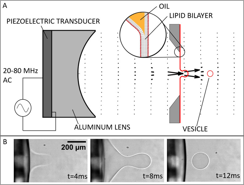
In this study, we demonstrate the ability to form and load GUVs using a focused jet induced by high-intensity ultrasound. This method, which we call “acoustic jetting,” takes advantage of full access to contents on both sides of the bilayer while eliminating stability and encapsulation challenges presented by the use of a nozzle. Acoustic jetting relies on the same local acceleration of liquid impinging on a planar lipid bilayer that has been shown to form and load GUVs in microfluidic jetting22 [Fig. 1(a)]. The ultrasonic transducer and its coupled plano–concave lens are shown at the left side of the image, millimeters away from the lipid bilayer. When the ultrasonic transducer is pulsed, the liquid is accelerated into the planar lipid bilayer, the planar lipid bilayer is deformed, and the GUV is formed [Fig. 1(b)].
FIG. 1.
Acoustic jetting. (a) Schematic of the acoustic jetting method. The focused flow generated by the ultrasonic transducer deforms a planar lipid bilayer to form GUVs. (b) Time series images of GUV formation.
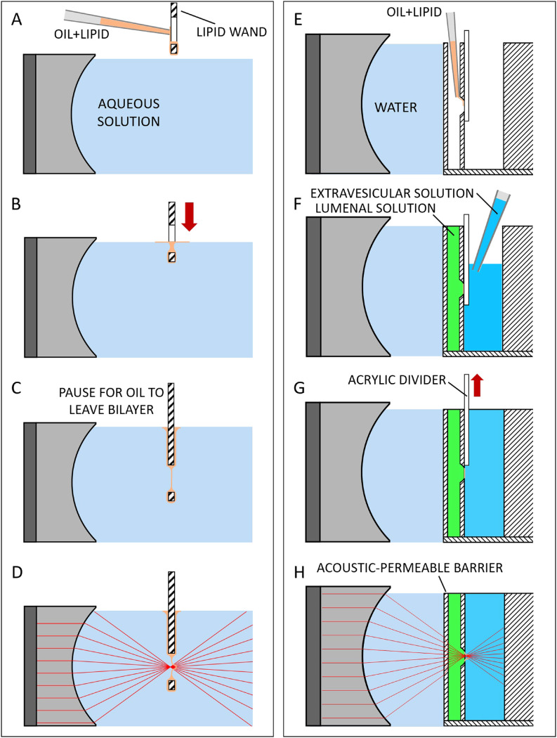
For acoustic jetting to successfully form GUVs, the planar lipid bilayer must be (i) positioned at the focus of the acoustic beam, (ii) not blocked by materials that will interfere with the acoustic beam, (iii) unaffected by pressure changes during focusing of the acoustic beam, and (iv) adjacent to a small (to conserve reagents) but sufficient volume of the solution to be loaded in the GUV. A simple approach to form the planar lipid bilayer that accomplishes the first three requirements is to use a supporting structure for the lipids, similar to the wands used to blow soap bubbles [technique 1, Figs. 2(a)–2(d)]. The lipid wand is cut from a thick acrylic and has an ovular hole where the planar lipid bilayer is formed. The wand is attached to a three-axis linear stage so that it can be centered and focused in the axis of the acoustic transducer. Before the wand is immersed in the aqueous solution, 10– of lipid in oil, typically diphytanoyl phosphatidylcholine (DPhPC) at 25 mg/ml in 80% decane and 20% squalene, is deposited onto the surfaces of the wand [Fig. 2(a)]. A large amount of the oil accumulates in a droplet at the bottom of the wand. Using the linear stage, the wand is slowly lowered into the aqueous solution. As this happens, buoyancy causes the oil to form a puddle on top of the aqueous solution around the wand. As the wand is lowered through this puddle, a layer of oil and lipid is formed over the orifice of the wand [Fig. 2(b)]. The wand with an oil film covering the orifice is allowed to rest fully immersed in the aqueous solution for 5–10 min, during which time the oil withdraws from the space between the two lipid leaflets formed at the oil–water interface, creating a clean planar lipid bilayer [Fig. 2(c)]. Squalene in the oil solution facilitates this process.23 After the incubation period, the acoustic transducer is ready to be actuated, causing a jet that pinches a vesicle off of the planar lipid bilayer [Fig. 2(d)].
FIG. 2.
Formation of planar lipid bilayers for acoustic jetting. Technique 1 (a)–(d): (a) While a lipid wand is held above the aqueous solution, a pipet is used to add a solution of oil and lipid to the surface of the acrylic. (b) The wand is lowered into the aqueous solution. Buoyancy of the oil causes it to pool at the air–water interface, pulling a thin layer over the ovular hole in the wand. (c) The membrane is positioned at the acoustic focus and oil evacuates the space between lipid leaflets. (d) The acoustic transducer is pulsed to form a vesicle from the planar lipid bilayer. Technique 2 (e)–(h): (e) A bilayer holder that physically separates solutions for inside and outside the GUV is submerged in water with the bilayer plane at the acoustic focal point. Oil and lipid are added to the bilayer orifice, which is initially covered on the opposite side with a thin sheet of acrylic. (f) The inner and outer aqueous solutions are pipeted into chambers to the same height. (g) The acrylic divider covering the orifice between the two solutions is slowly withdrawn, leaving a thin membrane of oil and lipid between the solutions. The system is left in this configuration while oil evacuates from the lipid bilayer. (h) The acoustic transducer is pulsed to form a vesicle from the planar lipid bilayer.
The lipid wand approach is sufficient for applications that do not require two separate solutions. However, for applications that require different solutions inside and outside the GUV, a modification must be made to the sample geometry. To accomplish this, we replace the lipid wand with an isolated holder for both the planar lipid bilayer and small volumes of the aqueous solutions [technique 2, Figs. 2(e)–2(h)]. The sample holder has a solid barrier, penetrable to acoustic waves, which is positioned adjacent to the aqueous solution to be encapsulated in the GUV. This solid barrier, when submerged in an additional solution (e.g., water) as an ultrasonic propagation medium, allows the focused acoustic beam to transmit from the propagation solution through the solution to be loaded into the GUV and to the bilayer, relatively unimpeded. To form the bilayer in this configuration, an acrylic divider is placed over the orifice between the proximal and distal aqueous solution chambers while they are still empty. The orifice and acrylic barrier are loaded with of lipid in oil [Fig. 2(e)]. The proximal chamber is filled with the solution to be loaded into the GUV, and the distal chamber is loaded with the solution that will surround the GUV, with the acrylic divider still in place to prevent hydrostatic pressure from rupturing the oil film between the chambers [Fig. 2(f)]. The acrylic divider is then removed from the orifice, and the device [Fig. 2(g)] is left undisturbed on the bench for 5–10 min while the nonpolar solvent withdraws from the film, leaving a clean planar lipid bilayer separating the intravesicular and extravesicular solutions.24 The sample is then ready for acoustic actuation and GUV formation.
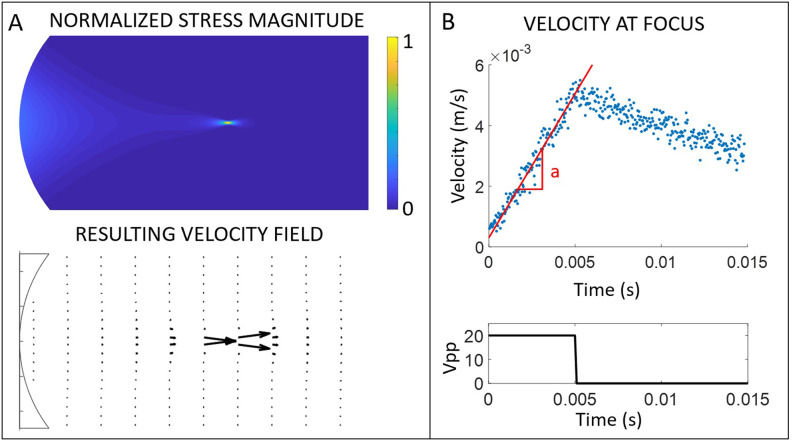
The acoustic transducers were fabricated by coupling a planar sheet of nickel-plated polycrystalline lead zirconate titanate (Navy Type II, 5A PZT) (Piezo Systems Inc., Woburn, MA) to a plano–concave lens using EpoTek 360T low viscosity epoxy (Epoxy Technology Inc., Billerica, MA). The 5 mm diameter plano–concave lens was machined from 6061 aluminum such that the concave face had a radius of curvature of 4 mm and the planar and concave faces were polished to a mirror finish before coupling to the piezo. Conductive silver epoxy was used to couple leads to the two electrodes of the piezo. To form GUVs, the transducers were excited using a square-enveloped sine wave pulse at the primary resonance frequency of the transducer, which is approximately 20 MHz for a transducer with thick PZT. The aluminum lens focuses the acoustic plane wave from the piezo to a beam with a focal point, millimeters from the concave face of the lens. Near the acoustic focal point, there is a large gradient in the acoustic amplitude. The gradient in amplitude induces a Reynolds stress and with it, a streaming flow near the focus25 [Fig. 3(a)]. The driving force for acoustic streaming in a focused acoustic device is described by Kamakura et al.26
FIG. 3.
Acoustic streaming generated by focused ultrasound. (a) A cartoon of the magnitude of axial force imposed by the focused ultrasound and the resulting acoustic streaming velocity field. (b) Particle image velocimetry was used to measure velocity as a function of time at the focal point of the acoustic transducer. At typical operating conditions, the acceleration, denoted by the slope “a,” is nearly constant during the pulse envelope, after which energy is dissipated to the surrounding fluid.
Because the induced flow relies on transmission of the acoustics to the focal point, care must be taken to choose a suitable material and thickness for the solid barrier as shown in Fig. 2(h). Acceleration at the focal point is measured using digital particle image velocimetry27 at 30 000 frames per second with polystyrene particles in water [Fig. 3(b)]. Barriers made of borosilicate glass, acrylic (PMMA), and polymethylpentene (PMP) are evaluated for their ability to transmit the acoustic beam by comparing the accelerations measured at a focal point past the barrier (Table I). PMMA and PMP, polymers that are well-matched to the acoustic impedance of water, reflect the beam significantly less than borosilicate glass, a mismatched material. Because the polymers have a higher Stokes attenuation than water, thinner barriers performed better at transmitting the acoustic beam.
TABLE I.
The acceleration at the acoustic focus during the ultrasonic pulse is compared for acoustic-penetrable solid barriers of varying material and thickness, normalized to acceleration with no barrier.
| Barrier material | Thickness (μm) | Normalized acceleration |
|---|---|---|
| None | 1.00 | |
| Glass | 150 | 0.06 |
| PMMA | 1500 | 0.06 |
| PMMA | 740 | 0.56 |
| PMMA | 200 | 0.96 |
| PMP | 500 | 0.56 |
The diameter of vesicles produced can be varied by changing the numerical aperture or acoustic wavelength, parameters that together control the size of the diffraction-limited focus of the acoustic waves (Fig. S1 of the supplementary material). In our device, the acoustic wavelength is dependent on the transducer resonance frequency, which in turn is dependent on the thickness of the piezoelectric material. Using nearly the thinnest commercial polycrystalline PZT and our smallest radius acoustic lens, we were able to form GUVs as small as in diameter, likely limited by spherical aberrations at high numerical apertures. Formation of vesicles over in diameter tended to burst the planar lipid bilayer, limiting the size of our largest GUVs. Using the transducers described here, the easily achievable range of vesicle diameters is from 100 to .
In addition to controlling resonance frequency and numerical aperture to set vesicle size, the acoustic energy delivered to the fluid must be controlled to enable vesicle formation. We find that there is a range of fluid acceleration magnitude and duration where vesicles are successfully formed. Values outside of this range will either result in the rupture of the planar lipid bilayer or deformation of the bilayer that is insufficient to produce a vesicle and relaxes back to a plane when the ultrasound pulse is stopped. In practice, the acceleration magnitude and duration are adjusted by varying the voltage amplitude across the transducer and the number of cycles in the pulse. For this study, we actuated the transducer with a sine wave of a single frequency within a square envelope, resulting in constant stress during the pulse.
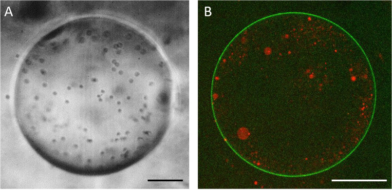
To demonstrate the ability of acoustic jetting to encapsulate particles, we suspended diameter polystyrene beads at in water and encapsulated the solution containing the particles in a GUV formed from 100% DPhPC [Fig. 4(a)]. The beads shown are significantly smaller than the acoustic wavelength such that scattered ultrasound does not significantly accelerate particles and the beads flow with the bulk fluid. As the diameter of beads approaches the acoustic wavelength, the encapsulation of acoustic-index-mismatched particles becomes more difficult, as scattering at the bead surfaces defocuses the acoustic waves and accelerates the particles relative to the fluid. As a second example, we suspended smaller vesicles in an isosmotic solution and encapsulated the solution containing the smaller vesicles in GUVs [Fig. 4(b)]. The smaller vesicles encapsulated in this example contain only lissamine–rhodamine for visualization, but they could be further functionalized with membrane proteins to investigate membrane–membrane contacts or lipid transport within GUVs.
FIG. 4.
Examples of GUVs formed with acoustic jetting. (a) Brightfield image of a DPhPC vesicle containing polystyrene beads and sucrose solution with osmolarity matched to the extravesicular saline solution to generate an optical index mismatch. . (b) Confocal image of an acoustic jetted GUV tagged with 7-nitro-2-1,3-benzoxadiazol-4-yl (NBD) encapsulating polydisperse vesicles tagged with lissamine Rhodamine B. .
During the formation of these GUVs, there are often smaller vesicles formed at the neck between the budding GUV and the planar lipid bilayer, which we refer to as secondary vesicles. Along with the pulse time and acceleration, an important operating condition that controls the formation of secondary vesicles is the position of the planar lipid bilayer relative to the focal plane of the transducer. When the bilayer is moved out of the acoustic focus plane, slightly closer to the transducer, a small part of the bilayer is caught in a high velocity region over a small area and pulled into the focal point, resulting in the formation of a slim membrane tube and subsequent secondary vesicles (Fig. S2 of the supplementary material). When the bilayer is moved farther from the transducer, there is often no vesicle formed, as in the examples shown at +150, +200, and . When the planar lipid bilayer is positioned at the acoustic focus, the secondary vesicle formation can be eliminated by carefully tuning the pulse time and acceleration.
In this paper, we demonstrate that GUVs can be formed with liquid jets accelerated into a planar lipid bilayer by an ultrasonic transducer. This allows for bulk solutions containing particles such as beads, vesicles, and even small cells to be directly encapsulated in the lumen of a GUV, avoiding the clogging and selectivity that can complicate some other methods. We show that vesicles in the range of can be formed with acoustic jetting, though this range could be expanded by the use of aspheric acoustic lenses to minimize aberrations and thinner, single-crystal PZT that expands the range of acoustic wavelengths available.
One of the motivations for developing acoustic jetting was to produce a tool that would enable the reconstitution of cell-like systems. Because biological membranes are naturally asymmetric, there are many biophysical applications of GUVs that benefit from independent access and control to the two sides of the membrane. The formation of a planar bilayer prior to acoustic jetting allows for asymmetric incorporation of lipids into the two leaflets and asymmetric insertion of transmembrane proteins, as well as the presence of different solutions for inside and outside of the GUV. Acoustic jetting offers a simple way to construct GUVs to study the vast number of pathways that entail both signal transduction across the plasma membrane and interaction with micrometer-scale structures or membrane-bound compartments within the cell.
While acoustic jetting presents benefits for some specialized applications, there are several practical difficulties associated with the technique that will need to be overcome to expand its usability. Chief among these is the stability of the planar lipid bilayers, which typically rupture before hundreds of vesicles can be formed and will be highly dependent on the lipid used in a given experiment. This issue persists despite efforts to reduce unsupported bilayer area and avoid geometries that might cause stress concentrations in the bilayer, suggesting that a bilayer composition optimized for stability is needed. In addition, slight mismatches in hydrostatic pressures between solutions on either side of the planar lipid bilayer resulting from pipeted solutions, differential evaporation, or vibrations can also cause membrane rupture. There is the added practical challenge of imaging the planar lipid bilayer after the oil, which conveniently provides brightfield contrast, has evacuated the area between the two lipid monolayers. Real-time imaging of the bilayer is essential to know when successful vesicles are formed and to ensure that the bilayer remains intact after vesicle formation. One way to avoid this problem is to use isotonic solutions with different refractive indices when the two solutions are completely separated, though this may not be an option for all experimental conditions.
Despite these practical challenges, our demonstration of acoustically formed GUVs expands the set of tools that can be used to form and load GUVs for a broad range of biomolecular and biophysical studies.
SUPPLEMENTARY MATERIAL
See the supplementary material for additional information about varying acoustic diffraction limit and focus scanning.
ACKNOWLEDGMENTS
This study was supported, in part, by the NIH (NIGMS Award No. GM114344) and by the Center for Cellular Construction, which is a Science and Technology Center funded by the National Science Foundation (NSF Award No. DBI-1548297).
DATA AVAILABILITY
The data that support the findings of this study are available from the corresponding author upon reasonable request.
REFERENCES
- 1.Veatch S. L. and Keller S. L., “Separation of liquid phases in giant vesicles of ternary mixtures of phospholipids and cholesterol,” Biophys. J. 85, 3074–3083 (2003). 10.1016/S0006-3495(03)74726-2 [DOI] [PMC free article] [PubMed] [Google Scholar]
- 2.Stachowiak J. C., Schmid E. M., Ryan C. J., Ann H. S., Sasaki D. Y., Sherman M. B., Geissler P. L., Fletcher D. A., and Hayden C. C., “Membrane bending by protein-protein crowding,” Nat. Cell Biol. 14, 944–949 (2012). 10.1038/ncb2561 [DOI] [PubMed] [Google Scholar]
- 3.Schmid E. M., Bakalar M. H., Choudhuri K., Weichsel J., Ann H. S., Geissler P. L., Dustin M. L., and Fletcher D. A., “Size-dependent protein segregation at membrane interfaces,” Nat. Phys. 12, 704–711 (2016). 10.1038/nphys3678 [DOI] [PMC free article] [PubMed] [Google Scholar]
- 4.Angelova M. I. and Dimitrov D. S., “Liposome electro formation,” Faraday Discuss. Chem. Soc. 81, 303–311 (1986). 10.1039/dc9868100303 [DOI] [Google Scholar]
- 5.Pott T., Bouvrais H., and Méléard P., “Giant unilamellar vesicle formation under physiologically relevant conditions,” Chem. Phys. Lipids 154, 115–119 (2008). 10.1016/j.chemphyslip.2008.03.008 [DOI] [PubMed] [Google Scholar]
- 6.Bouvrais H., Duelund L., and Ipsen J. H., “Buffers affect the bending rigidity of model lipid membranes,” Langmuir 30, 13–16 (2014). 10.1021/la403565f [DOI] [PubMed] [Google Scholar]
- 7.Li Q., Wang X., Ma S., Zhang Y., and Han X., “Electroformation of giant unilamellar vesicles in saline solution,” Colloids Surf. B 147, 368–375 (2016). 10.1016/j.colsurfb.2016.08.018 [DOI] [PubMed] [Google Scholar]
- 8.Kuroiwa T., Fujita R., Kobayashi I., Uemura K., Nakajima M., Sato S., Walde P., and Ichikawa S., “Efficient preparation of giant vesicles as biomimetic compartment systems with high entrapment yields for biomacromolecules,” Chem. Biodivers. 9, 2453–2472 (2012). 10.1002/cbdv.201200274 [DOI] [PubMed] [Google Scholar]
- 9.Dominak L. M. and Keating C. D., “Macromolecular crowding improves polymer encapsulation within giant lipid vesicles,” Langmuir 24, 13565–13571 (2008). 10.1021/la8028403 [DOI] [PubMed] [Google Scholar]
- 10.Witkowska A., Jablonski L., and Jahn R., “A convenient protocol for generating giant unilamellar vesicles containing SNARE proteins using electroformation,” Sci. Rep. 8, 9422 (2018). 10.1038/s41598-018-27456-4 [DOI] [PMC free article] [PubMed] [Google Scholar]
- 11.Jørgensen I. L., Kemmer G. C., and Pomorski T. G., “Membrane protein reconstitution into giant unilamellar vesicles: A review on current techniques,” Eur. Biophys. J. 46, 103–119 (2017). 10.1007/s00249-016-1155-9 [DOI] [PubMed] [Google Scholar]
- 12.Pautot S., Frisken B. J., and Weitz D. A., “Production of unilamellar vesicles using an inverted emulsion,” Langmuir 19, 2870–2879 (2003). 10.1021/la026100v [DOI] [Google Scholar]
- 13.Yandrapalli N., Seemann T., and Robinson T., “On-chip inverted emulsion method for fast giant vesicle production, handling, and analysis,” Micromachines 11, 285 (2020). 10.3390/mi11030285 [DOI] [PMC free article] [PubMed] [Google Scholar]
- 14.Shum H. C., Lee D., Yoon I., Kodger T., and Weitz D. A., “Double emulsion templated monodisperse phospholipid vesicles,” Langmuir 24, 7651–7653 (2008). 10.1021/la801833a [DOI] [PubMed] [Google Scholar]
- 15.Teh S.-Y., Khnouf R., Fan H., and Lee A. P., “Stable, biocompatible lipid vesicle generation by solvent extraction-based droplet microfluidics,” Biomicrofluidics 5, 044113 (2011). 10.1063/1.3665221 [DOI] [PMC free article] [PubMed] [Google Scholar]
- 16.Arriaga L. R., Datta S. S., Kim S.-H., Amstad E., Kodger T. E., Monroy F., and Weitz D. A., “Ultrathin shell double emulsion templated giant unilamellar lipid vesicles with controlled microdomain formation,” Small 10, 950–956 (2014). 10.1002/smll.201301904 [DOI] [PubMed] [Google Scholar]
- 17.Kim H. J., Decker E. A., and McClements D. J., “Impact of protein surface denaturation on droplet flocculation in hexadecane oil-in-water emulsions stabilized by -lactoglobulin,” J. Agric. Food Chem. 50, 7131–7137 (2002). 10.1021/jf020366q [DOI] [PubMed] [Google Scholar]
- 18.Reeves J. P. and Dowben R. M., “Formation and properties of thin-walled phospholipid vesicles,” J. Cell Physiol. 73, 49–60 (1969). 10.1002/jcp.1040730108 [DOI] [PubMed] [Google Scholar]
- 19.Szoka F. and Papahadjopoulos D., “Procedure for preparation of liposomes with large internal aqueous space and high capture by reverse-phase evaporation,” Proc. Natl. Acad. Sci. U. S. A. 75, 4194–4198 (1978). 10.1073/pnas.75.9.4194 [DOI] [PMC free article] [PubMed] [Google Scholar]
- 20.Karlsson M., Nolkrantz K., Davidson M. J., Stromberg A., Ryttsen F., Akerman B., and Orwar O., “Electroinjection of colloid particles and biopolymers into single unilamellar liposomes and cells for bioanalytical applications,” Anal. Chem. 72, 5857–5862 (2000). 10.1021/ac0003246 [DOI] [PubMed] [Google Scholar]
- 21.Olson F., Hunt C. A., Szoka F. C., Vail W. J., and Papahadjopoulos D., “Preparation of liposomes of defined size distribution by extrusion through polycarbonate membranes,” Biochim. Biophys. Acta Biomembr. 557, 9–23 (1979). 10.1016/0005-2736(79)90085-3 [DOI] [PubMed] [Google Scholar]
- 22.Stachowiak J. C., Richmond D. L., Li T. H., Liu A. P., Parekh S. H., and Fletcher D. A., “Unilamellar vesicle formation and encapsulation by microfluidic jetting,” Proc. Natl. Acad. Sci. U. S. A. 105, 4697–4702 (2008). 10.1073/pnas.0710875105 [DOI] [PMC free article] [PubMed] [Google Scholar]
- 23.White S. H., “Formation of ‘solvent-free’ black lipid bilayer membranes from glyceryl monooleate dispersed in squalene,” Biophys. J. 23, 337–347 (1978). 10.1016/S0006-3495(78)85453-8 [DOI] [PMC free article] [PubMed] [Google Scholar]
- 24.White S. H., Petersen D. C., Simon S., and Yafuso M., “Formation of planar bilayer membranes from lipid monolayers. A critique,” Biophys. J. 16, 481–489 (1976). 10.1016/S0006-3495(76)85703-7 [DOI] [PMC free article] [PubMed] [Google Scholar]
- 25.Lighthill S. J., “Acoustic streaming,” J. Sound Vib. 61, 315–471 (1978). 10.1016/0022-460X(78)90388-7 [DOI] [Google Scholar]
- 26.Kamakura T., Matsuda K., Kumamoto Y., and Breazeale M. A., “Acoustic streaming induced in focused Gaussian beams,” J. Acoust. Soc. Am. 97, 2740 (1995). 10.1121/1.411904 [DOI] [Google Scholar]
- 27.Willert C. E. and Gharib M., “Digital particle image velocimetry,” Exp. Fluids 10, 181–193 (1991). 10.1007/BF00190388 [DOI] [Google Scholar]
Associated Data
This section collects any data citations, data availability statements, or supplementary materials included in this article.
Supplementary Materials
See the supplementary material for additional information about varying acoustic diffraction limit and focus scanning.
Data Availability Statement
The data that support the findings of this study are available from the corresponding author upon reasonable request.


