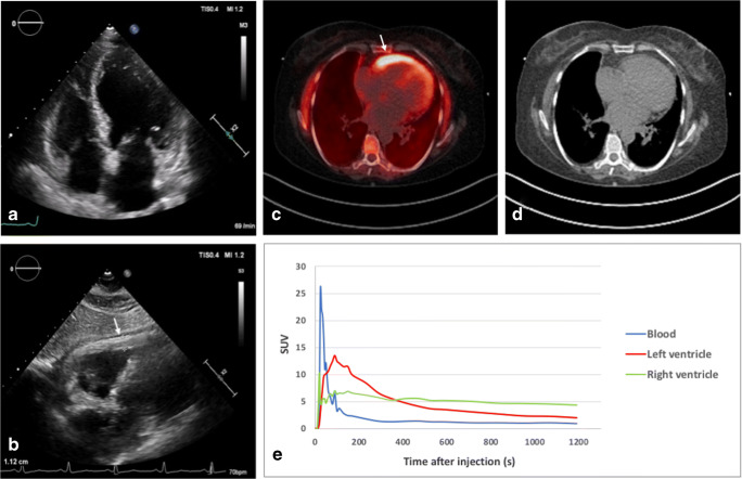
A 62-year-old female with known systemic serum amyloid A (AA) amyloidosis presented with signs and symptoms of new-onset heart failure. Echocardiography demonstrated mild left ventricular (LV) dilation, preserved ejection fraction, and grade II diastolic dysfunction without typical signs of cardiac amyloidosis (CA) but a profound focal hypertrophy of the free right ventricular (RV) wall (a, b). Positron emission tomography/computed tomography (PET/CT) with the amyloid-binding tracer 18F-florbetaben was subsequently performed since endomyocardial biopsy was not deemed justified due to localized hypertrophy of the free RV wall. As shown in c-e, a highly increased RV tracer uptake with only moderately increased LV tracer uptake was found (retention index for RV and LV is 0.0026 and 0.0016, respectively). Tracer uptake co-localized with the echocardiographic finding of RV hypertrophy, suggesting cardiac involvement of AA amyloidosis with predominant right-sided amyloid deposition.
Amyloid-binding radiotracers have already received approval for beta-amyloid brain imaging. Previous exploratory studies demonstrated their high diagnostic accuracy for CA of transthyretin or light-chain type [1–3], yet no study has evaluated their utility in an AA amyloidosis cohort. While endomyocardial biopsy is an established method for diagnosing CA, it may be prone to sampling error in the case of localized disease. This report demonstrates the sensitivity of 18F-florbetaben PET/CT to detect AA-CA. PET/CT provides high image quality and quantitative measures of tracer uptake, thus making detection, localization, and absolute quantification of amyloid feasible and has the potential to substitute or even outperform endomyocardial biopsy, particularly in the case of focal or early-stage disease.
Funding information
Open Access funding provided by Projekt DEAL. This work was supported by the Universitätsmedizin Essen Clinician Scientist Academy (UMEA)/German Research Foundation (DFG, Deutsche Forschungs-Gemeinschaft) research grant to MP (FU356/12-1), DFG grant to PL (LU2139/2-1), and DFG grant to TR (RA969/12-1).
Data availability
Clinical and image data are available for review upon request.
Compliance with ethical standards
Conflict of interest
The authors declare that they have no conflicts of interest.
Ethical approval
This article does not contain any studies with animals performed by any of the authors. All procedures performed involving human participants were in accordance with the ethical standards of the institutional and/or national research committee and with the principles of the 1964 Declaration of Helsinki and its later amendments or comparable ethical standards.
Consent to participate
Not applicable.
Consent for publication
Consent was obtained from the patient for the anonymous publication of clinical and imaging data for scientific purposes.
Footnotes
The original version of this article was revised. The correct word in the article title is emission and not computed.
This article is part of the Topical Collection on Cardiology
Publisher’s note
Springer Nature remains neutral with regard to jurisdictional claims in published maps and institutional affiliations.
Change history
6/15/2020
The correct title for this paper should be: 18F-florbetaben positron emission tomography detects cardiac involvement in systemic AA amyloidosis
References
- 1.Dorbala S, Vangala D, Semer J, et al. Imaging cardiac amyloidosis: a pilot study using 18F-florbetapir positron computed tomography. Eur J Nucl Med Mol Imaging. 2014;41:1652–1662. doi: 10.1007/s00259-014-2787-6. [DOI] [PubMed] [Google Scholar]
- 2.Lee SP, Lee ES, Choi H, et al. 11C-Pittsburgh B PET imaging in cardiac amyloidosis. JACC Cardiovasc Imaging. 2015;8:50–59. doi: 10.1016/j.jcmg.2014.09.018. [DOI] [PubMed] [Google Scholar]
- 3.Lee S-P, Suh H-Y, Park S, et al. Pittsburgh B compound positron emission tomography in patients with AL cardiac amyloidosis. J Am Coll Cardiol. 2020;75:380–390. doi: 10.1016/j.jacc.2019.11.037. [DOI] [PubMed] [Google Scholar]
Associated Data
This section collects any data citations, data availability statements, or supplementary materials included in this article.
Data Availability Statement
Clinical and image data are available for review upon request.


