
Fig. 2. Deletion of Slc7a11 in KPC mice induces tumor ferroptosis and extends survival.
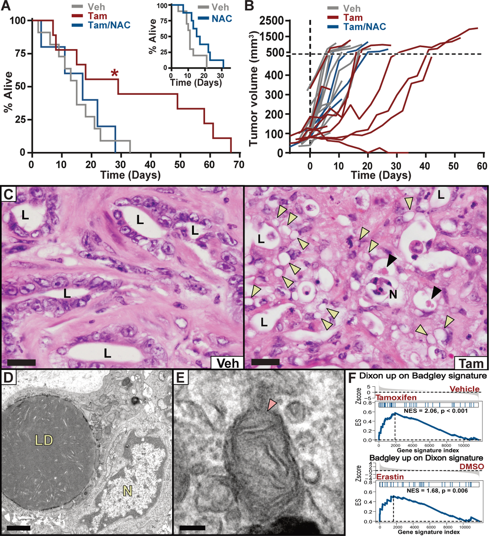
(A) Survival of KPFSR mice treated with vehicle (n = 11, median 15 days), tamoxifen (n = 9, median 29 days), or tamoxifen/NAC (n = 5, median 17 days). * p < 0.0295, log-rank. INSET: Survival of KPC mice treated with NAC alone (n = 8, median = 16 days) versus historical saline-treated controls (n = 10, median = 11 days). (B) Growth curves for each KPFSR tumor. (C) Hematoxylin and eosin (H&E) stained sections of tumor tissue from KPFSR mice treated with vehicle or tamoxifen. L = lumen of malignant epithelium, N = necrosis, yellow arrowheads = lipid droplets, black arrowheads = megamitochondria. Scale = 20 μm. (D and E) TEM images from tamoxifen-treated KPFSR tumors. LD = lipid droplets, N = nucleus, arrow indicates damaged mitochondrion. (D) scale = 1μm; (E) scale = 100 nm. (F) Gene set enrichment analysis. Top panel depicts enrichment of a published ferroptosis expression signature (Dixon) among genes differentially expressed in tamoxifen-treated KPFSR epithelia (Badgley) (p < 0.001). Bottom panel depicts the reciprocal comparison (p < 0.006).