Abstract
Background
Emerging evidence indicates that circular RNAs (circRNAs), which form as covalently closed loops, play a regulatory role in various types of cancer, including prostate cancer (PCa). CircSLC19A1, one kind of circRNA, was subjected to the study and its role in PCa was explored.
Methods
Expressions of circSLC19A1, miR-326 and MAPK1 in PCa tissues and cells were assessed by qRT-PCR. CircSLC19A1 was identified by RNase R treatment. The binding relations between circSLC19A1 and miR-326 and between miR-326 and MAPK1 were predicted by RegRNA2.0 or Targetscan7.2 and further confirmed by dual-luciferase reporter assay. Pearson correlation analysis of the correlation among circSLC19A1, miR-326 and MAPK1 was performed. CCK-8, cell colony formation, wound healing and Transwell assays were used to assess PCa cell viability, proliferation, migration and invasion, respectively.
Results
CircSLC19A1 expression was up-regulated in PCa tissue and cell cytoplasm. Silencing circSLC19A1 inhibited PCa cell viability, proliferation, migration, invasion and miR-326 expression. MiR-326 inhibitor promoted the luciferase activities of circSLC19A1 and MAPK1, increased MAPK1 expression and facilitated PCa cell progression. MiR-326 expression was down-regulated in PCa tissue and there was a negative correlation between miR-326 and circSLC19A1 expressions. MAPK1 expression was up-regulated in PCa tissue. There was a negative correlation between MAPK1 and miR-326 expressions as well as a positive correlation between MAPK1 and circSLC19A1 expressions. Silencing MAPK1 promoted the viability, proliferation, migration, and invasion of PCa cells co-transfected with siRNA-circSLC19A1a and miR-326 inhibitor.
Conclusion
CircSLC19A1 silencing inhibited PCa cell proliferation, migration and invasion through regulating miR-326/MAPK1 axis.
Keywords: circSLC19A1, miR-326, MAPK1, prostate cancer, proliferation, migration, invasion
Introduction
Prostate cancer (PCa), a most prevalent cancer in the urinary system, is the second most diagnosed cancer in men and is the fifth leading cause of cancer-related death for men.1 PCa is considered as a heterogeneous disease in either indolent or aggressive type.2 Around 70% of the newly diagnosed PCa patients in localized cancer stage or with intermediate-risk were regarded as potentially curable.3 However, a sharp decrease to merely 30% was shown in the five-year survival rate, as distant metastasis forms.3 Relapse occurs in up to 30% of patients in five to ten years of being cured of PCa and a higher incidence of mortality was found in PCa patients suffering from rapid biochemical failure and clinical recurrence.4 The standard treatment for localized PCa is prostatectomy, while metastatic PCa is counteracted by combinatorial treatment of androgen deprivation and abiraterone with prednisone.5,6 Unfortunately, there are limited treatment options for metastatic castrate-resistant PCa and the treatment effect is unsatisfactory. Given that, it prompts a new way to address the disease from molecular levels.
Circular RNAs (circRNAs) are one kind of endogenous non-coding RNAs that form covalently closed-loop structures through back-splicing pre-mRNA.7 RNA deep sequencing technology and bioinformatics have revealed that circRNAs are highly conserved genes that are stably and abundantly expressed in mammalian cells.8–11 CircRNAs have been found to function as transcriptional regulators to sponge miRNA expression,12 and post-transcriptional regulators in nucleus to promote expression of their parental genes.13 Increasing evidence demonstrate that circRNAs play important roles in the pathogenesis and progression of various types of cancer.14
Recent studies found that more than a thousand of circRNAs are differentially expressed in PCa and some of them can form circRNA-miRNA-mRNA networks to serve as biomarkers for PCa progression.2 Additionally, compared to single PSA detection, combinatorial detection of circSLC19A1 and prostate-specific antigen (PSA) could enhance the sensitivity and specificity of the area under the curve (AUC), thus being more helpful for the identification of PCa.2 Furthermore, according to Zheng, high-expressed circSLC19A1 was observed in PCa cells-secreted extracellular vesicles and its promotion effects on PCa cell proliferation and invasion were realized through a circRNA-miRNA-mRNA regulatory network. To be specific, circSLC19A1 exerted promotive effects through absorbing miR-497 to upregulate septin 2 expression.15
Thus, it is necessary for these kinds of networks that were conducive to modulating PCa progression to be uncovered. Our study tried to recognize a novel regulatory network of circSLC19A1 for PCa treatment.
Materials and Methods
Ethical Statement
The study obtained the approval of the ethics committee of Zhejiang Provincial People’s Hospital (approval number: DU201906043) and the written informed consents from all participants were obtained in any experimental work with humans.
Clinical Sample
Forty-eight pairs of the clinical samples of PCa and adjacent normal tissues were collected from PCa patients aged 56–73 years old undergoing radical prostatectomy at Zhejiang Provincial People’s Hospital in 2019 and were kept in liquid nitrogen at −80°C, after surgical resection, prior to use in experiments. Follow-up data of clinical T stage, lymph node metastasis and distant metastasis of PCa patients were collected. None of these patients had a history of chemotherapy, radiotherapy or other medical intervention. All patients had signed informed consent, and agreed that their tissues would be used for clinical research.
Cell Culture
Human prostate epithelial cell lines were purchased from Shanghai Cell Bank affiliated to Chinese Academy of Sciences (GNHu37, Shanghai, China).
PCa cell lines (DU145 cells, PC3 cells, LNCaP cells, and 22RV1 cells) were purchased from American Type Culture Collection (ATCC, HTB-81, CRL-1435, CRL-1740, CRL-2505, Manassas, VA, USA).
RWPE-1 cells were cultured in Defined Keratinocyte SFM medium (10,744,019, ThermorFisher, Waltham, MA, USA) at 37°C with 5% CO2.
LNCaP, PC3 and 22RV1 cells were cultured in ATCC-formulated RPMI-1640 medium (30–2001, ATCC, USA), supplemented with 10% fetal bovine serum (FBS, F8687, Sigma-Aldrich, St. Louis, MO, USA) and DU145 cells were cultured in ATCC-formulated Eagle’s Minimum Essential Medium (30–2003, ATCC, USA) supplemented with 10% FBS, in Cellbind culture bottles (3289, Corning, Corning, NY, USA), at 37°C with 5% CO2.
RNA Extraction
Total RNA was isolated from DU145 and PC3 cells and PCa tissues using TRIzol reagent (15,596,026, ThermorFisher, USA) and total miRNA was isolated using PureLink miRNA Isolation Kits (K157001, ThermoFisher, USA). Total cytoplasmic RNA and total nuclear RNA were separately isolated using Cytoplasmic & Nuclear RNA Purification Kits (21,000, NORGEN, Thorold, ON, Canada, https://norgenbiotek.com/).
RNase R Treatment
The extracted RNA from DU145 and PC3 cells were divided into RNase R (+) and RNase R (-) groups. In the RNase R (+) group, the RNA was incubated with RNase R (R6513, Sigma-Aldrich, USA), while the RNA was incubated with diethyl pyrocarbonate (DEPC) water (AM9906, ThermorFisher, USA) in the RNase R (-) group were incubated at 37°C for 10 min. The abundance of circSLC19A1 was determined by quantitative reverse-transcription polymerase chain reaction (qRT-PCR).
Transfection
siRNA-circSLC19A1 (sicircSLC19A1, 5ʹ-AGAUGUAGGAGGAAUAGGCGA-3ʹ) was synthesized by RIBOBIO, Guangzhou, China. siRNA-circSLC19A1, miR-326 inhibitor (3ʹ-GACCUCCUUCCCGGGUCUCC-5ʹ, miR20000756-1-5, RIBOBIO, China) and siRNA-mitogen-activated protein kinase 1 (MAPK1) (siMAPK1, 5ʹ-AAUGCUAUCGCUAGUUUGCUC-3ʹ, siB150423103920-1-5, RIBOBIO, China) were used to inhibit circSLC19A1, miR-326 and MAPK1 expressions in DU145 and PC3 cells via transfection using Lipofectamine 3000 Reagent (L3000015, ThermoFisher, USA). DU145 and PC3 cells at a density of 3×10⁴ cells/well were seeded in 96-well plates to reach a confluence of 80% (260,836, ThermoFisher, USA). sicircSLC19A1, siMAPK1 and siNC (5ʹ-AUUUGCCUAAUUUGGACGCUC-3ʹ, 1,022,076, QIAGEN, Hilden, Germany) were diluted using Opti-MEM media (31,985,088, ThermoFisher, USA). MiR-326 inhibitor and inhibitor control (3ʹ-GCCCUAUCUCCCGCGCGUUC-5ʹ, 1,027,271, QIAGEN, Germany) were diluted using Opti-MEM media, supplemented with P3000 reagent. After dilution, genes of interest were incubated with Lipofectamine 3000 reagent for 5min at room temperature and then added into 10µL Lipid complex. DU145 or PC3 cells were incubated with the genes-added Lipid complex and 0.2µL P3000 reagent for 3 days at 37°C.
Dual-Luciferase Reporter Assay
The binding sites between circSLC19A1 and miR-326 and between miR-326 and MAPK1 were predicted by RegRNA2.0 (http://regrna2.mbc.nctu.edu.tw/) and Targetscan7.2 (http://www.targetscan.org/vert_72/), respectively. Sequences of circSLC19A1 (Wild-Type: 5ʹ-GAGGCCTGCAGCCAGCCCAGAGC-3ʹ, Mutant-Type: 5ʹ-GAGGCCTGCAGCCAGTAGGACTC-3ʹ) and sequences of MAPK1 (Wild-Type: 5ʹ-AAGAATTTCTTCAGTCCAGAGAA-3ʹ, Mutant-Type: 5ʹ-AAGAATTTCTTCAGTTGCATAAA-3ʹ) were separately inserted into pmirGLO vectors (E1330, Promega, Madison, WI, USA). DU145 or PC3 cells at a density of 2×10⁵ cells/well were cultured in 12-well plates (140,656, ThermoFisher, USA). When the cells reached 70% confluence, DU145 or PC3 cells were co-transfected with pmirGLO vectors containing circSLC19A1 or MAPK1 sequences and miR-326 inhibitor, or with pmirGLO vectors containing circSLC19A1 or MAPK1 sequences and inhibitor control using Lipofectamine 3000 Reagent. After transfection, the binding specificity was reflected by Dual-Luciferase Reporter Assay System (E1980, Promega, USA). After lysed by Passive Lysis Buffer, DU145 or PC3 cells were added with Luciferase Assay Reagent II to measure firefly luciferase activity and with Stop & Glo Reagent to measure firefly renilla luciferase activity.
Cell Counting Kit (CCK)-8 Assay
Following transfection, DU145 or PC3 cells at a density of 5×10³ per well were seeded in 96-well plates. DU145 or PC3 cells in each well were treated with 10µL CCK-8 reagent (CA1210, Solarbio, Beijing, China) and incubated in the dark for 2 h at 37°C. The absorbance of DU145 or PC3 cells was measured on a microplate reader (ELx808, BioTek, Winooski, VT, USA, https://www.biotek.com) at 450nm.
Cell Colony Formation Assay
Following transfection, DU145 or PC3 cells at a density of 1×10³ per well were seeded in a 6-well plate (A1098201, ThermoFisher, USA). DU145 or PC3 cells were cultured in corresponding culture medium supplemented with 30% FBS for 7 days. The culture medium was refreshed every three days. After the culture medium was aspirated, DU145 or PC3 cells were washed with PBS (P5493, Sigma-Aldrich, USA), fixed by 4% paraformaldehyde (P6148, Sigma-Aldrich, USA) and stained with 0.2% crystal violet (C0775, Sigma-Aldrich, USA) for 5min. The state of DU145 or PC3 cells was observed by a microscope (Axioskop 40, Carl Zeiss AG, Dresden, Germany).
Wound Healing Assay
Following transfection, DU145 or PC3 cells at a density of 5×10⁵ per well were seeded in culture-Insert 2 Well 24 (80,241, ibidi, Munich, Germany) packed in 24 well plates (82,406, ibidi, Munich, Germany). When cell monolayers were formed, wounds were made manually using a sterilized steel rule. DU145 or PC3 cell monolayers were washed with PBS for removing unattached cells and serum-free culture media were then added into the cell and incubated for 48 h. The area occupied by migratory DU145 or PC3 cells were estimated by a microscope (Axioskop 40, Carl Zeiss AG, Dresden, Germany) at 100× magnification. The photographs taken at 0 h and 48 h were analyzed using an image analysis system (Wound Healing ACAS, ibidi, Germany).
Transwell Assay
24mm Transwell chambers with 8.0µm Pore Polycarbonate Membrane Insert (3428, Corning, Corning, NY, USA) was used to assess the invasive capacity of DU145 or PC3 cells. One hundred-microliter diluted Matrigel (BD Biosciences, Franklin Lakes, NJ, USA) was laid in the upper chamber and incubated in 100µL serum-free culture media for 5 h at 37°C. Following transfection, DU145 or PC3 cells were adjusted to 2×10⁵/mL in serum-free culture media. One hundred-microliter DU145 or PC3 cell solution was added into the upper chamber. Five hundred-microliter culture media containing 10% FBS as chemoattractant to induce cell invasion was added into the lower chamber. After incubation, the unattached cells in the upper chamber were removed and the invasive cells were fixed with 4% paraformaldehyde for and stained with 0.1% crystal violet. The invasive cells were counted in randomly selected fields under a microscope (Axioskop 40, Carl Zeiss AG, Dresden, Germany) at 250× magnification.
qRT-PCR
PCa tissues as well as DU145 or PC3 cells were used for qRT-PCR validation. The isolated cytoplasmic RNA, nuclear RNA, total RNA and total miRNA were stratified by chloroform (151,858, Sigma-Aldrich, USA). Isopropanol precipitation (I9516, Sigma-Aldrich, USA) was used to precipitate the RNA or miRNA in water phase. Then, the RNA or miRNA was washed by 75% ethanol (E7023, Sigma-Aldrich, USA) and dissolved in RNase-free water (10,977,023, ThermoFisher, USA). cDNA was synthesized by SuperScript IV reverse transcriptase (18,090,010, ThermoFisher, USA). PowerUp SYBR Green Master Mix (A25742, ThermoFisher, USA) was used to amplify cDNA with primers listed in Table 1. Real-time PCR reaction was initiated at 95°C pre-denaturation for 10min, 95°C denaturation for 15s and 40 circles of 95°C annealing for 15 s and 60°C elongation for 60s, on a Real-Time PCR detection System (CFX Connect, Bio-Rad, Philadelphia, PA, USA). The relative changes in the circSLC19A1, SLC19A1, miR-326 and MAPK1 expression levels were calculated by 2−ΔΔCt method.16
Table 1.
Primers Used in qPCR for the Target Genes
| Gene | Species | Forward | Reverse |
|---|---|---|---|
| circSLC19A1 | Human | 5ʹ-AGCTTCATCACCCCCTACCT-3’ | 5ʹ-GATCTCGTTCGTGACCTGCT-3’ |
| SLC19A1 | Human | 5ʹ-CTGCATCCTGGAAACTCTC-3’ | 5ʹ-CGGGTTGAGACCTAGAGCTG-3’ |
| miR-326 | Human | 5ʹ-TTCCTCCAGTGTCGTATCCA-3’ | 5ʹ-CGTATCCAGTGCGTGTCGT-3’ |
| MAPK1 | Human | 5ʹ-GGGCAGACATGCTAAATGGT-3’ | 5ʹ-TCCAGTGGAGGGAGTGTACC-3’ |
| GAPDH | Human | 5ʹ-GAAGGTCGGAGTCAACGGAT-3’ | 5ʹ-CCTGGAAGATGGTGATGGG-3’ |
| U6 | Human | 5ʹ-CTCGCTTCGGCAGCAGCACATATA-3’ | 5ʹ-AAATATGGAACGCTTCACGA-3’ |
Statistical Analysis
Data from three independent experiments were presented as the mean ± standard deviation (SD). One-way (ANOVA) and Student’s t-test were used to analyze the differences among multiple groups and between two groups, respectively. The correlations between any two of circSLC19A1, miR-326 and MAPK1 were reflected by Pearson correlation analysis. Statistical analysis was conducted using the SPSS software (version 20.0, IBM-SPSS Inc., Chicago, IL, USA). p <0.05 was considered as statistically significant.
Results
CircSLC19A1 Was Strikingly Up-Regulated in PCa Tissue and Cells
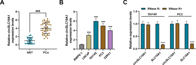
To investigate the role of circSLC19A1 in PCa, its expression pattern in PCa tissue was observed. A high expression of circSLC19A1 was observed in PCa tissue in comparison to the adjacent normal tissue (Figure 1A, P <0.001). To uncover the relationship between circSLC19A1 and clinical manifestations of PCa, clinical characteristics in PCa patients with high circSLC19A1 expression and low circSLC19A1 expression were recorded, respectively. Statistical analysis revealed that among PCa patients in T3-T4 stage, or having lymph node and distant metastasis, more of them were with high circSLC19A1 expression (P <0.05), while less of them were with low circSLC19A1 expression (P <0.01, Table 2). Next, in vitro experiments showed that circSLC19A1 expression was differentially up-regulated in PCa cell lines (LNCaP, DU145, PC3 and 22RV1 cells), compared to that in human prostate epithelial cells (RWPE-1 cells) (P <0.001, Figure 1B). Due to their closed structure and highly conservative sequence, circRNAs are resistant to RNase digestion.17 RNase R resistance analysis showed that after RNase R treatment, SLC19A1 expression was declined (P <0.001) in DU145 and PC3 cells, while no obvious alteration of circSLC19A1 expression was observed, confirming the aforementioned finding that circSLC19A1 was high-expressed in PCa cells (Figure 1C).
Figure 1.
CircSLC19A1 was significantly up-regulated in PCa tissues and cells. (A) CircSLC19A1 expression in PCa tissues and the adjacent normal tissues was assessed by qRT-PCR, with GAPDH serving as a reference gene. (B) circSLC19A1 expression in RWPE-1, LNCaP, DU145, PC3 and 22RV1 cells was assessed by qRT-PCR, with GAPDH serving as a reference gene. (C) The expressions of circSLC19A1 and SLC19A1 in DU145 and PC3 cells with or without RNase R treatment were assessed by qRT-PCR, with GAPDH serving as a reference gene. ###P<0.001 vs ANT; ***P<0.001 vs RWPE-1; ^^^P<0.001 vs RNase R.
Abbreviations: PCa, prostate cancer; ANT, adjacent normal tissue; qRT-PCR, quantitative reverse-transcription polymerase chain reaction.
Table 2.
Correlations Between circSLC19A1 Expression and Clinicopathological Features
| Characteristics | Low Expression (n=20) |
High Expression (n=28) |
P value |
|---|---|---|---|
| Age ≤65 | 10 | 15 | 0.807 |
| >65 | 10 | 13 | |
| Clinical T stage T1-T2 | 13 | 10 | 0.048 |
| T3-T4 | 7 | 18 | |
| Lymph node metastasis NO | 14 | 9 | 0.010 |
| YES | 6 | 19 | |
| Distant metastasis NO | 15 | 11 | 0.014 |
| YES | 5 | 17 |
Silencing of circSLC19A1 Inhibited Viability, Proliferation, Migration and Invasion in PCa Cells
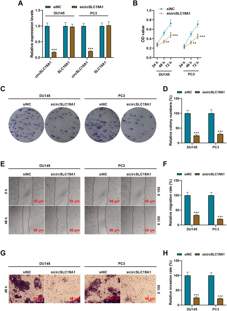
Since high circSLC19A1 expression was associated with PCa, its specific relation to PCa progression was explored by introducing sicircSLC19A1. sicircSLC19A1 transfection in DU145 and PC3 cells was successfully established, as evidenced by decreased circSLC19A1 expression (Figure 2A, P <0.001). The viability of DU145 and PC3 cells was inhibited by sicircSLC19A1 transfection at 48 h and 72 h in comparison with untreated PCa cells (Figure 2B, P <0.01). Meanwhile, sicircSLC19A1 transfection significantly suppressed cell colony formation in DU145 and PC3 cells (Figure 2C and D, P <0.001). Furthermore, the migratory and invasive abilities were all attenuated by sicircSLC19A1 transfection (Figure 2E–H, P <0.001), as evidenced by less migratory distant and less cell invasion after sicircSLC19A1 transfection. Collectively, these findings indicated that silencing circSLC19A1 expression inhibited DU145 and PC3 cell viability, migration and invasion.
Figure 2.
Silencing of circSLC19A1 inhibited viability, proliferation, migration and invasion in PCa cells. (A) The expressions of circSLC19A1 and SLC19A1 in DU145 and PC3 cells transfected with or without sicircSLC19A1 were assessed by qRT-PCR, with GAPDH serving as a reference gene. (B) The viability of DU145 and PC3 cells transfected with or without sicircSLC19A1 was measured by Cell Counting Kit-8 assay. (C) Representative photos of the colony formation of DU145 and PC3 cells transfected with or without sicircSLC19A1. (D) The colony formation of DU145 and PC3 cells transfected with or without sicircSLC19A1 was evaluated by cell colony formation assay. (E) Representative photos of the migration of DU145 and PC3 cells transfected with or without sicircSLC19A1. (F) The migration of DU145 and PC3 cells transfected with or without sicircSLC19A1 was evaluated by wound healing assay. (G) Representative photos of the invasive state of DU145 and PC3 cells transfected with or without sicircSLC19A1. (H) The invasive state of DU145 and PC3 cells transfected with or without sicircSLC19A1 was evaluated by Transwell assay. ^^P<0.01 and ^^^P<0.001 vs siNC.
Abbreviations: sicircSLC19A1, siRNA-circSLC19A1; siNC, siRNA-negative control; qRT-PCR, quantitative reverse-transcription polymerase chain reaction.
CircSLC19A1 Directly Targeted miR-326, a Down-Regulated Gene in PCa Tissue
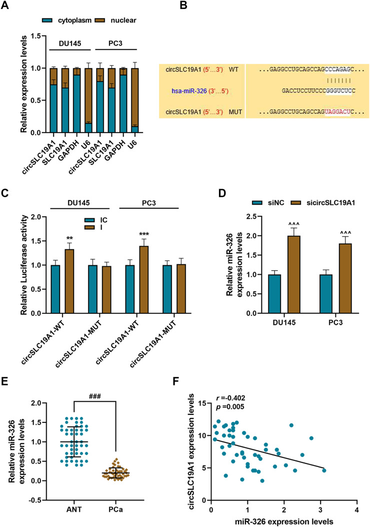
Intracellular localization is crucial for circRNAs to perform their functions.18 Thus, for obtaining the insights of the function of circSLC19A1 in PCa, the localization of circSLC19A1 and SLC19A1 was detected in DU145 and PC3 cells. The abundances of circSLC19A1 and SLC19A1 were both much higher in the cytoplasm than those in the nucleus (Figure 3A). Next, bioinformatics analysis conducted by RegRNA2.0 predicted that circSLC19A1 wild-type could directly bind to miR-326 (Figure 3B). To verify the result from RegRNA2.0, dual-luciferase report assay was performed, showing that transfection of miR-326 inhibitor increased the luciferase activity of circSLC19A1 wild-type, compared to circSLC19A1 mutant type (Figure 3C, P <0.01). SicircSLC19A1 transfection up-regulated miR-326 expression in PCa cells (Figure 3D, P <0.001). Given the existence of the binding relation between circSLC19A1 and miR-326, the expression pattern and the relation to circSLC19A1 of miR-326 were investigated. MiR-326 expression was evidently low-expressed in PCa tissue, compared to the adjacent normal tissue (Figure 3E, P <0.001). Pearson correlation analysis illustrated that miR-326 expression was negatively correlated to circSLC19A1 expression in PCa tissue (Figure 3F). All these results suggested that the inhibitory effect of silencing circSLC19A1 expression in PCa cells might be resulted from up-regulation of miR-326 expression.
Figure 3.
CirSLC19A1 directly targeted miR-326, a down-regulated gene in PCa tissue. (A) The abundances of circSLC19A1 and SLC19A1 in the cytoplasm and nucleus of DU145 or PC3 cells were assessed by qRT-PCR, with GAPDH and U6 serving as reference genes. (B) RegRNA2.0 predicted the mutual binding sites between circSLC19A1 and miR-326. (C) Dual-luciferase reporter assay confirmed the interaction between circSLC19A1 and miR-326. (D) miR-326 expression in DU145 and PC3 cells transfected with or without sicircSLC19A1 was assessed by qRT-PCR, with U6 serving as a reference gene. (E) miR-326 expression in PCa tissues and the adjacent normal tissue tissues was assessed by qRT-PCR, with U6 serving as a reference gene. (F) Spearman correlation analysis reflected the correlation between circSLC19A1 and miR-326. **P<0.01 and ***P<0.001 vs IC; ^^^P<0.001 vs siNC; ###P<0.001 vs ANT.
Abbreviations: PCa, prostate cancer; sicircSLC19A1, siRNA-specific target circSLC19A1; siNC, siRNA-negative control; WT, wild type; MUT, mutant type; ANT, adjacent normal tissue; qRT-PCR, quantitative reverse-transcription polymerase chain reaction.
MAPK1, a Target Gene of miR-326, Was Up-Regulated in PCa Tissue and Had Positive Correlation with circSLC19A1
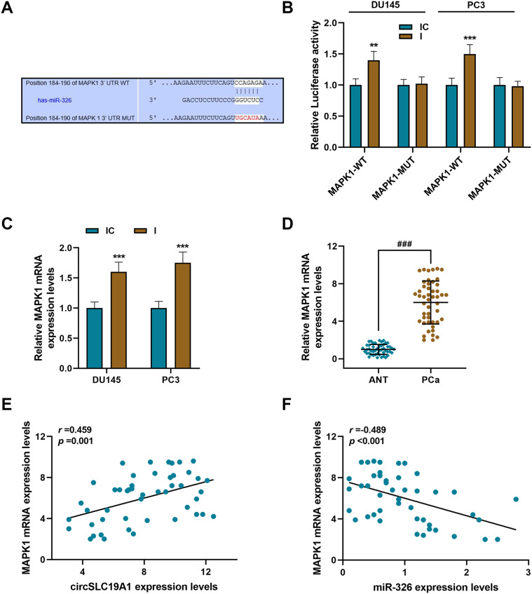
To further determine the regulatory mechanism of miR-326 in PCa, the downstream target of miR-326 was analyzed. Bioinformatics analysis conducted by Targetscan7.2 predicted that miR-326 could bind to the 3ʹUTR of MAPK1 wild-type (Figure 4A). The subsequent dual-luciferase reporter assay showed that transfection of miR-326 inhibitor increased the luciferase activity of MAPK1 wild-type, compared to MAPK1 mutant-type (Figure 4B, P <0.01). MAPK1 mRNA expression was up-regulated, after transfection of miR-326 inhibitor (Figure 4C, P <0.001). Since the binding relation between miR-326 and MAPK1 was verified, the expression patterns of and the relation between miR-326 and MAPK1 in PCa were investigated. MAPK1 expression was high-expressed in PCa tissue, as compared with the adjacent normal tissue (Figure 4D, P <0.001). Pearson correlation analysis illustrated that MAPK1 expression was positively correlated to circSLC19A1 expression, while negatively correlated to miR-326 expression in PCa tissue, suggesting a competing endogenous (ceRNA) regulatory network of circSLC19A1 in PCa (Figure 4E and F).
Figure 4.
MAPK1, a target gene of miR-326, was up-regulated in PCa tissue and there is positive correlation between MAPK1 and circSLC19A1. (A) Targetscan7.2 predicted the mutual binding sites between miR-326 and MAPK1. (B) Dual-luciferase reporter assay confirmed the interaction between miR-326 and MAPK1. (C) MAPK1 expression in DU145 and PC3 cells transfected with miR-326 inhibitor or inhibitor control was assessed by qRT-PCR, with GAPDH serving as a reference gene. (D) MAPK1 expression in PCa tissues and the adjacent normal tissue tissues was assessed by qRT-PCR, with GAPDH serving as a reference gene. (E) Pearson’s correlation analysis reflected the correlation between circSLC19A1 and MAPK1. (F) Pearson’s correlation analysis reflected the correlation between miR-326 and MAPK1. **P <0.01 and ***P<0.001 vs IC; ###P<0.001 vs ANT.
Abbreviations: PCa, prostate cancer; IC, inhibitor control; I, miR-326 inhibitor; WT, wild type; MUT, mutant type; ANT, adjacent normal tissue; qRT-PCR, quantitative reverse-transcription polymerase chain reaction.
Silencing of circSLC19A1 Inhibited Viability, Proliferation, Migration and Invasion in PCa Cells by Regulating miR-326/MAPK1 Axis
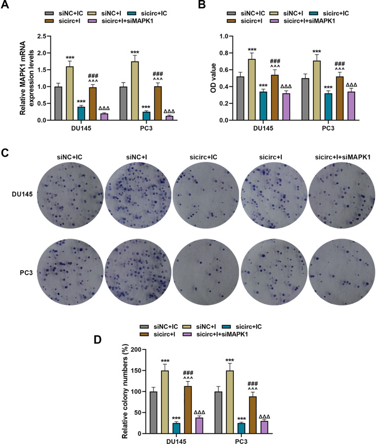
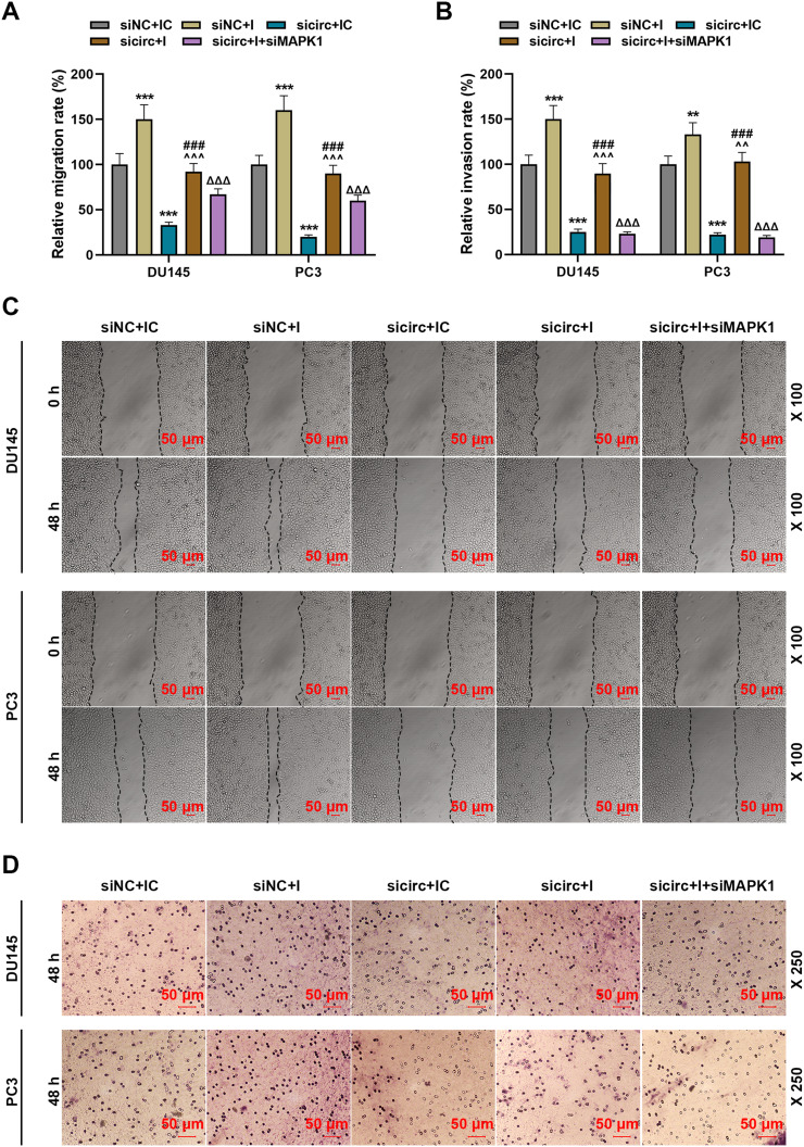
To further ascertain the mechanism of the ceRNA regulatory network of circSLC19A1 in PCa progression regulation, phenotype changes of DU145 and PC3 cells were observed under transfection of miR-326 inhibitor, sicircSLC19A1 or siMAPK1. Transfection of miR-326 inhibitor increased MAPK1 expression, while silencing circSLC19A1 decreased MAPK1 expression (Figure 5A, P <0.001). MAPK1 expression in sicircSLC19A1 and miR-326 inhibitor co-transfected PCa cells was markedly lower than that in miR-326 inhibitor-transfected PCa cells, but was pronouncedly higher than that in sicircSLC19A1-transfected PCa cells (Figure 5A, P <0.001). Significantly increases of Viability and colony number of PCa cells were shown under the treatment of miR-326 inhibition, whereas remarkably decreases of those were shown under the treatment of silencing circSLC19A1 (Figure 5B–D, P <0.001). Silencing circSLC19A1 expression decreased the viability and colony number of miR-326 inhibitor-transfected PCa cells, while transfection of miR-326 inhibitor increased the viability and colony number of sicircSLC19A1-transfected PCa cells (Figure 5B–D, P <0.001). Silencing MAPK1 decreased the viability and colony number of sicircSLC19A1 and miR-326 inhibitor co-transfected PCa cells (Figure 5B–D, P <0.001). Likewise, PCa cell migration and invasion were promoted by miR-326 inhibition, but were inhibited by silencing circSLC19A1 (Figure 6B, P <0.001). Silencing circSLC19A1 expression inhibited the migration and invasion of miR-326 inhibitor-transfected PCa cells, while transfection of miR-326 inhibitor promoted the migration and invasion of sicircSLC19A1-transfected PCa cells (Figure 6A–D, P <0.001). Silencing MAPK1 expression inhibited the migration and invasion of sicircSLC19A1 and miR-326 inhibitor co-transfected PCa cells (Figure 6A–D, P <0.001). These results showed that the effects of miR-326 inhibitor and silenced circSLC19A1 were mutually antagonized, and silenced MAPK1 further inhibited PCa cell progression, corroborating a silenced circSLC19A1-initiated ceRNA network of circSLC19A1/miR-326/MAPK1 for PCa cell progression inhibition.
Figure 5.
Silencing circSLC19A1 expression inhibited viability and proliferation in PCa cells by regulating miR-326/MAPK1 axis. (A) MAPK1 expression in DU145 and PC3 cells transfected with miR-326 inhibitor, sicircSLC19A1, siMAPK1 or miR-326 inhibitor plus sicircSLC19A1 was assessed by qRT-PCR, with GAPDH serving as a reference gene. (B) The viability of DU145 and PC3 cells transfected with miR-326 inhibitor, sicircSLC19A1, siMAPK1 or miR-326 inhibitor plus sicircSLC19A1 was measured by Cell Counting Kit-8 assay. (C) Representative photos of the colony formation of DU145 and PC3 cells transfected with miR-326 inhibitor, sicircSLC19A1, siMAPK1 or miR-326 inhibitor plus sicircSLC19A1. (D) The colony formation of DU145 and PC3 cells transfected with miR-326 inhibitor, sicircSLC19A1, siMAPK1 or miR-326 inhibitor plus sicircSLC19A1 was evaluated by cell colony formation assay. ***P<0.001 vs siNC+IC; ^^^P<0.001 vs siNC+I; ###P<0.001 vs sicirc+IC; ΔΔΔP<0.001 vs sicirc+I.
Abbreviations: sicircSLC19A1, siRNA-specific target circSLC19A1; siNC, siRNA-negative control; siMAPK1, siRNA-MAPK1; IC, inhibitor control; I, miR-326 inhibitor; qRT-PCR, quantitative reverse-transcription polymerase chain reaction.
Figure 6.
Silencing circSLC19A1 expression inhibited migration and invasion in PCa cells by regulating miR-326/MAPK1 axis. (A) The migration of DU145 and PC3 cells transfected with miR-326 inhibitor, sicircSLC19A1, siMAPK1 or miR-326 inhibitor plus sicircSLC19A1 was evaluated by wound healing assay. (B) The invasive state of DU145 and PC3 cells transfected with miR-326 inhibitor, sicircSLC19A1, siMAPK1 or miR-326 inhibitor plus sicircSLC19A1 was evaluated by Transwell assay. (C) Representative photos of the migration of DU145 and PC3 cells transfected with miR-326 inhibitor, sicircSLC19A1, siMAPK1 or miR-326 inhibitor plus sicircSLC19A1. (D) Representative photos of the invasive state of DU145 and PC3 cells transfected with or without sicircSLC19A1. **P<0.01 or ***P<0.001 vs siNC+IC; ^^P<0.01 and ^^^P<0.001 vs siNC+I; ###P<0.001 vs sicirc+IC; ΔΔΔP<0.001 vs sicirc+I.
Abbreviations: sicircSLC19A1, siRNA-specific target circSLC19A1; siNC, siRNA-negative control; siMAPK1, siRNA-MAPK1; IC, inhibitor control; I, miR-326 inhibitor; qRT-PCR, quantitative reverse-transcription polymerase chain reaction.
Discussion
PCa is the second most common cancer in men, especially leading to high morbidity and mortality in elderly men.2,19 Although progress has been continuously made in treatment of PCa, there are still the challenges preventing recurrence and poor prognosis to be faced.22,23 There have been numerous molecules documented contributing to the progression of tumor.20,21 With the development of high-throughput technologies, more and more circRNAs have been found to exert important regulatory roles in different types of treatment resistance in human tumors.24,25 In 2020, CircHIPK3 was reported to be able to facilitate the G2/M transition in PCa Cells via sponging miR-338-3p.25 circFMN2 was found to sponge miR-1238 to enhance the proliferation ability of the Expression of PCa cells through regulating the expression of LIM-Homeobox Gene 2.26 In our study, circSLC19A1 was found high-expressed in PCa tissues and cells and silencing of circSLC19A1 significantly inhibited PCa cell colony formation, migration and invasion by releasing miR-326 to inhibit MAPK1 expression.
Accumulating evidence demonstrates that aberrant expression of circRNA is associated with PCa cell proliferation, metastasis and treatment resistance.30,31 Recent studies highlighted the clinical diagnosis indicative function of circRNAs and the possibility of circRNAs serving as therapeutic targets.14 According to Cao, 13 circRNAs that produced from androgen receptor genes have been identified to promote PCa progression.31 Xia investigated circRNA expression profiling in PCa. Building on the finding that circRNAs were transcribed from parental gene SLC19A1, a differentially expressed gene in PCa, she suggested that circRNAs might be utilized as novel biomarkers for PCa.2 Based on Xia’s findings, our study focused on exploring the underlying mechanism of circSLC19A1 affecting PCa progression. According to one previous study by Zheng, circSLC19A1 was high-expressed in extracellular vesicles secreted by PCa cells.15 Similarly, we found circSLC19A1 expression was up-regulated in PCa tissues and cell lines. To further confirm the result, DU145 and PC3 cells which exhibited relatively higher circSLC19A1 expression were subjected to RNase R digestion. Our result showed an unchanged circSLC19A1 expression and a decreased SLC19A1 expression in DU145 and PC3 cells, indicating the existence of circSLC19A1 expression in PC3 cells. Besides, Zheng’s study showed that high circSLC19A1 expression was associated with promotion of PCa cell migration and invasion.15 Consistent with this, we found that forced decrease of circSLC19A1 expression inhibited PCa cell proliferation, colony formation, migration and invasion, reaffirming the oncogenic role of circSLC19A1.
CircRNAs are well known for its indirect regulation on mRNA through impeding the miRNA-mediated inhibition of mRNA expression.32 Such regulatory mechanism has been found useful to therapeutic intervention in PCa progression.33 He’s study discovered a great amount of sequence binding-based interaction between circRNAs and miR-145 (a tumor suppressor) in the pathogenesis of PCa.33 Dai’s study recognized circMYLK (Myosin Light Chain Kinase) in PCa, which targets miR-29a to promote PCa progression.34 Xiang’s study showed that circUCK2 plays an inhibitory role in PCa cell invasion and proliferation by sponging miR-767-5p to increase TET1 expression.35 In Zheng’s study, high circSLC19A1 expression resulted in promoted PCa cell migration and invasion, accompanied with down-regulation of miR-497 expression and up-regulation of septin 2 and sequence analysis proposed a potential regulatory network in which circSLC19A1 competitively binds to miR-497, unleashing septin 2.15 Our study identified miR-326as a novel binding target of circSLC19A1, which was low-expressed in PCa tissue and regained its expression by silenced circSLC19A1. MiR-326, as its down-regulation correlates with poor survival rate of PCa patients, has therefore been previously proposed as a prognostic maker for PCa.36 Consistent with this view, in our study, we found that forced inhibition on miR-326 expression caused promoted PCa cell progression and abolished the inhibitory effects of silenced circSLC19A1 on PCa cell progression. In the study of Liang, he made clarification on the tumor-suppressive function of miR-326 that miR-326 targets Mucin1 to suppress human prostatic carcinoma.37 According to Jiang, MAPK1, a target gene of miR-326, was found to promote cervical cancer cell proliferation, migration and invasion.38 Likewise, our study reaffirmed MAPK1 as a target gene of miR-326. MAPK1 is involved with various biological processes, such as synaptic and structural plasticity, and the initiation and progression of inflammatory processes related to depression. Previous study showed that MAPK1 expression was up-regulated in several types of cancer including PCa and could exert an oncogenic role. Our study again validated that MAPK1 was also high-expressed in PCa tissue, and silencing of MAPK1 inhibited PCa cell progression.
In conclusion, our results reaffirmed that circSLC19A1 is up-regulated in PCa tissues and cells and silencing of circSLC19A1 inhibited the proliferation, colony formation, migration and invasion of PCa cells. More importantly, the inhibitory effect of silencing circSLC19A1 on PCa cell progression may be attributed to suppression of superior binding between circSLC19A1 and miR-326, leading to binding between miR-326 and MAPK1. Counterevidence in our study indicates that circSLC19A1 can function as a sponge for miR-326 to increase MAPK1 expression. Therefore, our study proposed a novel circRNA/miRNA/mRNA regulatory network of circSLC19A1 for the management of PCa.
Funding Statement
This work was supported by the Zhejiang Provincial Basic Public Welfare Research Project [GF18H160064].
Abbreviation
circRNAs, circular RNAs; PCa, prostate cancer; PSA, prostate-specific antigen; AUC, area under the curve; ATCC, American Type Culture Collection; DEPC, diethyl pyrocarbonate; qRT-PCR, quantitative reverse-transcription polymerase chain reaction; MAPK1, mitogen-activated protein kinase 1; CCK-8, Cell Counting Kit-8; SD, standard deviation; ceRNA, competing endogenous RNA.
Disclosure
All authors report no conflicts of interest in this work.
References
- 1.Bray F, Ferlay J, Soerjomataram I, Siegel RL, Torre LA, Jemal A. Global cancer statistics 2018: GLOBOCAN estimates of incidence and mortality worldwide for 36 cancers in 185 countries. CA Cancer J Clin. 2018;68(6):394–424. [DOI] [PubMed] [Google Scholar]
- 2.Xia Q, Ding T, Zhang G, et al. Circular RNA expression profiling identifies prostate cancer- specific circRNAs in prostate cancer. Cell Physiol Biochem. 2018;50(5):1903–1915. doi: 10.1159/000494870 [DOI] [PubMed] [Google Scholar]
- 3.Schymura MJ, Sun L, Percy-Laurry A. Prostate cancer collaborative stage data items–their definitions, quality, usage, and clinical implications: a review of SEER data for 2004–2010. Cancer. 2014;120(Suppl 23):3758–3770. doi: 10.1002/cncr.29052 [DOI] [PubMed] [Google Scholar]
- 4.Buyyounouski MK, Pickles T, Kestin LL, Allison R, Williams SG. Validating the interval to biochemical failure for the identification of potentially lethal prostate cancer. J Clin Oncol. 2012;30(15):1857–1863. doi: 10.1200/JCO.2011.35.1924 [DOI] [PubMed] [Google Scholar]
- 5.Masson S, Bahl A. Metastatic castrate-resistant prostate cancer: dawn of a new age of management. BJU Int. 2012;110(8):1110–1114. doi: 10.1111/j.1464-410X.2012.11076.x [DOI] [PubMed] [Google Scholar]
- 6.Sartor O, de Bono JS, Longo DL. Metastatic prostate cancer. N Engl J Med. 2018;378(7):645–657. doi: 10.1056/NEJMra1701695 [DOI] [PubMed] [Google Scholar]
- 7.Qu S, Yang X, Li X, et al. Circular RNA: a new star of noncoding RNAs. Cancer Lett. 2015;365(2):141–148. doi: 10.1016/j.canlet.2015.06.003 [DOI] [PubMed] [Google Scholar]
- 8.Salzman J, Gawad C, Wang PL, Lacayo N, Brown PO, Preiss T. Circular RNAs are the predominant transcript isoform from hundreds of human genes in diverse cell types. PLoS One. 2012;7(2):e30733. doi: 10.1371/journal.pone.0030733 [DOI] [PMC free article] [PubMed] [Google Scholar]
- 9.Jeck WR, Sorrentino JA, Wang K, et al. Circular RNAs are abundant, conserved, and associated with ALU repeats. RNA. 2013;19(2):141–157. doi: 10.1261/rna.035667.112 [DOI] [PMC free article] [PubMed] [Google Scholar]
- 10.Memczak S, Jens M, Elefsinioti A, et al. Circular RNAs are a large class of animal RNAs with regulatory potency. Nature. 2013;495(7441):333–338. doi: 10.1038/nature11928 [DOI] [PubMed] [Google Scholar]
- 11.Guo JU, Agarwal V, Guo H, Bartel DP. Expanded identification and characterization of mammalian circular RNAs. Genome Biol. 2014;15(7):409. doi: 10.1186/s13059-014-0409-z [DOI] [PMC free article] [PubMed] [Google Scholar]
- 12.Panda AC. Circular RNAs act as miRNA sponges. Adv Exp Med Biol. 2018;1087:67–79. [DOI] [PubMed] [Google Scholar]
- 13.Li Z, Huang C, Bao C, et al. Exon-intron circular RNAs regulate transcription in the nucleus. Nat Struct Mol Biol. 2015;22(3):256–264. doi: 10.1038/nsmb.2959 [DOI] [PubMed] [Google Scholar]
- 14.Kristensen LS, Hansen TB, Venø MT, Kjems J. Circular RNAs in cancer: opportunities and challenges in the field. Oncogene. 2018;37(5):555–565. doi: 10.1038/onc.2017.361 [DOI] [PMC free article] [PubMed] [Google Scholar]
- 15.Zheng Y, Li JX, Chen CJ, Lin ZY, Liu JX, Lin FJ. Extracellular vesicle-derived circ_SLC19A1 promotes prostate cancer cell growth and invasion through the miR-497/septin 2 pathway. Cell Biol Int. 2020;44(4):1037–1045. doi: 10.1002/cbin.11303 [DOI] [PubMed] [Google Scholar]
- 16.Livak KJ, Schmittgen TD. Analysis of relative gene expression data using real-time quantitative PCR and the 2(-delta delta C(T)) Method. Methods. 2001;25(4):402–408. doi: 10.1006/meth.2001.1262 [DOI] [PubMed] [Google Scholar]
- 17.Werfel S, Nothjunge S, Schwarzmayr T, Strom TM, Meitinger T, Engelhardt S. Characterization of circular RNAs in human, mouse and rat hearts. J Mol Cell Cardiol. 2016;98:103–107. doi: 10.1016/j.yjmcc.2016.07.007 [DOI] [PubMed] [Google Scholar]
- 18.Salzman J. Circular RNA expression: its potential regulation and function. Trends Genet. 2016;32(5):309–316. doi: 10.1016/j.tig.2016.03.002 [DOI] [PMC free article] [PubMed] [Google Scholar]
- 19.Torre LA, Bray F, Siegel RL, Ferlay J, Lortet-Tieulent J, Jemal A. Global cancer statistics, 2012. CA Cancer J Clin. 2015;65(2):87–108. [DOI] [PubMed] [Google Scholar]
- 20.Cai F, Li J, Zhang J, Huang S. Knockdown of Circ_CCNB2 sensitizes prostate cancer to radiation through repressing autophagy by the miR-30b-5p/KIF18A axis. Cancer Biother Radiopharm. 2020. doi: 10.1089/cbr.2019.3538 [DOI] [PubMed] [Google Scholar]
- 21.Gao Y, Liu J, Huan J, Che F. Downregulation of circular RNA hsa_circ_0000735 boosts prostate cancer sensitivity to docetaxel via sponging miR-7. Cancer Cell Int. 2020;20(1):334. doi: 10.1186/s12935-020-01421-6 [DOI] [PMC free article] [PubMed] [Google Scholar]
- 22.Miller K, Nogueira L, Mariotto A, et al. Cancer treatment and survivorship statistics, 2019. CA Cancer J Clin. 2019;69. [DOI] [PubMed] [Google Scholar]
- 23.Andersen S, Richardsen E, Moi L, et al. Fibroblast miR-210 overexpression is independently associated with clinical failure in prostate cancer - a multicenter (in situ hybridization) study. Sci Rep. 2016;6(1):36573. doi: 10.1038/srep36573 [DOI] [PMC free article] [PubMed] [Google Scholar]
- 24.Shi J, Liu C, Chen C, et al. Circular RNA circMBOAT2 promotes prostate cancer progression via a miR-1271-5p/mTOR axis. Aging. 2020;12(13):13255–13280. doi: 10.18632/aging.103432 [DOI] [PMC free article] [PubMed] [Google Scholar]
- 25.Liu F, Fan Y, Ou L, et al. CircHIPK3 facilitates the G2/M transition in prostate cancer cells by sponging miR-338-3p. Onco Targets Ther. 2020;13:4545–4558. doi: 10.2147/OTT.S242482 [DOI] [PMC free article] [PubMed] [Google Scholar]
- 26.Shan G, Shao B, Liu Q, et al. circFMN2 sponges miR-1238 to promote the expression of LIM-homeobox gene 2 in prostate cancer cells. Mol Ther Nucleic Acids. 2020;21:133–146. doi: 10.1016/j.omtn.2020.05.008 [DOI] [PMC free article] [PubMed] [Google Scholar] [Retracted]
- 27.Kong Z, Wan X, Zhang Y, et al. Androgen-responsive circular RNA circSMARCA5 is up-regulated and promotes cell proliferation in prostate cancer. Biochem Biophys Res Commun. 2017;493(3):1217–1223. doi: 10.1016/j.bbrc.2017.07.162 [DOI] [PubMed] [Google Scholar]
- 28.Cao S, Ma T, Ungerleider N, et al. Circular RNAs add diversity to androgen receptor isoform repertoire in castration-resistant prostate cancer. Oncogene. 2019;38(45):7060–7072. doi: 10.1038/s41388-019-0947-7 [DOI] [PMC free article] [PubMed] [Google Scholar]
- 29.Greene J, Baird AM, Brady L, et al. Circular RNAs: biogenesis, function and role in human diseases. Front Mol Biosci. 2017;4:38. doi: 10.3389/fmolb.2017.00038 [DOI] [PMC free article] [PubMed] [Google Scholar]
- 30.He JH, Han ZP, Zhou JB, et al. MiR-145 affected the circular RNA expression in prostate cancer LNCaP cells. J Cell Biochem. 2018;119(11):9168–9177. doi: 10.1002/jcb.27181 [DOI] [PMC free article] [PubMed] [Google Scholar]
- 31.Dai Y, Li D, Chen X, et al. Circular RNA myosin light chain kinase (MYLK) promotes prostate cancer progression through modulating Mir-29a expression. Med Sci Monit. 2018;24:3462–3471. doi: 10.12659/MSM.908009 [DOI] [PMC free article] [PubMed] [Google Scholar]
- 32.Xiang Z, Xu C, Wu G, Liu B, Wu D. CircRNA-UCK2 increased TET1 inhibits proliferation and invasion of prostate cancer cells via sponge MiRNA-767-5p. Open Med. 2019;14(1):833–842. doi: 10.1515/med-2019-0097 [DOI] [PMC free article] [PubMed] [Google Scholar]
- 33.Moya L, Meijer J, Schubert S, Matin F, Batra J. Assessment of miR-98-5p, miR-152-3p, miR-326 and miR-4289 expression as biomarker for prostate cancer diagnosis. Int J Mol Sci. 2019;20(5):1154. doi: 10.3390/ijms20051154 [DOI] [PMC free article] [PubMed] [Google Scholar]
- 34.Liang X, Li Z, Men Q, Li Y, Li H, Chong T. miR-326 functions as a tumor suppressor in human prostatic carcinoma by targeting Mucin1. Biomed Pharmacother. 2018;108:574–583. doi: 10.1016/j.biopha.2018.09.053 [DOI] [PubMed] [Google Scholar]
- 35.Jiang H, Liang M, Jiang Y, et al. The lncRNA TDRG1 promotes cell proliferation, migration and invasion by targeting miR-326 to regulate MAPK1 expression in cervical cancer. Cancer Cell Int. 2019;19(1):152. doi: 10.1186/s12935-019-0872-4 [DOI] [PMC free article] [PubMed] [Google Scholar]
- 36.Calati R, Crisafulli C, Balestri M, et al. Evaluation of the role of MAPK1 and CREB1 polymorphisms on treatment resistance, response and remission in mood disorder patients. Prog Neuropsychopharmacol Biol Psychiatry. 2013;44:271–278. doi: 10.1016/j.pnpbp.2013.03.005 [DOI] [PubMed] [Google Scholar]
- 37.García-Fuster MJ, Díez-Alarcia R, Ferrer-Alcón M, La Harpe R, Meana JJ, García-Sevilla JA. FADD adaptor and PEA-15/ERK1/2 partners in major depression and schizophrenia postmortem brains: basal contents and effects of psychotropic treatments. Neuroscience. 2014;277:541–551. doi: 10.1016/j.neuroscience.2014.07.027 [DOI] [PubMed] [Google Scholar]
- 38.Zunszain PA, Hepgul N, Pariante CM. Inflammation and depression. Curr Top Behav Neurosci. 2013;14:135–151. [DOI] [PubMed] [Google Scholar]
- 39.Zhu L, Yang S, Wang J. miR-217 inhibits the migration and invasion of HeLa cells through modulating MAPK1. Int J Mol Med. 2019;44(5):1824–1832. [DOI] [PMC free article] [PubMed] [Google Scholar]
- 40.Zhang Y, Meng L, Xiao L, Liu R, Li Z, Wang YL. The RNA-binding protein PCBP1 functions as a tumor suppressor in prostate cancer by inhibiting mitogen activated protein kinase 1. Cell Physiol Biochem. 2018;48(4):1747–1754. doi: 10.1159/000492315 [DOI] [PubMed] [Google Scholar]


