PLOS ONE logoLink to PLOS ONE
. 2020 Nov 23;15(11):e0242533. doi: 10.1371/journal.pone.0242533

Low prevalence of bloodstream infection and high blood culture contamination rates in patients with COVID-19

David Yu 1,2,¤, Karolina Ininbergs 1,3, Karolina Hedman 3, Christian G Giske 1,3, Kristoffer Strålin 4,5, Volkan Özenci 1,3,¤,*
Editor: Surbhi Leekha6
PMCID: PMC7682817  PMID: 33226995

Abstract

Purpose

In the management of COVID-19, knowledge is lacking on the frequency of secondary bacterial infections and on how empirical antibiotic therapy should be used. In the present study, we aimed to compare blood culture (BC) results of a COVID-19 patient cohort with two cohorts of patients without detected COVID-19.

Methods

Using a retrospective cohort study design of patients subjected to BC in six tertiary care hospitals, SARS-CoV-2 positive patients from March 1 to April 30 in 2020 (COVID-19 group) were compared to patients without confirmed SARS-CoV-2 during the same period (control group-2020) and with patients sampled March 1 to April 30 in 2019 (control group-2019). The outcomes studied were proportion of BC positivity, clinically relevant growth, and contaminant growth.

Results

In total 15,103 patients and 17,865 BC episodes were studied. Clinically relevant growth was detected in 197/3,027 (6.5%) BC episodes in the COVID-19 group compared to 717/6,663 (10.8%) in control group-2020 (p<0.0001) and 850/8,175 (10.4%) in control group-2019 (p<0.0001). Contamination was present in 255/3,027 (8.4%) BC episodes in the COVID-19 group compared to 330/6,663 (5.0%) in control group-2020 (p<0.0001) and 354/8,175 (4.3%) in control group-2019 (p<0.0001).

Conclusion

In COVID-19 patients, the prevalence of bloodstream bacterial infection is low and the contamination rate of BC is high. This knowledge should influence guidelines regarding blood culture sampling and empirical antibiotic therapy in COVID-19 patients.

Introduction

Secondary bacterial infections are a major clinical problem in patients with influenza virus infections and have previously been reported to be associated with poor disease outcome [1, 2]. Bloodstream infections (BSIs) remain one of the most common and life-threatening complications in patients with severe viral infections. Epidemiological data of secondary BSIs might therefore play a significant role in reducing mortality and morbidity rates due to COVID-19. A retrospective study from USA reported that patients with COVID-19 have low bacteremia rates than controls [3]. The blood culture routines and the characteristics of COVID-19 patients differ significantly between centers and geographic locations. Therefore, there is an imminent need for studies on BSIs in COVID-19 to understand their importance for disease outcome.

The aim of the present study is to analyze blood culture data of a cohort of COVID-19 patients and compare it with two cohorts of patients without COVID-19.

Materials and methods

Setting

The study was performed between 1 March 2020 and 30 April 2020 at Karolinska University Hospital, which serves a population of 2,436,767. The Karolinska University Laboratory receives blood culture specimens from six tertiary care hospitals in the greater Stockholm area and surrounding cities and suburbs. Historically, we have a yearly 10% increase in numbers of our blood culture bottles at Karolinska University Laboratory without any change in contamination rates.

Study design

Blood cultures collected from patients with COVID-19 and controls were analyzed retrospectively. The blood culture data was retrieved from the laboratory information system (wwLab/ADBakt, Autonik AB, Nykoping, Sweden) using QlikView (Qlik, King of Prussia, PA, USA).

Study population

Patients with COVID-19

Patients were considered to have COVID-19 if they were positive for SARS-CoV-2 RNA by reverse transcriptase PCR in respiratory secretions. Blood culture results from the COVID-19 patients registered between 1 March and 30 April in 2020 and were included and referred to as “COVID-19 group” herein after.

Control groups

The study included two control groups, i.e. one historical control group with blood culture results registered between 1 March and 30 April in 2019 (referred to as “control group-2019”) and one contemporary control group with blood culture results registered between 1 March and 30 April in 2020, and with no confirmed PCR-positivity for SARS-CoV-2 (referred to as “control group-2020”). In the beginning of the pandemic, testing was only done in patients with symptoms consistent with COVID-19 and not all admitted patients. In the present study, the control group-2020 therefore consisted of a mix of negative patients and patients not tested. However, all patients with COVID-19-like symptoms were tested. Therefore, it is reasonable to assume that the patients not tested for SARS-COV-2 did not have clinical findings of COVID-19.

Laboratory methods

Blood cultures

Three different blood culture bottles were used in the study; BacT/Alert FA Plus aerobic, BacT/ALERT-PF Plus pediatric and BacT/Alert FN Plus anaerobic plus bottles. Bottles were incubated in BacT/ALERT Virtuo (bioMérieux, Durham, NC, USA) blood culture system until they signaled positive or for a maximum of five days.

The Gram stains were done directly from positive blood culture bottles. According to the result from the staining, specimen from the positive bottles were subcultured onto relevant agar plates. The microorganisms grown on the agar plates were identified by Bruker MALDI-TOF MS. Antimicrobial susceptibility testing was performed by disc diffusion and the results were interpreted following EUCAST recommendations (www.eucast.org).

SARS-CoV-2 RT-PCR

Testing for SARS-CoV-2 was performed using three different RT-PCR assays: cobas SARS-CoV-2 (Roche Molecular Systems, Inc., Branchburg, NJ), Xpert Xpress SARS-CoV-2 (XPRSARS-COV2-10) (Cepheid, Sunnyvale, CA) or an in-house developed assay based on Corman et al. [4] targeting the E- and RdRP-genes, with modifications of primers according to Edén A et al. (Neurology, in revision); (Supplementary methods).

Data analysis

The data on blood cultures were presented as individual BSI episodes, from here on called only “episode”. More than one episode from the same patient could be included, however, to be defined as a new episode a minimum of 72 h had to pass between sampling of the same patient. In case of more than 4 bottles taken in a single episode, only the first four bottles were considered in the analysis. Following isolates were considered as contaminants if they grew in less than 3 out of 4 blood culture bottles: Bacillus spp., Corynebacterium spp., Cutibacterium spp., coagulase negative staphylococci (CoNS), Micrococcus spp., Cellulomonas spp., Lactobacillus spp., Dermabacter spp., Facklamia spp., Rothia spp., Exiguobacterium spp., Brevibacterium spp., and Trueperella spp.

Statistical analysis

The statistical analyses were performed with GraphPad Prism 5.0 (GraphPad Software, San Diego, CA). The blood culture results in patients with COVID-19, control group-2020 and control group-2019 were compared using the Pearson’s chi-square test. Values of P <0.05 were considered as statistically significant.

Results

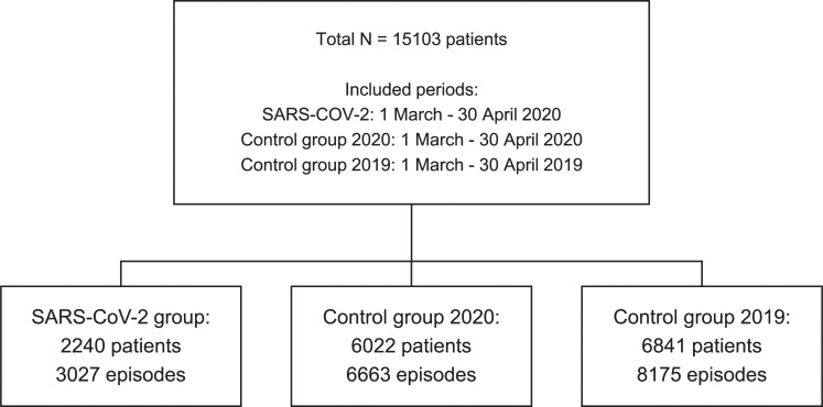
In total, 58,704 blood culture bottles from 17,865 episodes in 15,103 patients were studied. The study flow chart is depicted in Fig 1. The patients of the study groups had the following characteristics; COVID-19 group, 790/2,240 (35.3%) female, mean (Standard Deviation [SD]) age 64 (18) years; control group-2020, 2,789/6,022 (46.3%) female, mean (SD) age 57 (26) years; and control group-2019, 3,322/6,841 (48.6%) female, mean (SD) age 60 (26) years.

Fig 1. Flow chart of the study population.

Fig 1

Overall blood culture positivity

The COVID-19 group consisted of 2240/8262 (27%) of all patients sampled for blood cultures during the study period in 2020. The total number of blood cultured patients during the 2020 study period, 8262, is an increase of 1421 (20%), compared to the same period last year (total 6841 patients). In 511/2,240 (22.8%) patients in the COVID-19 group, there were two or more episodes during the study period. In the control groups, BCs were obtained from 6022 and 6841 patients in control group-2020 and control group-2019, respectively. In control group-2020, 459 (7.6%) patients had two or more episodes. In control group-2019, 910 (13.3%) patients had two or more episodes. In total 3,027 episodes in the COVID-19 group, 6,663 in control group-2020 and 8,175 in control group-2019 were studied. Considering episodes, growth of microorganisms in BC was detected in 433/3,027 (14.3%) episodes in the COVID-19 group, compared with 1,015/6,663 (15.2%) in control group-2020 (non-significant) and 1,153/8,175 (14.1%) in control group-2019 (non-significant) (Table 1).

Table 1. Bloodstream infection episode data for patients with COVID-19 and both control groups.

Episode type COVID-19 Control group-2020 Control group-2019
Included episodes, N 3027 6663 8175
Episodes with growth, n (%) 433 (14.3) 1015 (15.2) 1153 (14.1)
Episodes with clinically relevant growth, n (%) 197 (6.5) 717 (10.8) 851 (10.4)
    • Gram positive* 116 (3.8) 344 (5.2) 420 (5.1)
    • Gram negative* 64 (1.7) 306 (4.6) 351 (4.3)
    • Yeast* 2 (0.07) 11 (0.17) 8 (0.10)
    • Polymicrobial episodes** 27 (0.89) 56 (0.84) 72 (0.88)
Episodes with contaminant growth, n (%) 255 (8.4) 330 (5.0) 354 (4.3)
    • Only contaminant growth 236 (7.8) 298 (4.5) 302 (3.7)
    • Both contaminant and clinically relevant growth 19 (0.63) 32 (0.48) 52 (0.64)

*Monomicrobial episodes.

**Polymicrobial episode is defined as an episode with occurrence of more than one clinically relevant isolate.

Clinically relevant growth

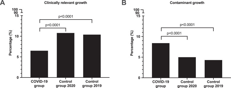
Clinically relevant growth was detected in 197/3,027 (6.5%) of episodes in the COVID-19 group, compared with 717/6,663 (10.8%) in control group-2020 (p<0.0001) and 851/8,175 (10.4%) in control group-2019 (p<0.0001) (Table 1, Fig 2 [Panel A]).

Fig 2.

Fig 2

Blood culture episodes with clinically relevant growth (Panel A) and with contaminant growth (Panel B). Total number of episodes included for analysis were COVID-19 group: 3,027, Control group-2020: 6,663, Control group-2019: 8,175.

When blood cultures with polymicrobial bacteremia were analyzed there was no difference among the three groups studied (Table 1).

Contaminant growth

Contamination in blood cultures were detected in 255/3,027 (8.4%) episodes in patients with COVID-19, compared with 330/6,663 (4.95%) episodes in control group-2020 (p<0.0001) and 354/8,175 (4.33%) episodes in control group-2019 (p<0.0001). The two control groups had similar numbers of episodes with contaminant growth (non-significant) (Table 1, Fig 2 [Panel B]).

When relationship between the frequency of contaminants and the hospital localization was analyzed, control group-2019 had higher contamination rates in ICUs than in emergency departments and other clinics. In contrast, the contamination rates were similar in all hospital locations for control group-2020. In the COVID-19 group, contamination rates were higher in all hospital locations compared to the control groups, but more so in the emergency departments and ICUs (Table 2).

Table 2. Numbers and proportions of contamination in blood cultures from different hospital locations.

COVID-19 Control group 2020 Control group 2019
Hospital location Contaminant (% of total) Total episodes (n) Contaminant (% of total) Total episodes (n) Contaminant (% of total) Total episodes (n)
Emergency department 112 (9.2) 1221 130 (4.8) 2705 110 (4.0) 2769
Intensive care unit 70 (10.6) 659 18 (5.6) 319 16 (7.9) 202
Other hospital locations 73 (6.4) 1147 182 (5.0) 3639 228 (4.4) 5204

Similar results were observed when numbers of bottles with contaminants were analyzed. In total 337/10,504 (3.2%) bottles in the COVID-19 group were contaminated, compared to 433/21,261 (2.0%) in control group-2020 (p<0.0001) and 470/26,939 (1.7%) in control group-2019 (p<0.0001).

Diversity of microorganisms in blood cultures

There was a significant diversity in microorganisms detected from blood cultures among the three groups studied. Gram-positive growth was significantly higher in patients with COVID-19, 150/226 (66%) isolates, than in control group-2020 and -2019, 385/781 (49%) 487/940 (52%) isolates respectively (p<0.0001 for both comparisons) (Table 3). The two control groups had similar levels of numbers of Gram-positive growth (non-significant). In contrast, Gram-negative isolates was significantly fewer in patients with COVID-19, 66/226 (29%) isolates, than in control group-2020 353/781 (45%) isolates and -2019 398/940 (42%) isolates (p<0.0001 and p<0.001 respectively). The two control groups had similar levels of numbers of Gram-negative isolates (non-significant).

Table 3. Distribution of microorganisms isolated from blood cultures in patients with COVID-19 and both control groups.

COVID-19 Control group-2020 Control group-2019
All isolates, N 226 781 940
Gram-positive bacteria* n (%) 150 (66) 385 (49) 487 (52)
    • Coagulase negative staphylococci** 49 (22) 55 (7.1) 58 (6.2)
    • Staphylococcus aureus 44 (19) 124 (16) 162 (17)
    • Enterococcus spp. 23 (10) 46 (5.9) 75 (8.0)
    • Viridans group streptococci 22 (9.7) 64 (8.2) 63 (6.7)
    • Beta-hemolytic streptococci 6 (2.7) 47 (6.0) 50 (5.3)
    • Streptococcus pneumoniae 3 (1.3) 21 (1.4) 51 (1.6)
    • Other Gram-positive bacteria 3 (1.3) 28 (3.6) 28 (3.0)
Gram-negative bacteria* n (%) 66 (29) 352 (45) 397 (42)
    • Escherichia coli 34 (15) 213 (27) 242 (26)
    • Other Enterobacterales 24 (11) 111 (14) 117 (13)
    • Pseudomonas aeruginosa 7 (3.1) 13 (1.7) 14 (1.5)
    • Other Gram-negative bacteria 1 (0.4) 15 (1.9) 24 (2.6)
Anaerobic bacteria n (%) 7 (3.1) 32 (4.1) 40 (4.3)
Yeast n (%) 3 (1.3) 11 (1.4) 15 (1.6)

*Not including anaerobic bacteria

**Including Staphylococcus epidermidis

†Other bacteria and yeast are shown in S1 Table.

The most common three microorganisms detected in blood cultures from COVID-19 patients were CoNS, Staphylococcus aureus and Escherichia coli. In contrast, for both control groups the three most common isolates were E. coli, S. aureus and other Enterobacterales.

Patients with COVID-19 had higher numbers of episodes with clinically relevant CoNS than in control group-2020 and -2019 (p<0.0001 for both comparisons). There was no difference in numbers of episodes with CoNS between the two control groups (non-significant). In contrast, lower numbers of Escherichia coli were observed in patients with COVID-19 (15%) compared to control group-2020 (27%) and -2019 (26%) (p = 0.0001 and p = 0.0005, respectively). There was no significant difference for S. aureus, anaerobes and yeasts among the three groups studied. A detailed list of all microorganisms can be found in supplements (S1 Table).

Time to detection in blood cultures

The time to detection (TTD) in positive blood culture bottles differed between the three groups analyzed. The mean (SD) TTD was 22.8 (17.6) in COVID-19 group whereas control group-2020 and control group-2019 had 18.4 (15.3) and 18.2 (17.2) h (Table 4).

Table 4. Time to detection of microorganisms in positive blood cultures in patients with COVID-19 and both control groups.

Time to detection COVID-19 Control group Control group
(n = 820) 2020 (n = 2185) 2019 (n = 2663)
Mean (SD [h]) 22.8 (17.6) 18.4 (15.3) 18.2 (17.2)
Median (IQR [h]) 18.5 (11.5) 14.0 (10.7) 13.2 (10.4)

SD: standard deviation. IQR: interquartile range.

When the incubation period for blood cultures were analyzed in a total of 5568 bottles, we observed that the vast majority (93–96%) of the bottles signaled positive in 48 h. An additional 4–5% signaled positive in 4 days. The remaining BC that signaled positive in 5 days were 2% in COVID-19 group and 1% in both control groups (Table 5).

Table 5. Proportions of positive blood cultures signaling positive during given time intervals in the blood culture system.

Time to detection COVID-19 Control group 2020 Control group 2019
(n = 820) (n = 2185) (n = 2663)
Day 1–2 93% 96% 95%
Day 3 4% 3% 3%
Day 4 1% 1% 1%
Day 5 2% 1% 1%

Discussion

Blood culture is the gold standard for detection of microorganisms in patients with BSI. We presented the blood culture findings in patients with COVID-19 and in other patients with suspected BSIs from a contemporary and a historical control group.

In the present study the overall blood culture positivity rate was similar in all three groups analyzed. However, the proportion of episodes with clinically relevant growth was significantly lower in in patients with COVID-19 than both control groups. Although in our study the true incidence of bacteremia was not known, the proportion of episodes with clinically relevant growth correlates with previous data on clinical characteristics in COVID-19, where bacteremia was observed in 5.6% of cases [5] and septic shock in 4% of cases [6]. The reason for lower bacteremia rates in patient with COVID-19 is largely unknown. Patients with severe COVID-19 fulfill the sepsis-3 criteria for sepsis [7] and the term viral sepsis has been introduced [8]. In patients who are hospitalized for COVID-19, it is thus difficult to use clinical and laboratory parameters to differentiate between the viral component and a potential bacterial component. The low rate of relevant bacteremia indicates that the viral component is predominant in COVID-19. There was a higher proportion of patients with more than one suspected BSI episode in the COVID-19 group compared to the control groups. The reason for this is not known.

In a recent study, from New York, USA, it is reported that only 3.8% of COVID-19 patients had positive blood cultures which was significantly lower than the controls in that study [3]. The present data differs from the NY study since the overall BC positivity rate in our COVID-19 group was 14.3% and did not differ from controls. Moreover, clinically relevant growth in the present COVID-19 cohort was 6.5% in comparison to 1.8% in COVID-19 patients reported by Sepulveda et al. [3]. The underlying reason for these differences is unknown. It is reasonable to assume that the blood culture routines and the characteristics of COVID-19 patients were different. However, both studies showed that the clinically relevant growth is lower in patients with COVID-19 than in controls.

The present results show that bacterial and fungal BSIs are uncommon in patients with COVID-19 and it is warranted to establish stringent clinical criteria for empiric antibiotic treatment for BSI in these patients.

The species composition of microorganisms isolated from blood cultures from patients with COVID-19 and controls differed. When the three most common microorganisms isolated from the positive bottles were considered, patients with COVID-19 had significantly higher rates of clinically relevant CoNS than both control groups (p<0.0001 for both comparisons). In contrast, both control groups had higher rates of E. coli compared to COVID-19 patients (p = 0.0001 and p = 0.0005, respectively). All three groups had similar levels of S. aureus (non-significant). The underlying differences in occurrence of BSI with CoNS and E. coli in patients with COVID-19 and controls might be important in empiric antibiotic treatment of these patients. Overall, our results emphasize the importance of antimicrobial stewardship in the treatment of COVID-19 patient to minimize the threat of superinfections [9].

Contamination is a major problem in blood cultures. It was recently reported that COVID-19 patients had higher proportion of cultures that likely represented contamination with normal skin microbiota than controls [3]. However, the study did not analyze the growth of contaminants further. Growth of normal skin microbiota might be clinically relevant. The present study analyzed the contaminants in detail both at episode and BC bottle level, by using an algorithm to discriminate possible clinically relevant growth of normal skin flora bacteria [10, 11]. Patients with COVID-19 had 3.2% blood culture bottles (8.4% episodes) contaminated as compared to 2.04% (5.0% episodes) and 1.74% (4.2% episodes) in the two control groups, respectively. Under normal circumstances, blood cultures received in our center has low contamination rates as shown in the two control groups studied. In contrast, the blood culture bottles in the COVID-19 group exceeded the recommended rate of contamination of <3% according to CLSI guidelines [12], which may lead to unnecessary antibiotic use and longer hospital stay for these patients. The underlying reason for high contamination rates in patients with COVID-19 is not known. In the present study, contamination rates were higher in emergency departments and ICUs compared to other units in the COVID-19 group. The relationship between high stress environments and BC contamination rates has been previously reported [13, 14]. The patient characteristics and the work pace in emergency departments and ICUs differs from the other units. It is reasonable to suggest that the stressful working environment in these two units with well-known risk to be exposed to SARS-CoV-2 might play an important role in higher contamination rates observed in COVID-19 group.

TTD of positive blood cultures might be a relevant parameter in comparing different patient populations [15]. In the present study we showed that the mean TTD in COVID-19 was approximately 20% longer compared to controls. It is reasonable to suggest that the longer TTD in COVID-19 group is based on higher rates of contaminants in this group.

The recommended incubation period for blood cultures is 5 days. In line with a recently published study, we observed that 98–99% of the BC bottles in all three groups signaled positive in 4 days [3]. The present data support the assumption that BC can be incubated for a maximum of 4 days when it is necessary.

To our knowledge, this is the first European study analyzing blood culture data from patients with COVID-19 and has several strengths. The inclusion of over 15,000 patients from six tertiary care hospital is an important strength of the study design. The study analyzed the clinically relevant growth and contaminants in detail and had relatively high positive blood culture rates in all three groups studied.

The present study also has important limitations. First, we did not have access to baseline clinical data such as comorbidity, disease duration, length of hospital stay and treatment of patients in the three study groups. Therefore, assessment of the impact of differences in patient characteristics on the BC results could not be analyzed. Second, we did not include a control group with another viral respiratory infection, such as influenza, during the same season. Third, as the present study focused on BSI in COVID-19, data regarding other culture results were not analyzed, which precluded the analysis of other secondary infections such as pneumonia.

Conclusions

The present study shows that patients with COVID-19 have low prevalence of BSI and a higher rate of contamination in blood cultures. Further clinical studies are warranted in order to improve blood culture-based diagnostics in patients with COVID-19.

Supporting information

S1 Table. Distribution of all microorganisms isolated from blood cultures.

(DOCX)

Data Availability

All relevant data are within the manuscript and its Supporting Information files.

Funding Statement

The authors received no specific funding for this work.

References

  • 1.Liderot K, Ahl M, Ozenci V (2013) Secondary bacterial infections in patients with seasonal influenza A and pandemic H1N1. Biomed Res Int 2013:376219 10.1155/2013/376219 [DOI] [PMC free article] [PubMed] [Google Scholar]
  • 2.Morens DM, Taubenberger JK, Fauci AS (2008) Predominant role of bacterial pneumonia as a cause of death in pandemic influenza: implications for pandemic influenza preparedness. J Infect Dis 198 (7):962–970 10.1086/591708 [DOI] [PMC free article] [PubMed] [Google Scholar]
  • 3.Sepulveda J, Westblade LF, Whittier S, Satlin MJ, Greendyke WG, Aaron JG, et al. (2020) Bacteremia and Blood Culture Utilization During COVID-19 Surge in New York City. J Clin Microbiol 10.1128/JCM.00875-20 [DOI] [PMC free article] [PubMed] [Google Scholar]
  • 4.Corman VM, Landt O, Kaiser M, Molenkamp R, Meijer A, Chu DK, et al. (2020) Detection of 2019 novel coronavirus (2019-nCoV) by real-time RT-PCR. Euro Surveill 25 (3) [DOI] [PMC free article] [PubMed] [Google Scholar]
  • 5.Goyal P, Choi JJ, Pinheiro LC, Schenck EJ, Chen R, Jabri A, et al. (2020) Clinical Characteristics of Covid-19 in New York City. New England Journal of Medicine 10.1056/NEJMc2010419 [DOI] [PMC free article] [PubMed] [Google Scholar]
  • 6.Chen N, Zhou M, Dong X, Qu J, Gong F, Han Y, et al. (2020) Epidemiological and clinical characteristics of 99 cases of 2019 novel coronavirus pneumonia in Wuhan, China: a descriptive study. Lancet 395 (10223):507–513 10.1016/S0140-6736(20)30211-7 [DOI] [PMC free article] [PubMed] [Google Scholar]
  • 7.Singer M, Deutschman CS, Seymour CW, Shankar-Hari M, Annane D, Bauer M, et al. (2016) The Third International Consensus Definitions for Sepsis and Septic Shock (Sepsis-3). JAMA 315 (8):801–810 10.1001/jama.2016.0287 [DOI] [PMC free article] [PubMed] [Google Scholar]
  • 8.Li H, Liu L, Zhang D, Xu J, Dai H, Tang N, et al. (2020) SARS-CoV-2 and viral sepsis: observations and hypotheses. Lancet 395 (10235):1517–1520 10.1016/S0140-6736(20)30920-X [DOI] [PMC free article] [PubMed] [Google Scholar]
  • 9.Clancy CJ, Nguyen MH (2020) COVID-19, superinfections and antimicrobial development: What can we expect? Clin Infect Dis [DOI] [PMC free article] [PubMed] [Google Scholar]
  • 10.Yu D, Larsson A, Parke A, Unge C, Henning C, Sunden-Cullberg J, et al. (2020) Single-Sampling Strategy vs. Multi-Sampling Strategy for Blood Cultures in Sepsis: A Prospective Non-inferiority Study. Front Microbiol 11:1639 10.3389/fmicb.2020.01639 [DOI] [PMC free article] [PubMed] [Google Scholar]
  • 11.Leyssene D, Gardes S, Vilquin P, Flandrois JP, Carret G, Lamy B (2011) Species-driven interpretation guidelines in case of a single-sampling strategy for blood culture. Eur J Clin Microbiol Infect Dis 30 (12):1537–1541 10.1007/s10096-011-1257-3 [DOI] [PubMed] [Google Scholar]
  • 12.Dargere S, Cormier H, Verdon R (2018) Contaminants in blood cultures: importance, implications, interpretation and prevention. Clin Microbiol Infect 24 (9):964–969 10.1016/j.cmi.2018.03.030 [DOI] [PubMed] [Google Scholar]
  • 13.Self WH, Mickanin J, Grijalva CG, Grant FH, Henderson MC, Corley G, et al. (2014) Reducing blood culture contamination in community hospital emergency departments: a multicenter evaluation of a quality improvement intervention. Acad Emerg Med 21 (3):274–282 10.1111/acem.12337 [DOI] [PMC free article] [PubMed] [Google Scholar]
  • 14.Self WH, Speroff T, Grijalva CG, McNaughton CD, Ashburn J, Liu D, et al. (2013) Reducing blood culture contamination in the emergency department: an interrupted time series quality improvement study. Acad Emerg Med 20 (1):89–97 10.1111/acem.12057 [DOI] [PMC free article] [PubMed] [Google Scholar]
  • 15.Lamy B (2019) Reprint of: Blood culture time-to-positivity: making use of the hidden information. Clin Microbiol Infect</References> 10.1016/j.cmi.2019.03.005 [DOI] [PubMed] [Google Scholar]

Decision Letter 0

Surbhi Leekha

21 Aug 2020

PONE-D-20-20448

Low prevalence of bloodstream infection and high blood culture contamination rates in patients with COVID-19

PLOS ONE

Dear Dr. Özenci,

Thank you for submitting your manuscript to PLOS ONE. After careful consideration, we feel that it has merit but does not fully meet PLOS ONE’s publication criteria as it currently stands. Therefore, we invite you to submit a revised version of the manuscript that addresses the points raised during the review process.

My evaluation of this manuscript is noted below under "Additional Editor Comments". Please be sure to address these in addition to the comments provided by the reviewers. I have highlighted key points from reviewers in my evaluation as well.

Please submit your revised manuscript by Oct 05 2020 11:59PM. If you will need more time than this to complete your revisions, please reply to this message or contact the journal office at plosone@plos.org. When you're ready to submit your revision, log on to https://www.editorialmanager.com/pone/ and select the 'Submissions Needing Revision' folder to locate your manuscript file.

Please include the following items when submitting your revised manuscript:

  • A rebuttal letter that responds to each point raised by the academic editor and reviewer(s). You should upload this letter as a separate file labeled 'Response to Reviewers'.

  • A marked-up copy of your manuscript that highlights changes made to the original version. You should upload this as a separate file labeled 'Revised Manuscript with Track Changes'.

  • An unmarked version of your revised paper without tracked changes. You should upload this as a separate file labeled 'Manuscript'.

If you would like to make changes to your financial disclosure, please include your updated statement in your cover letter. Guidelines for resubmitting your figure files are available below the reviewer comments at the end of this letter.

If applicable, we recommend that you deposit your laboratory protocols in protocols.io to enhance the reproducibility of your results. Protocols.io assigns your protocol its own identifier (DOI) so that it can be cited independently in the future. For instructions see: http://journals.plos.org/plosone/s/submission-guidelines#loc-laboratory-protocols

We look forward to receiving your revised manuscript.

Kind regards,

Surbhi Leekha

Academic Editor

PLOS ONE

Additional Editor Comments:

1. In the absence of clinical data, the length of the manuscript should be shortened and the lack of clinical data should be acknowledged as an important limitation.

2. Include numbers and proportions of patients admitted during the study time-frame (in 2020) that underwent SARS-CoV-2 testing. Also state whether testing for SARS-CoV-2 was done on all admitted patients or only those who presented with symptoms, to help understand if the control group represented SARS-CoV2 negative patients, or a mix of tested-negative and patients not tested. If possible, briefly describe the COVID-19 care model and infection control practice in the study period. This would help better understand the context for higher contamination rates.

3. Please include the blood culturing frequency (relative to the number of patients or patient-days) for the three cohorts. The number of contaminated blood cultures may also be related to more blood cultures being obtained.

4. Related to the point above, please be careful with the use of terms “positivity” and “bacteremia rate” both when describing your own results and when comparing with the published literature (e.g., New York data). I believe that in this context, positivity is the positive blood cultures/total number of episodes whereas bacteremia rates are positive blood cultures/total patients. These terms should be described and used consistently.

5. Both reviewers have commented on the lack of relevant clinical data. While including clinical data might be out of the scope of your study, are you able to include data on two important variables: 1) hospital location, and 2) time from admission as relevant clinical variables that would provide important information. Specifically, blood cultures obtained in the ED setting are well described to have high rates of contamination so location where blood cultures were obtained particularly ED vs inpatient should be reported. Second, as pointed out be reviewer 2, it would be clinically useful to compare positivity for contaminants and non-contaminants by time since admission – you could report this stratified by within one week from admission, and after one week from admission.

6. Was any statistical testing done for time to positivity, and proportion positive by time period?

Journal requirements:

When submitting your revision, we need you to address these additional requirements.

1. Please ensure that your manuscript meets PLOS ONE's style requirements, including those for file naming. The PLOS ONE style templates can be found at

https://journals.plos.org/plosone/s/file?id=wjVg/PLOSOne_formatting_sample_main_body.pdf and

https://journals.plos.org/plosone/s/file?id=ba62/PLOSOne_formatting_sample_title_authors_affiliations.pdf

2. Please include the date(s) on which you accessed the laboratory information system to obtain the data used in your study.

3. PLOS ONE requires experimental methods to be described in enough detail to allow suitably skilled investigators to fully replicate and evaluate your study.

See https://journals.plos.org/plosone/s/submission-guidelines#loc-materials-and-methods for more information. To comply with PLOS ONE submission guidelines, in your Methods section, please provide a more detailed description of microorganism identification and antimicrobial susceptibility methodology.

4. We note that you have included the phrase “data not shown” in your manuscript. Unfortunately, this does not meet our data sharing requirements. PLOS does not permit references to inaccessible data. We require that authors provide all relevant data within the paper, Supporting Information files, or in an acceptable, public repository. Please add a citation to support this phrase or upload the data that corresponds with these findings to a stable repository (such as Figshare or Dryad) and provide and URLs, DOIs, or accession numbers that may be used to access these data. Or, if the data are not a core part of the research being presented in your study, we ask that you remove the phrase that refers to these data.

5. Please include your tables as part of your main manuscript and remove the individual files.

Please note that supplementary tables should be uploaded as separate "supporting information" files.

[Note: HTML markup is below. Please do not edit.]

Reviewers' comments:

Reviewer's Responses to Questions

Comments to the Author

1. Is the manuscript technically sound, and do the data support the conclusions?

The manuscript must describe a technically sound piece of scientific research with data that supports the conclusions. Experiments must have been conducted rigorously, with appropriate controls, replication, and sample sizes. The conclusions must be drawn appropriately based on the data presented.

Reviewer #1: Yes

Reviewer #2: Yes

**********

2. Has the statistical analysis been performed appropriately and rigorously?

Reviewer #1: Yes

Reviewer #2: Yes

**********

3. Have the authors made all data underlying the findings in their manuscript fully available?

The PLOS Data policy requires authors to make all data underlying the findings described in their manuscript fully available without restriction, with rare exception (please refer to the Data Availability Statement in the manuscript PDF file). The data should be provided as part of the manuscript or its supporting information, or deposited to a public repository. For example, in addition to summary statistics, the data points behind means, medians and variance measures should be available. If there are restrictions on publicly sharing data—e.g. participant privacy or use of data from a third party—those must be specified.

Reviewer #1: Yes

Reviewer #2: Yes

**********

4. Is the manuscript presented in an intelligible fashion and written in standard English?

PLOS ONE does not copyedit accepted manuscripts, so the language in submitted articles must be clear, correct, and unambiguous. Any typographical or grammatical errors should be corrected at revision, so please note any specific errors here.

Reviewer #1: Yes

Reviewer #2: Yes

**********

5. Review Comments to the Author

Please use the space provided to explain your answers to the questions above. You may also include additional comments for the author, including concerns about dual publication, research ethics, or publication ethics. (Please upload your review as an attachment if it exceeds 20,000 characters)

Reviewer #1: a bit lengthy and wordy for the data being presented, and could be presented much more concisely

while writing is grammatically accurate, the English is a little clunky

clinical data would be useful given some of the demographic discrepancies (greater M > F ratio in COVID group)

would favor medians instead of means

would be consistent with abbreviations, especially with bacteria names

line 181 - Enterobacterales isn't a species

might be helpful to note numbers of repeat episodes

any difference in pathogens isolated by hospital day? ie were contaminants more likely to be isolated on admission vs later in the hospitalization which may have represented a clinically relevant pathogen?

Reviewer #2: Thank you for the opportunity to review this interesting and well written manuscript on secondary bacterial BSI in COVD-19 patients. This study adds important information to the literature by providing comparison to non-COVID patients both during the pandemic and historically. In my opinion, this is an important comparison that has not yet been made properly and begins to address some of the missing gaps in knowledge surrounding COVID-19 and bacterial / fungal infections. One area that could be stronger in the discussion is the nature of secondary infection in COVID-19 and an exploration of whether it is directly attributable to COVID-19 or a consequence of increased healthcare exposure and pressures on services.

Abstract

No comments

Introduction

1. Secondary bacterial infections in influenza often refers to respiratory tract infection. The authors of this study have focused on blood culture results. This makes comparison between influenza and COVID-19 in this case challenging.

Method:

1. Were all patients admitted during March and April tested for SARS-CoV-2? If patients with high clinical suspicion were not routinely tested for example this may have led to overlap in the SARS-CoV-2 and negative control group. Similarly, false negatives / positive results may have occurred. This should be acknowledged as a limitation of the study.

2. How were contaminant organisms determined?

3. Were control groups matched in any way? If not how were the control samples selected?

Results:

1. What was the total number of COVID-19 positive patients from which the blood culture pool were selected during the study time period?

2. Doe the authors have data to describe the setting where blood cultures were taken for these patients? For example, were line associated cultures more common in COVID patients? Were more cultures performed in critical care?

3. Can the authors provide any information of antimicrobial prescribing trends during the time periods described?

Discussion:

1. An important point is the reason for contamination of blood cultures. This should be explored further. Is this the nature of PPE use, reduced hand hygiene, or due to staffing issues during expanded critical care capacity?

2. Furthermore, could the shift in observed species be due to reductions in elective intra-abdominal and urinary operations / procedures being performed during this period?

3. It would also be interesting to known whether the absolute number of patients with COVID in the healthcare system during this time. For example, could it be that Gram-negative infections were missed as fever was put down to COVID and less cultures were actually performed?

4. Another important limitation of this study that should be acknowledged and discussed is that the authors have not looked at other microbiological cultures. For example, a major concern during COVID-19 has been the reporting of bacterial/fungal respiratory infection. The authors are unable to comment on secondary infections such as HAP and VAP given their focus on blood cultures only.

5. Furthermore, by not providing clinical details, it is not possible to know whether patients with and without COVID-19 are matched or different cohorts (e.g. number of critically unwell patients, number of line days per patient, number of patients with respiratory versus GI pathology etc.).

6. One area that could come through stronger in the discussion is the nature of secondary infection in COVID-19 and exploration of whether it is directly attributable to COVID-19 or a consequence of increased healthcare exposure and pressures on services. For example, are these infections similar to routine HCAI’s associated with ICU / increased line days? And are we seeing more of them due to expanded ICU capacity during this period?

**********

6. PLOS authors have the option to publish the peer review history of their article (what does this mean?). If published, this will include your full peer review and any attached files.

If you choose “no”, your identity will remain anonymous but your review may still be made public.

Do you want your identity to be public for this peer review? For information about this choice, including consent withdrawal, please see our Privacy Policy.

Reviewer #1: No

Reviewer #2: No

[NOTE: If reviewer comments were submitted as an attachment file, they will be attached to this email and accessible via the submission site. Please log into your account, locate the manuscript record, and check for the action link "View Attachments". If this link does not appear, there are no attachment files.]

While revising your submission, please upload your figure files to the Preflight Analysis and Conversion Engine (PACE) digital diagnostic tool, https://pacev2.apexcovantage.com/. PACE helps ensure that figures meet PLOS requirements. To use PACE, you must first register as a user. Registration is free. Then, login and navigate to the UPLOAD tab, where you will find detailed instructions on how to use the tool. If you encounter any issues or have any questions when using PACE, please email PLOS at figures@plos.org. Please note that Supporting Information files do not need this step.

PLoS One. 2020 Nov 23;15(11):e0242533. doi: 10.1371/journal.pone.0242533.r002

Author response to Decision Letter 0


17 Sep 2020

Reply to the Editor and Reviewers’ Comments

Additional Editor Comments:

1. In the absence of clinical data, the length of the manuscript should be shortened and the lack of clinical data should be acknowledged as an important limitation.

As the Editor suggested the original version of the manuscript is shortened and the limitation with the lack of clinical data is included in the discussion section as follows: “The present study has important limitations. First, we did not have access to base-line clinical data such as co-morbidity, disease duration and treatment of patient in all three groups. Therefore, assessment of the impact of differences in patient characteristics on the blood culture results could not be analyzed….”

2. Include numbers and proportions of patients admitted during the study time-frame (in 2020) that underwent SARS-CoV-2 testing. Also state whether testing for SARS-CoV-2 was done on all admitted patients or only those who presented with symptoms, to help understand if the control group represented SARS-CoV2 negative patients, or a mix of tested-negative and patients not tested. If possible, briefly describe the COVID-19 care model and infection control practice in the study period. This would help better understand the context for higher contamination rates.

The contemporary group in 2020 was heterogenous in regard to SARS-COV-2 testing. The number of patients admitted to the hospital during the study periods is not known to us, and therefore the proportion of tested patients is unknown. The testing routine for SARS-COV-2 varied during the study period. In the beginning of the pandemic, testing was only done in patients with symptoms consistent with COVID-19 and not all admitted patients. In the present study, the control group therefore consisted of a mix of negative patients and patients not tested. However, all patients with COVID-19-like symtoms were tested. Therefore, it is reasonable to assume that the patients not tested for SARS-COV-2 did not have clinical findings of COVID-19.

The question on the COVID-19 care model and infection control practice in the study period is very relevant. However, the care of the COVID-19 patients was not consistent during the early phase of the pandemic in Sweden as in other countries and it is difficult to describe shortly in the current manuscript. We believe it is also out of the scope of the current study.

3. Please include the blood culturing frequency (relative to the number of patients or patient-days) for the three cohorts. The number of contaminated blood cultures may also be related to more blood cultures being obtained.

The study included patient samples from six tertiary care hospitals. Therefore, it is rather difficult to obtain exact numbers of patients treated during this period.

The COVID-19 patients were in total 2240 (27%), of all patients (8262) sampled for blood cultures during the study period. The total number of blood cultured patient during the study period, 8262, is an increase of 1421 (20%), compared to the same period last year (total 6841 patients). Normally we have a yearly 10% increase in numbers of our blood culture bottles at Karolinska without any change in contamination rates. Therefore it is highly unlikely that the increase in number of contaminated blood cultures is related to more blood cultures being obtained.

4. Related to the point above, please be careful with the use of terms “positivity” and “bacteremia rate” both when describing your own results and when comparing with the published literature (e.g., New York data). I believe that in this context, positivity is the positive blood cultures/total number of episodes whereas bacteremia rates are positive blood cultures/total patients. These terms should be described and used consistently.

In the present study, a previous established algorithm was used to determine isolates as clinically relevant or contamination. Therefore, we used “positivity” to denote overall positive blood culture episodes, where as “clinically relevant” is used to describe isolates which represent true bacteremia. As the analyses in our study is based on episodes, we avoid the term bacteremia and instead use “positive episodes” and “episodes with clinically relevant growth” in the revised version.

5. Both reviewers have commented on the lack of relevant clinical data. While including clinical data might be out of the scope of your study, are you able to include data on two important variables: 1) hospital location, and 2) time from admission as relevant clinical variables that would provide important information. Specifically, blood cultures obtained in the ED setting are well described to have high rates of contamination so location where blood cultures were obtained particularly ED vs inpatient should be reported. Second, as pointed out be reviewer 2, it would be clinically useful to compare positivity for contaminants and non-contaminants by time since admission – you could report this stratified by within one week from admission, and after one week from admission.

We acknowledge the lack of clinical data. As the reviewers suggested, the data on blood culture results in relation to hospital locations is included and the data on the following three groups are presented in the revised version: Emergency department, intensive care unit, and others. The data is presented in a new table (Table 3) as follows:

Table 3. Positive episodes and proportion of contamination in blood cultures from different hospital locations.

Hospital location COVID-19 Control group 2020 Control group 2019

Positive episodes Contaminant (% of positive) Positive episodes Contaminant (% of positive) Positive episodes Contaminant (% of positive)

Emergency department 183 112 (61.2) 415 130 (31.3) 352 110 (31.2)

Intensive care unit 108 70 (64.8) 32 18 (56.2) 30 16 (53.3)

Other hospital locations 142 73 (58.9) 568 182 (32.0) 771 228 (29.6)

The information on time since admission is unfortunately not available, as we did not have access to the patient records including time that the patient was admitted to the hospital. We had only the time of BC sampling as it is available in our laboratory information system.

6. Was any statistical testing done for time to positivity, and proportion positive by time period?

No statistical testing was done for time to positivity and proportion positive by time period.

Journal requirements:

When submitting your revision, we need you to address these additional requirements.

1. Please ensure that your manuscript meets PLOS ONE's style requirements, including those for file naming. The PLOS ONE style templates can be found at

https://journals.plos.org/plosone/s/file?id=wjVg/PLOSOne_formatting_sample_main_body.pdf and

https://journals.plos.org/plosone/s/file?id=ba62/PLOSOne_formatting_sample_title_authors_affiliations.pdf

We followed the PLOS ONE's style requirements.

2. Please include the date(s) on which you accessed the laboratory information system to obtain the data used in your study.

All data was retrieved from the LIS on 30th of April 2020.

3. PLOS ONE requires experimental methods to be described in enough detail to allow suitably skilled investigators to fully replicate and evaluate your study.

The study is based on data collection from the LIS. The study can easily be reproduced by a similar search in any LIS including ours.

See https://journals.plos.org/plosone/s/submission-guidelines#loc-materials-and-methods for more information. To comply with PLOS ONE submission guidelines, in your Methods section, please provide a more detailed description of microorganism identification and antimicrobial susceptibility methodology.

4. We note that you have included the phrase “data not shown” in your manuscript. Unfortunately, this does not meet our data sharing requirements. PLOS does not permit references to inaccessible data. We require that authors provide all relevant data within the paper, Supporting Information files, or in an acceptable, public repository. Please add a citation to support this phrase or upload the data that corresponds with these findings to a stable repository (such as Figshare or Dryad) and provide and URLs, DOIs, or accession numbers that may be used to access these data. Or, if the data are not a core part of the research being presented in your study, we ask that you remove the phrase that refers to these data.

As the data that were not shown was not a part of the core results, we have decided to remove it from the revised version of the manuscript.

5. Please include your tables as part of your main manuscript and remove the individual files.

Please note that supplementary tables should be uploaded as separate "supporting information" files.

Thank you, we will submit the tables and supplementary tables as suggested.

Reply to Reviewers’ comments

Comments to the Author

1. Is the manuscript technically sound, and do the data support the conclusions?

The manuscript must describe a technically sound piece of scientific research with data that supports the conclusions. Experiments must have been conducted rigorously, with appropriate controls, replication, and sample sizes. The conclusions must be drawn appropriately based on the data presented.

Reviewer #1: Yes

Reviewer #2: Yes

________________________________________

2. Has the statistical analysis been performed appropriately and rigorously?

Reviewer #1: Yes

Reviewer #2: Yes

________________________________________

3. Have the authors made all data underlying the findings in their manuscript fully available?

The PLOS Data policy requires authors to make all data underlying the findings described in their manuscript fully available without restriction, with rare exception (please refer to the Data Availability Statement in the manuscript PDF file). The data should be provided as part of the manuscript or its supporting information, or deposited to a public repository. For example, in addition to summary statistics, the data points behind means, medians and variance measures should be available. If there are restrictions on publicly sharing data—e.g. participant privacy or use of data from a third party—those must be specified.

Reviewer #1: Yes

Reviewer #2: Yes

________________________________________

4. Is the manuscript presented in an intelligible fashion and written in standard English?

PLOS ONE does not copyedit accepted manuscripts, so the language in submitted articles must be clear, correct, and unambiguous. Any typographical or grammatical errors should be corrected at revision, so please note any specific errors here.

Reviewer #1: Yes

Reviewer #2: Yes

________________________________________

5. Review Comments to the Author

Please use the space provided to explain your answers to the questions above. You may also include additional comments for the author, including concerns about dual publication, research ethics, or publication ethics. (Please upload your review as an attachment if it exceeds 20,000 characters)

Reviewer #1: a bit lengthy and wordy for the data being presented, and could be presented much more concisely while writing is grammatically accurate, the English is a little clunky clinical data would be useful given some of the demographic discrepancies (greater M > F ratio in COVID group) would favor medians instead of means would be consistent with abbreviations, especially with bacteria names

As the Reviewer suggested the revised version of the manuscript is shortened and medians were used instead of means.

line 181 - Enterobacterales isn't a species

Corrected

might be helpful to note numbers of repeat episodes

Done

any difference in pathogens isolated by hospital day? ie were contaminants more likely to be isolated on admission vs later in the hospitalization which may have represented a clinically relevant pathogen?

Unfortunately, we did not have access to patient journals. The revised manuscript includes data on hospital location when BC sampling was performed. As it is shown in Table 3, both control groups had higher contamination rates in ICU than in emergency departments and other clinics. However, the contamination rates were similar among emergency departments, ICU and other clinics for patients with COVID-19 which indicates that the high rate of contamination in patients with COVID-19 is probably related to difficulties in optimal blood culture sampling with the protective equipment and stress level.

Reviewer #2: Thank you for the opportunity to review this interesting and well written manuscript on secondary bacterial BSI in COVD-19 patients. This study adds important information to the literature by providing comparison to non-COVID patients both during the pandemic and historically. In my opinion, this is an important comparison that has not yet been made properly and begins to address some of the missing gaps in knowledge surrounding COVID-19 and bacterial / fungal infections.

We thank the Reviewer for acknowledging the importance of our study.

One area that could be stronger in the discussion is the nature of secondary infection in COVID-19 and an exploration of whether it is directly attributable to COVID-19 or a consequence of increased healthcare exposure and pressures on services.

The question of the underlying cause for lower bacteremia rates and higher contamination rates in COVID-19 is highly relevant. However, the results of the present study are not sufficient to determine the underlying cause as clinical parameters were lacking.

Abstract

No comments

Introduction

1. Secondary bacterial infections in influenza often refers to respiratory tract infection. The authors of this study have focused on blood culture results. This makes comparison between influenza and COVID-19 in this case challenging.

We agree with the Reviewer that bacterial LRTI are the major secondary bacterial infections after viral RTI. However, the aim of the current study was to analyze the association between COVID-19 and BSI.

Method:

1. Were all patients admitted during March and April tested for SARS-CoV-2? If patients with high clinical suspicion were not routinely tested for example this may have led to overlap in the SARS-CoV-2 and negative control group. Similarly, false negatives / positive results may have occurred. This should be acknowledged as a limitation of the study.

This relevant question was also raised by the Editor. In the present study, during the 2020 study period, the testing routine for SARS-COV-2 was not consistent. During the pandemic, SARS-COV-2 testing was initially performed only for patients with clinical signs of COVID-19. Therefore, there is a possibility that the negative control group might have included patients that had been positive if a ”test all” strategy had been employed. However, as virtually all patients with clinical signs were tested, it is possible to assume that the control group-2020 represent patients without clinical significant COVID-19.

2. How were contaminant organisms determined?

There is no gold standard definition of contaminants in blood cultures. We used a modified version of a previously published algorithm by our group as well as others (Yu et al 2020; Bekeris et al., 2005; Dawson, 2014).

3. Were control groups matched in any way? If not how were the control samples selected?

Control groups were not matched. Control samples 2020 were taken during the same study period as SARS-COV-2 samples.

Results:

1. What was the total number of COVID-19 positive patients from which the blood culture pool were selected during the study time period?

The total numbers of COVID-19 patients under the study period were 7617.

2. Doe the authors have data to describe the setting where blood cultures were taken for these patients? For example, were line associated cultures more common in COVID patients? Were more cultures performed in critical care?

Whether sampling from peripheral or central vein catheters were not documented consistently. However, we described the numbers of positive samples from the three hospital locations namely emergency department, intensive care unit, and others in the revised version of the manuscript.

3. Can the authors provide any information of antimicrobial prescribing trends during the time periods described?

This is an important question. The study included six tertiary care hospitals and the data on antimicrobial prescribing trends during the time periods was not available since we did not have access to patient journals.

Discussion:

1. An important point is the reason for contamination of blood cultures. This should be explored further. Is this the nature of PPE use, reduced hand hygiene, or due to staffing issues during expanded critical care capacity?

The underlying reasons for higher contamination rates are probably multifactorial and consist of all of the above-mentioned problems. Detailed analysis of these factors could not be made, and they are out of the scope of the study.

2. Furthermore, could the shift in observed species be due to reductions in elective intra-abdominal and urinary operations / procedures being performed during this period?

The patient profile in these six hospitals and in almost all hospitals in Sweden and in other countries has indeed changed dramatically as the Reviewer indicated. It is possible that the reduction of elective patients reflects the microorganisms isolated from BC. However, this factor was not analyzed in the current study.

3. It would also be interesting to known whether the absolute number of patients with COVID in the healthcare system during this time. For example, could it be that Gram-negative infections were missed as fever was put down to COVID and less cultures were actually performed?

The COVID-19 patients were in total 2240/8262 (27%), of all patients sampled for blood cultures during the study period. The total number of blood cultured patients during the study period were 8262 and 20% higher compared to the same period last year (total 6841 patients in 2019).

The total number of COVID-19 positive patients during the study period was 7617, which includes also non-hospitalized patients with mild symptoms. It is reasonable to assume that the results are influenced by the shift in patient profiles, as mentioned above. However, our data precludes conclusions regarding potential effects from clinical sampling strategies.

4. Another important limitation of this study that should be acknowledged and discussed is that the authors have not looked at other microbiological cultures. For example, a major concern during COVID-19 has been the reporting of bacterial/fungal respiratory infection. The authors are unable to comment on secondary infections such as HAP and VAP given their focus on blood cultures only.

We thank the reviewer for acknowledging this limitation, which have been added to the discussion section in the revised manuscript: “Third, as the present study focused on BSI in COVID-19, data regarding other cultures were not analyzed, which precluded the analysis of other secondary infections such as pneumonia. “

5. Furthermore, by not providing clinical details, it is not possible to know whether patients with and without COVID-19 are matched or different cohorts (e.g. number of critically unwell patients, number of line days per patient, number of patients with respiratory versus GI pathology etc.).

The lack of clinical data has been emphasized as an important limitation in the revised manuscript.

6. One area that could come through stronger in the discussion is the nature of secondary infection in COVID-19 and exploration of whether it is directly attributable to COVID-19 or a consequence of increased healthcare exposure and pressures on services. For example, are these infections similar to routine HCAI’s associated with ICU / increased line days? And are we seeing more of them due to expanded ICU capacity during this period?

This is an important point, however the cause of secondary infection in COVID-19 is difficult to elucidate from the parameters included in the present study. Most likely, it is multifactorial and requires an analysis on both a microbiological, clinical, and logistical level which is out of the scope of this study.

________________________________________

6. PLOS authors have the option to publish the peer review history of their article (what does this mean?). If published, this will include your full peer review and any attached files.

If you choose “no”, your identity will remain anonymous but your review may still be made public.

Do you want your identity to be public for this peer review? For information about this choice, including consent withdrawal, please see our Privacy Policy.

Reviewer #1: No

Reviewer #2: No

[NOTE: If reviewer comments were submitted as an attachment file, they will be attached to this email and accessible via the submission site. Please log into your account, locate the manuscript record, and check for the action link "View Attachments". If this link does not appear, there are no attachment files.]

While revising your submission, please upload your figure files to the Preflight Analysis and Conversion Engine (PACE) digital diagnostic tool, https://pacev2.apexcovantage.com/. PACE helps ensure that figures meet PLOS requirements. To use PACE, you must first register as a user. Registration is free. Then, login and navigate to the UPLOAD tab, where you will find detailed instructions on how to use the tool. If you encounter any issues or have any questions when using PACE, please email PLOS at figures@plos.org. Please note that Supporting Information files do not need this step.

________________________________________

In compliance with data protection regulations, you may request that we remove your personal registration details at any time. (Remove my information/details). Please contact the publication office if you have any questions.

När du skickar e-post till Karolinska Institutet (KI) innebär detta att KI kommer att behandla dina personuppgifter. Här finns information om hur KI behandlar personuppgifter.

Sending email to Karolinska Institutet (KI) will result in KI processing your personal data. You can read more about KI’s processing of personal data here.

Attachment

Submitted filename: LAST Reply to the Editor and Reviewers 200917 .docx

Decision Letter 1

Surbhi Leekha

30 Sep 2020

PONE-D-20-20448R1

Low prevalence of bloodstream infection and high blood culture contamination rates in patients with COVID-19

PLOS ONE

Dear Dr. Özenci,

Thank you for submitting your manuscript to PLOS ONE. After careful consideration, we feel that it has merit but does not fully meet PLOS ONE’s publication criteria as it currently stands. Therefore, we invite you to submit a revised version of the manuscript that addresses the points raised during the review process.

==============================

  1. Please add part of your response to the question about which patient population was tested for SARS-CoV-2 to the manuscript:

“The contemporary group in 2020 was heterogenous in regard to SARS-COV-2 testing. The number of patients admitted to the hospital during the study periods is not known to us, and therefore the proportion of tested patients is unknown. The testing routine for SARS-COV-2 varied during the study period. In the beginning of the pandemic, testing was only done in patients with symptoms consistent with COVID-19 and not all admitted patients. In the present study, the control group therefore consisted of a mix of negative patients and patients not tested. However, all patients with COVID-19-like symtoms were tested. Therefore, it is reasonable to assume that the patients not tested for SARS-COV-2 did not have clinical findings of COVID-19.”

  1. Below is your response to the previous question on blood culturing frequency. Please include part of this response in the results and discussion.

“The study included patient samples from six tertiary care hospitals. Therefore, it is rather difficult to obtain exact numbers of patients treated during this period. The COVID-19 patients were in total 2240 (27%), of all patients (8262) sampled for blood cultures during the study period. The total number of blood cultured patient during the study period, 8262, is an increase of 1421 (20%), compared to the same period last year (total 6841 patients). Normally we have a yearly 10% increase in numbers of our blood culture bottles at Karolinska without any change in contamination rates. Therefore it is highly unlikely that the increase in number of contaminated blood cultures is related to more blood cultures being obtained.

  1. Additionally in the results, you report that: ”BCs were obtained from 2,240 patients with COVID-19. In 511/2,240 (22.8%) patients, there were two or more episodes during the study period. In the control groups, BCs were obtained from 6022 and 6841 patients in control group-2020 and control group-2019, respectively. In control group-2020, 459 (7.6%) patients had two or more episodes. In control group-2019, 910 (13.3%) patients had two or more episodes.”

The above suggests that the frequency of repeat culturing was higher in the COVID-19 group. I would add that to the discussion.

  1. Thank you for adding Table 2 and distribution of blood cultures by hospital location. However, the comparison of blood culture contamination rates across locations requires looking at contaminated blood cultures as a proportion of the total blood cultures obtained at that location (and not the total positives). Typically ED contamination rates higher than inpatient locations. You want to assess whether that difference was similar for COVID-19 patients vs controls. Please make changes to Table 2 to reflect that, and add to discussion as appropriate.

  1. Once again, in the discussion you are comparing your “positivity” rate to bacteremia incidence in published studies. I would clearly state that while you don’t have true incidence in your study, the positivity rate is similar to incidence of bacteremia reported in other studies.

“In the present study the overall blood culture positivity rate was similar in all three groups analyzed. However, the proportion of episodes with clinically relevant growth was significantly lower in in patients with COVID-19 than both control groups. This correlates with previous data on clinical characteristics in COVID-19, where bacteremia was observed in 5.6% of cases and septic shock in 4% of cases and septic shock in 4% of cases”

==============================

Please submit your revised manuscript by Nov 14 2020 11:59PM. If you will need more time than this to complete your revisions, please reply to this message or contact the journal office at plosone@plos.org. When you're ready to submit your revision, log on to https://www.editorialmanager.com/pone/ and select the 'Submissions Needing Revision' folder to locate your manuscript file.

Please include the following items when submitting your revised manuscript:

  • A rebuttal letter that responds to each point raised by the academic editor and reviewer(s). You should upload this letter as a separate file labeled 'Response to Reviewers'.

  • A marked-up copy of your manuscript that highlights changes made to the original version. You should upload this as a separate file labeled 'Revised Manuscript with Track Changes'.

  • An unmarked version of your revised paper without tracked changes. You should upload this as a separate file labeled 'Manuscript'.

If you would like to make changes to your financial disclosure, please include your updated statement in your cover letter. Guidelines for resubmitting your figure files are available below the reviewer comments at the end of this letter.

If applicable, we recommend that you deposit your laboratory protocols in protocols.io to enhance the reproducibility of your results. Protocols.io assigns your protocol its own identifier (DOI) so that it can be cited independently in the future. For instructions see: http://journals.plos.org/plosone/s/submission-guidelines#loc-laboratory-protocols

We look forward to receiving your revised manuscript.

Kind regards,

Surbhi Leekha

Academic Editor

PLOS ONE

[Note: HTML markup is below. Please do not edit.]

[NOTE: If reviewer comments were submitted as an attachment file, they will be attached to this email and accessible via the submission site. Please log into your account, locate the manuscript record, and check for the action link "View Attachments". If this link does not appear, there are no attachment files.]

While revising your submission, please upload your figure files to the Preflight Analysis and Conversion Engine (PACE) digital diagnostic tool, https://pacev2.apexcovantage.com/. PACE helps ensure that figures meet PLOS requirements. To use PACE, you must first register as a user. Registration is free. Then, login and navigate to the UPLOAD tab, where you will find detailed instructions on how to use the tool. If you encounter any issues or have any questions when using PACE, please email PLOS at figures@plos.org. Please note that Supporting Information files do not need this step.

PLoS One. 2020 Nov 23;15(11):e0242533. doi: 10.1371/journal.pone.0242533.r004

Author response to Decision Letter 1


8 Oct 2020

PONE-D-20-20448R1

Low prevalence of bloodstream infection and high blood culture contamination rates in patients with COVID-19

PLOS ONE

Dear Dr. Özenci,

Thank you for submitting your manuscript to PLOS ONE. After careful consideration, we feel that it has merit but does not fully meet PLOS ONE’s publication criteria as it currently stands. Therefore, we invite you to submit a revised version of the manuscript that addresses the points raised during the review process.

==============================

1. Please add part of your response to the question about which patient population was tested for SARS-CoV-2 to the manuscript:

“The contemporary group in 2020 was heterogenous in regard to SARS-COV-2 testing. The number of patients admitted to the hospital during the study periods is not known to us, and therefore the proportion of tested patients is unknown. The testing routine for SARS-COV-2 varied during the study period. In the beginning of the pandemic, testing was only done in patients with symptoms consistent with COVID-19 and not all admitted patients. In the present study, the control group therefore consisted of a mix of negative patients and patients not tested. However, all patients with COVID-19-like symtoms were tested. Therefore, it is reasonable to assume that the patients not tested for SARS-COV-2 did not have clinical findings of COVID-19.”

In the revised “Materials and methods” section the following information is added as the Editor suggested (L94-99): “In the beginning of the pandemic, testing was only done in patients with symptoms consistent with COVID-19 and not all admitted patients. In the present study, the control group therefore consisted of a mix of negative patients and patients not tested. However, all patients with COVID-19-like symptoms were tested. Therefore, it is reasonable to assume that the patients not tested for SARS-COV-2 did not have clinical findings of COVID-19.”

2. Below is your response to the previous question on blood culturing frequency. Please include part of this response in the results and discussion.

“The study included patient samples from six tertiary care hospitals. Therefore, it is rather difficult to obtain exact numbers of patients treated during this period. The COVID-19 patients were in total 2240 (27%), of all patients (8262) sampled for blood cultures during the study period. The total number of blood cultured patient during the study period, 8262, is an increase of 1421 (20%), compared to the same period last year (total 6841 patients). Normally we have a yearly 10% increase in numbers of our blood culture bottles at Karolinska without any change in contamination rates. Therefore, it is highly unlikely that the increase in number of contaminated blood cultures is related to more blood cultures being obtained.

In the revised “Materials and methods” section the following information has been added as the Editor suggested (L75-77): Historically, we have a yearly 10% increase in numbers of our blood culture bottles at Karolinska University Laboratory without any significant change in contamination rates.

In the revised “Results” section the following information has been added as the Editor suggested (L144-147): “The COVID-19 group consisted of 2,240/8,262 (27%), of all patients sampled for blood cultures during the study period in 2020. The total number of blood cultured patients during the 2020 study period, 8,262, is an increase of 1,421 (20%), compared to the same period in 2019 (total 6,841 patients).”

3. Additionally in the results, you report that: ”BCs were obtained from 2,240 patients with COVID-19. In 511/2,240 (22.8%) patients, there were two or more episodes during the study period. In the control groups, BCs were obtained from 6022 and 6841 patients in control group-2020 and control group-2019, respectively. In control group-2020, 459 (7.6%) patients had two or more episodes. In control group-2019, 910 (13.3%) patients had two or more episodes.”

The above suggests that the frequency of repeat culturing was higher in the COVID-19 group. I would add that to the discussion.

In the revised “Discussion” section the following information has been added as the Editor suggested (L250-253): There were higher numbers of patients with two or more suspected BSI episodes in the COVID-19 group compared to the two control groups. The underlying reason for this difference might be longer overall hospital stay and/or prolonged ICU stay in patients with COVID-19.

4. Thank you for adding Table 2 and distribution of blood cultures by hospital location. However, the comparison of blood culture contamination rates across locations requires looking at contaminated blood cultures as a proportion of the total blood cultures obtained at that location (and not the total positives). Typically ED contamination rates higher than inpatient locations. You want to assess whether that difference was similar for COVID-19 patients vs controls. Please make changes to Table 2 to reflect that, and add to discussion as appropriate.

We thank the Editor for this very relevant feedback. Table 2 has been revised with the inclusion of contamination rates as proportion of the total BSI episodes as suggested. In addition, the data is discussed as follows: (L287-294) “. In the present study, contamination rates were higher in emergency departments and ICUs compared to other units in the COVID-19 group. The relationship between high stress environments and BC contamination rates has been previously reported [13, 14]. The patient characteristics and the work pace in emergency departments and ICUs differs from the other units. It is reasonable to suggest that the stressful working environment in these two units with well-known risk to be exposed with SARS-CoV-2 might play an important role in higher contamination rates observed in COVID-19 group.”

5. Once again, in the discussion you are comparing your “positivity” rate to bacteremia incidence in published studies. I would clearly state that while you don’t have true incidence in your study, the positivity rate is similar to incidence of bacteremia reported in other studies.

“In the present study the overall blood culture positivity rate was similar in all three groups analyzed. However, the proportion of episodes with clinically relevant growth was significantly lower in in patients with COVID-19 than both control groups. This correlates with previous data on clinical characteristics in COVID-19, where bacteremia was observed in 5.6% of cases and septic shock in 4% of cases”

In the present “Discussion” section the comparison of positivity rate to previous studies has been revised as the editor suggested. The present sections are as follows:

(L241-244): Although in our study the true incidence of bacteremia was not known, the proportion of episodes with clinically relevant growth correlates with previous data on clinical characteristics in COVID-19, where bacteremia was observed in 5.6% of cases [5] and septic shock in 4% of cases [6].

(L256-260): The present data differs from the New York study since the overall BC positivity rate in our COVID-19 group did not differ from controls. The underlying reason for this difference is unknown. It is reasonable to assume that the blood culture routines and the characteristics of COVID-19 patients were different. However, both studies showed that clinically relevant growth was lower in patients with COVID-19 than in controls.

Decision Letter 2

Surbhi Leekha

30 Oct 2020

PONE-D-20-20448R2

Low prevalence of bloodstream infection and high blood culture contamination rates in patients with COVID-19

PLOS ONE

Dear Dr. Özenci,

Thank you for submitting your manuscript to PLOS ONE. After careful consideration, we feel that it has merit but does not fully meet PLOS ONE’s publication criteria as it currently stands. Therefore, we invite you to submit a revised version of the manuscript that addresses the points raised during the review process.

==============================

Thank you for the edits thus far. I have one other recommendation with respect to the results text associated with Table 2. The text of the results should be changed to reflect Table 2 edits. Currently it reads as follows:

"When relationship between the frequency of contaminants and the hospital localization was analyzed, both control groups had higher contamination rates in ICU than in emergency departments and other clinics. In contrast, the contamination rates were similar among emergency departments, ICU, and other clinics for patients with COVID-19 (Table 2)." 

Relative to control groups, contamination rates are higher in all locations but more so in ED and ICU locations. It also appears that the contamination rates are not different by location in control group 2020.​

==============================

Please submit your revised manuscript by Dec 14 2020 11:59PM. If you will need more time than this to complete your revisions, please reply to this message or contact the journal office at plosone@plos.org. When you're ready to submit your revision, log on to https://www.editorialmanager.com/pone/ and select the 'Submissions Needing Revision' folder to locate your manuscript file.

Please include the following items when submitting your revised manuscript:

  • A rebuttal letter that responds to each point raised by the academic editor and reviewer(s). You should upload this letter as a separate file labeled 'Response to Reviewers'.

  • A marked-up copy of your manuscript that highlights changes made to the original version. You should upload this as a separate file labeled 'Revised Manuscript with Track Changes'.

  • An unmarked version of your revised paper without tracked changes. You should upload this as a separate file labeled 'Manuscript'.

If you would like to make changes to your financial disclosure, please include your updated statement in your cover letter. Guidelines for resubmitting your figure files are available below the reviewer comments at the end of this letter.

If applicable, we recommend that you deposit your laboratory protocols in protocols.io to enhance the reproducibility of your results. Protocols.io assigns your protocol its own identifier (DOI) so that it can be cited independently in the future. For instructions see: http://journals.plos.org/plosone/s/submission-guidelines#loc-laboratory-protocols

We look forward to receiving your revised manuscript.

Kind regards,

Surbhi Leekha

Academic Editor

PLOS ONE

[Note: HTML markup is below. Please do not edit.]

[NOTE: If reviewer comments were submitted as an attachment file, they will be attached to this email and accessible via the submission site. Please log into your account, locate the manuscript record, and check for the action link "View Attachments". If this link does not appear, there are no attachment files.]

While revising your submission, please upload your figure files to the Preflight Analysis and Conversion Engine (PACE) digital diagnostic tool, https://pacev2.apexcovantage.com/. PACE helps ensure that figures meet PLOS requirements. To use PACE, you must first register as a user. Registration is free. Then, login and navigate to the UPLOAD tab, where you will find detailed instructions on how to use the tool. If you encounter any issues or have any questions when using PACE, please email PLOS at figures@plos.org. Please note that Supporting Information files do not need this step.

PLoS One. 2020 Nov 23;15(11):e0242533. doi: 10.1371/journal.pone.0242533.r006

Author response to Decision Letter 2


2 Nov 2020

We thank the Editor for clarifying the findings presented in Table 2. The differences in contamination rates are important to clarify. A correction has been made to reflect the Editor’s remarks. The revised paragraph commenting Table 2 is as follows:

”When relationship between the frequency of contaminants and the hospital localization was analyzed, control group-2019 had higher contamination rates in ICUs than in emergency departments and other clinics. In contrast, the contamination rates were similar in all hospital locations for control group-2020. In the COVID-19 group, contamination rates were higher in all hospital locations compared to the control groups, but more so in the emergency departments and ICUs (Table 2).”

Decision Letter 3

Surbhi Leekha

5 Nov 2020

Low prevalence of bloodstream infection and high blood culture contamination rates in patients with COVID-19

PONE-D-20-20448R3

Dear Dr. Özenci,

We’re pleased to inform you that your manuscript has been judged scientifically suitable for publication and will be formally accepted for publication once it meets all outstanding technical requirements.

Within one week, you’ll receive an e-mail detailing the required amendments. When these have been addressed, you’ll receive a formal acceptance letter and your manuscript will be scheduled for publication.

An invoice for payment will follow shortly after the formal acceptance. To ensure an efficient process, please log into Editorial Manager at http://www.editorialmanager.com/pone/, click the 'Update My Information' link at the top of the page, and double check that your user information is up-to-date. If you have any billing related questions, please contact our Author Billing department directly at authorbilling@plos.org.

If your institution or institutions have a press office, please notify them about your upcoming paper to help maximize its impact. If they’ll be preparing press materials, please inform our press team as soon as possible -- no later than 48 hours after receiving the formal acceptance. Your manuscript will remain under strict press embargo until 2 pm Eastern Time on the date of publication. For more information, please contact onepress@plos.org.

Kind regards,

Surbhi Leekha

Academic Editor

PLOS ONE

Additional Editor Comments (optional):

Reviewers' comments:

Acceptance letter

Surbhi Leekha

12 Nov 2020

PONE-D-20-20448R3

Low prevalence of bloodstream infection and high blood culture contamination rates in patients with COVID-19

Dear Dr. Özenci:

I'm pleased to inform you that your manuscript has been deemed suitable for publication in PLOS ONE. Congratulations! Your manuscript is now with our production department.

If your institution or institutions have a press office, please let them know about your upcoming paper now to help maximize its impact. If they'll be preparing press materials, please inform our press team within the next 48 hours. Your manuscript will remain under strict press embargo until 2 pm Eastern Time on the date of publication. For more information please contact onepress@plos.org.

If we can help with anything else, please email us at plosone@plos.org.

Thank you for submitting your work to PLOS ONE and supporting open access.

Kind regards,

PLOS ONE Editorial Office Staff

on behalf of

Dr. Surbhi Leekha

Academic Editor

PLOS ONE

Associated Data

    This section collects any data citations, data availability statements, or supplementary materials included in this article.

    Supplementary Materials

    S1 Table. Distribution of all microorganisms isolated from blood cultures.

    (DOCX)

    Attachment

    Submitted filename: LAST Reply to the Editor and Reviewers 200917 .docx

    Data Availability Statement

    All relevant data are within the manuscript and its Supporting Information files.


    Articles from PLoS ONE are provided here courtesy of PLOS

    RESOURCES