. 2020 Nov 6;9:e54835. doi: 10.7554/eLife.54835

Figure 6. Reducing the excitatory drive onto prefrontal FSINs impairs the transmission of hippocampal inputs.

(A) Computational model schematic. Both a model pyramidal neuron (triangle) and a model FSIN (circle) receive simulated hippocampal input (which is rhythmically modulated at 8 Hz), and additional input which represents noise. (B) The correlation between the pyramidal neuron output spike rate and the rate of either noise inputs (dark blue) or hippocampal spikes (turquoise), as functions of a single parameter which represents how strongly hippocampal and noise inputs excite the model FSIN. (C) The spike rate of the model pyramidal neuron (turquoise) and FSIN (dark blue) as functions of a single parameter representing how strongly hippocampal and noise inputs excite the model FSIN. (D) The ratio of the correlation between pyramidal neuron output spikes and either hippocampal input or noise input.

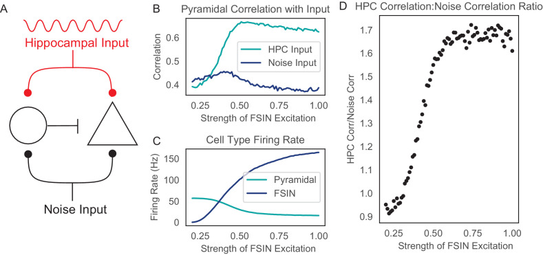
Figure 6.

Figure 6—figure supplement 1. Adding feedforward disinhibition does not change the relationship between inhibitory strength and hippocampal correlation.

Figure 6—figure supplement 1.

(A) Schematic of the computational model including cells and input sources. In comparison to the original model (Figure 6), this model includes an additional interneuron (ellipse) which receives feedforward excitation representing noise or hippocampal input. This new interneuron inhibits the first interneuron (circle), providing disinhibition. (B) The correlation between the pyramidal neuron output spike rate and the rate of either noise inputs (dark blue) or hippocampal spikes (turquoise), as functions of a single parameter which represents how strongly hippocampal and noise inputs excite the model FSIN. (C) The spike rate of the model pyramidal neuron (turquoise) and FSIN (dark blue) as functions of a single parameter representing how strongly hippocampal and noise inputs excite the model FSIN. (D) The ratio of the correlation between pyramidal neuron output spikes and either hippocampal input or noise input.
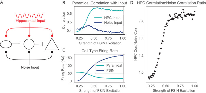
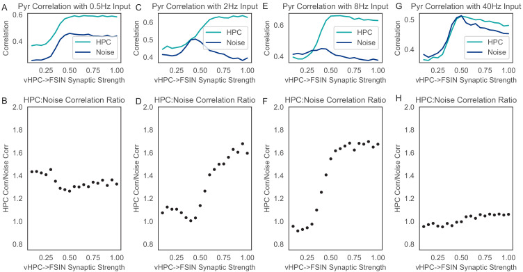
Figure 6—figure supplement 2. The effect of reducing inhibition on the transmission of signals across hippocampal-prefrontal synapses depends on the frequency of hippocampal input.

Figure 6—figure supplement 2.

We simulated the same model shown in Figure 6 using non-rhythmic noise together with hippocampal input that varied sinusoidally at various-frequencies: 0.5 Hz (A, B), 2 Hz (C, D), 8 Hz (E, F), or 40 Hz (G, H). Similar to Figure 6 and Figure 6—figure supplement 1, we plotted the correlation between the pyramidal neuron output spike rate and the rate of either noise inputs (dark blue) or hippocampal spikes (turquoise), as functions of how strongly hippocampal and noise inputs excite the model FSIN. Inhibition serves to enhance the signal-to-noise ratio when hippocampal input is modulated at 2 or 8 Hz, but not for higher (40 Hz) or lower (0.5 Hz) frequencies.