

Anhedonia is a primary feature of depression and other psychiatric illnesses and refers to a loss of pleasure in activities that were previously enjoyable – social interaction, sex, hobbies. “Motivational anhedonia” refers to difficulties in volitionally acting for outcomes known to be reinforcing based on prior experiences. Motivational anhedonia can be debilitating, interfering with even day-to-day activities. The neurobiology of motivational anhedonia is not well-understood, in part because traditional behavioral assays for model organisms require very little “motivation” or goal seeking. Historically, mice or rats have been presented with a palatable food, such as sugar water. The organism need only approach and ingest, providing little insight into whether a rodent is translating prior experience into reward-seeking action. A second limitation is that mice or rats are typically tested following some period of stressor exposure or other insult, leaving opaque the processes by which anhedonic-like behavior forms.
Investigations using elaborate operant conditioning procedures in which rodents must work for reward indicate that stress and stress hormones cause amotivation and failures in goal- directed action (1,2) – resembling motivational anhedonia in depression – but the emergence of these behaviors remains under-studied. In this issue of Biological Psychiatry, Barthas et al. (3) present a procedure for rapidly measuring aspects of anhedonic-like behavior in mice. The authors dissociate motivational from consummatory anhedonic-like behaviors (figure 1), longitudinally characterize their development upon repeated threat, and interestingly, reveal that they emerge and recover on different time scales, allowing for the identification of potential causal factors.
Figure 1. Separating seeking and taking.
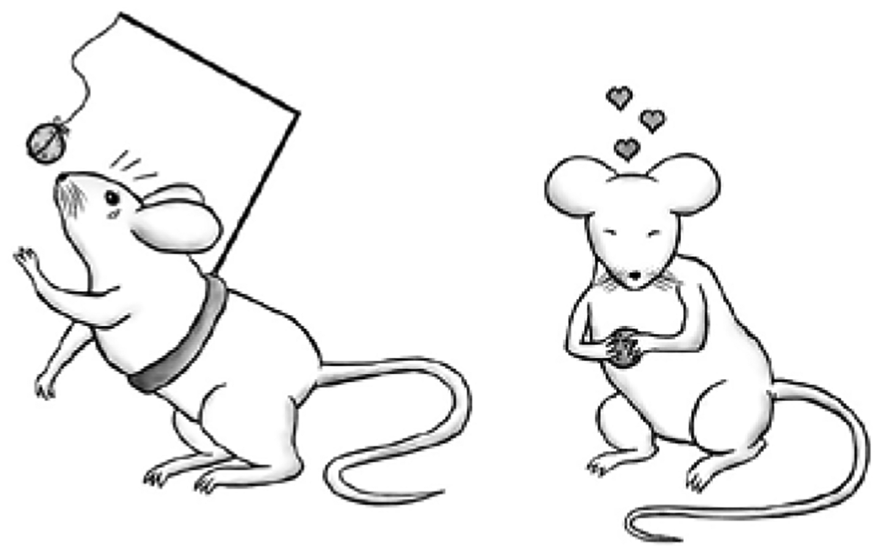
Barthas and colleagues (3) develop a procedure that rapidly captures – and dissociates – the mouse’s propensity to work for palatable foods and consume those foods. The task provides a platform for quantifying stress burden on motivated behavior over time. Illustration credit: Aylet Allen.
Fluid-restricted mice were trained to lick a spout for sucrose. Effectively, 10 licks triggered sucrose delivery, followed by a 5-second timeout while mice ingested the sucrose. Then, the response requirement was reinstated, and the mouse had to lick 10 more times to access the sucrose, and so on. Reinforcer delivery cycled between 0, 3, and 10% sucrose concentrations in predictable testing blocks, allowing authors to measure response sensitivity to absolute outcome value – the 10% solution being most highly “valued” – as well as relative positive and negative contrast. A stress-related decrement in the number of reinforcers earned or sensitivity to the value of future reinforcement (inferred by energized responding ahead of higher sucrose concentrations) would be interpreted as evidence of motivational anhedonia. Meanwhile, a stress-related decrement in consummatory licks is interpreted as “appetitive anhedonia,” as it is directly reflects the ingestion of a palatable food.
To trigger a recurring stress response, mice were subjected to repeated attack and threat in a “social defeat” procedure. The experimental mouse is placed in the home cage of an aggressive male, experiencing periods of both direct physical aggression and indirect sensory interaction. Conspecific-directed attack occurs in most animal species and is thus an ethologically-relevant threat (4). In recent years, defeated mice have routinely been classified as “susceptible” vs. “resilient,” generally referring to those that avoid novel conspecifics vs. those that resemble non-stressed control animals following defeat, revealing specific brain circuits and molecular factors involved in stressor vulnerability and resilience [e.g., ref. (5)].
Here, Barthas et al. (3) repeatedly tested mice as they underwent the social defeat procedure. Control mice obtained more reinforcers as the sucrose concentration increased, as expected, while mice ultimately categorized as susceptible developed insensitivity to sucrose concentration – responding equivalently for “low” and “high” concentrations. This insensitivity persisted even after the stressor exposure period. Mice ultimately categorized as resilient exhibited similar impairments, but this apparent reward insensitivity was transient, recovering once the defeat procedure ended. Interestingly, consummatory licks also dropped with repeated defeat in the susceptible mice, but the deviation from baseline was transient, recovering upon cessation of the defeat procedure. Thus, the authors dissociated persistent motivational anhedonic-like behavior from briefer appetitive anhedonic-like behavior in susceptible mice and revealed that resilient mice were not identical to control mice, but rather, appeared to “bounce back” from repeated threat.
The authors next developed a computational model of “response vigor”, taking into account the “cost” of licking (the energetic requirement of physical actions performed in quick succession), the “opportunity cost” (incurred when rewards are delayed due to inaction), and an estimation of satiety. Their efforts revealed a loss of motivational vigor in the “opportunity cost” parameter, indicating that susceptible mice were less able to discriminate between the opportunity cost of “losing out” on a high vs. low sucrose concentration, and this loss emerged relatively late in the testing procedure, presumably representing cumulative effects of repeated duress.
To begin to understand cellular predictors of behavior, Barthas et al. (3) turned to Cg1 and M2, subregions of the anterior cingulate and motor cortices respectively, involved in reward-related behavior. Equipped with a bicistronic expression vector permitting the visualization of somatic calcium transients in pyramidal neurons, the authors found that a single acute stressor elevated spontaneous activity in layer II/III neurons in mice ultimately categorized as susceptible, but not those categorized as resilient. Meanwhile, resilient mice exhibited a more dispersed distribution of Cg1/M2 activity change following a single social defeat – potentially a signature of resilience-related activity representing acute adaptation to stress.
A more nuanced picture arose in response to repeated defeat, with longitudinal Cg1/M2 activity tracking revealing marked cellular heterogeneity. Using hierarchical clustering to categorize this heterogeneity, four predominant profiles of cellular activity emerged, which the authors term cell types 1–4. Of these profiles, type 2 cells exhibited a monotonic decline in spontaneous activity and represented a greater percentage of cells in response to chronic social defeat in resilient mice. The other 3 cell clusters appeared in roughly equivalent percentages between groups, suggesting that type 2 cell activity confers stressor resilience. Thus, Barthas et al. (3) differentiate between cellular responses to repeated stress in susceptible vs. resilient mice, and they reveal a cellular activity profile that may underlie behavioral adaptation over time.
Interestingly, some stress-related changes in neural activity were detectable prior to the emergence of behavioral modifications. If they are causally related to behavior, some process presumably bridges Cg1/M2 neuronal activity and stress-related behavioral (mal)adaptations. Determining the distinguishing features of type 2 cells in the future will be informative. Are they positioned within specific neural circuits? Do they communicate with the hypothalamic-pituitary axis, or so-called reward circuitry – regions like the ventral striatum or ventral tegmental area?
A longitudinal analysis of the molecular/genomic profile of type 2 neurons may also shed light onto divergent factors in stressor vulnerability and resilience. Presumably, the activity of certain intracellular signaling pathways in type 2 neurons changes and evolves with repeated stressor exposure. Understanding these modifications could reveal molecular “building blocks” of susceptibility and resilience. Famously, chronic stress disrupts neurotrophin systems in many regions of the brain, including the prefrontal cortex (6), and stimulating Brain-derived Neurotrophic Factor-Tropomyosin receptor kinase B (BDNF-TrkB) systems can correct amotivation and failures in goal-oriented action in mice exposed to excess stress hormones (2,7). Thus, neurotrophin systems could provide an entryway into investigating individual differences in stress vulnerabilities; these investigations could be complemented by discovery-based strategies to reveal previously unappreciated factors.
Of note, stressors modify the structure of excitatory neurons throughout the cortex (8), and neuron structure (dendritic spine densities) in some regions correlates with stress-related insensitivity to goals (9). A future challenge will be to determine whether stress-related structural modifications are causally associated with stressor vulnerability or resilience.
One final point is that the stressor-exposed mice in the report of Barthas et al. (3) did not consume less sucrose than control mice – rather, response patterns changed as stressor-exposed mice failed to differentiate between “low” and “high” sucrose concentrations. Thus, what is referred to as anhedonic-like behavior can be described in simple terms as insensitivity to the value of future reward. This definition differs significantly from what we might typically conceptualize as anhedonic-like behavior in rodents – in which a mouse or rat neglects a sucrose solution or generates less effort for reinforcers relative to non-stressed control animals [e.g., ref. (2)]. Hopefully, pairing new, diverse behavioral assays with traditional procedures will ultimately lead to meaningful insights into different aspects of multi-faceted disease symptoms like anhedonia.
Repeated stress has long-term physical and emotional consequences at both individual and societal levels. The clinical understanding that stressful life events can precipitate neuropsychiatric disorders such as depression (10) has invigorated decades of translational research aimed at better understanding the stressed brain. Although an acute stressor activates neural efforts presumed to maintain homeostasis, continued exposure to stress over time may instead bias the brain towards maladaptation. This series of adaptations is perhaps best characterized by the concept of allostatic load, or a cascading series of adaptations resulting in heightened reactivity and an accumulation of putatively deleterious changes in the brain. How and when successive stress episodes result in such maladaptive responses is not well understood. Barthas et al. (3) make important in-roads in resolving this issue by revealing the progressive emergence of behavioral impairments (or resiliencies) and heterogeneous adaptations in cortical activity through successive episodes of social stress.
Acknowledgements:
The authors are supported in part by NIH MH117103, MH100023, and MH117878. The Yerkes National Primate Research Center is supported by NIH OD011132.
Footnotes
Disclosures: The authors have no disclosures.
References
- 1.Dias-Ferreira E, Sousa JC, Melo I, Morgado P, Mesquita AR, Cerqueira JJ, Costa RM, Sousa N. Chronic stress causes frontostriatal reorganization and affects decision-making. Science. 2009;325(5940):621–5. [DOI] [PubMed] [Google Scholar]
- 2.Gourley SL, Swanson AM, Jacobs AM, Howell JL, Mo M, Dileone RJ, Koleske AJ, Taylor JR. Action control is mediated by prefrontal BDNF and glucocorticoid receptor binding. Proc Natl Acad Sci U S A. 2012;109(50):20714–9. [DOI] [PMC free article] [PubMed] [Google Scholar]
- 3.Barthas F, Hu MY, Siniscalchi MJ, Ali F, Mineur YS, Picciotto MR, Kwan AC. Cumulative Effects of Social Stress on Reward-Guided Actions and Prefrontal Cortical Activity. Biol Psychiatry. 2020. [DOI] [PMC free article] [PubMed] [Google Scholar]
- 4.Covington HE 3rd, Newman EL, Leonard MZ, Miczek KA. Translational models of adaptive and excessive fighting: an emerging role for neural circuits in pathological aggression. F1000Res. 2019;8. [DOI] [PMC free article] [PubMed] [Google Scholar]
- 5.Krishnan V, Han MH, Graham DL, Berton O, Renthal W, Russo SJ, Laplant Q, Graham A, Lutter M, Lagace DC, Ghose S, Reister R, Tannous P, Green TA, Neve RL, Chakravarty S, Kumar A, Eisch AJ, Self DW, Lee FS, Tamminga CA, Cooper DC, Gershenfeld HK, Nestler EJ. Molecular adaptations underlying susceptibility and resistance to social defeat in brain reward regions. Cell. 2007;131(2):391–404. [DOI] [PubMed] [Google Scholar]
- 6.Gerhard DM, Duman RS. Rapid-Acting Antidepressants: Mechanistic Insights and Future Directions. Curr Behav Neurosci Rep. 2018;5(1):36–47. [PMC free article] [PubMed] [Google Scholar]
- 7.Barfield ET, Gerber KJ, Zimmermann KS, Ressler KJ, Parsons RG, Gourley SL. Regulation of actions and habits by ventral hippocampal trkB and adolescent corticosteroid exposure. PLoS Biol. 2017;15(11):e2003000. [DOI] [PMC free article] [PubMed] [Google Scholar]
- 8.McEwen BS, Morrison JH. The brain on stress: vulnerability and plasticity of the prefrontal cortex over the life course. Neuron. 2013;79(1):16–29. [DOI] [PMC free article] [PubMed] [Google Scholar]
- 9.Barfield ET, Gourley SL. Glucocorticoid-sensitive ventral hippocampal-orbitofrontal cortical connections support goal-directed action - Curt Richter Award Paper 2019. Psychoneuroendocrinology. 2019;110:104436. [DOI] [PMC free article] [PubMed] [Google Scholar]
- 10.Kessler RC. The effects of stressful life events on depression. Annu Rev Psychol. 1997;48:191–214. [DOI] [PubMed] [Google Scholar]