Abstract
Background:
Creatinine values are unreliable within the first weeks of life; however, creatinine is used most commonly to assess kidney function. Controversy remains surrounding the time required for neonates to clear maternal creatinine.
Methods:
Eligible infants had multiple creatinine lab values and were admitted to the NICU. A mathematical model was fit to the lab data to estimate the filtration onset delay, creatinine filtration half-life, and steady-state creatinine concentration for each subject. Infants were grouped by gestational age (GA) [(1)22-27, (2)>27-32, (3)>32-37, and (4)>37-42 weeks].
Results:
4,808 neonates with mean GA 34.4 ± 5 weeks and birth weight 2.34 ± 1.1 kg were enrolled. Median (95% CI) filtration onset delay for Group 1 was 4.3 (3.71,4.89) days and was significantly different than all other groups (p<0.001). Creatinine filtration half-life of Groups 1, 2, and 3 were significantly different from each other (p<0.001). There was no difference in steady-state creatinine concentration amongst the groups.
Conclusion:
We quantified the observed kidney behavior in a large NICU population as a function of day of life and GA using creatinine lab results. These results can be used to interpret individual creatinine labs for infants to detect those most at-risk for acute kidney injury.
Introduction
Creatinine is a waste product formed as the body supplies muscles with energy and is eventually cleared by the kidneys. In adults, creatinine clearance is used to approximate the glomerular filtration rate (GFR) to assess kidney function. It is defined as the volume of blood that is cleared of creatinine per unit time. Accurate measurements of creatinine clearance require stable creatinine levels and timed urine and serum sample collections. This process proves difficult in the neonatal intensive care unit (NICU) where it is widely accepted that neonates do not have stable creatinine levels. Additionally, term, developmentally appropriate neonates lack bowel and bladder function which contributes to difficulties with timed sample collections. In preterm infants, it is further complicated by incomplete nephrogenesis(1) and abnormal glomeruli.(2)
Neonates are born with a relatively high creatinine level for body mass, in part due to creatinine equilibration in utero. Some studies have shown initial creatinine levels to be equal to or higher than maternal creatinine levels without definitive correlation between the two values.(3-6) Multiple studies have shown increased initial creatinine lab values and variable ranges in the first days to weeks of life.(7-12) Studies have also demonstrated a critical value for creatinine from 1.0 to 1.6 mg/dL depending on gestational age (GA), above which patients have a higher risk for mortality and neurodevelopmental delay.(13) On the other hand, Miall et al. concluded that two- to three-fold increases in creatinine level within the first 48 hours of life in premature infants up to 35 weeks’ gestation can be expected and should not be used to diagnose renal failure.(14) This leaves a critical gap in our assessment of optimal renal function and GFR in the neonatal population.
During the neonatal period, the inability to monitor creatinine clearance as a surrogate for GFR contributes to a lack of a consensus for the definition of acute kidney injury (AKI).(15) This can be concerning for infants who may experience AKI in that time period but go undiagnosed.(16) Weintraub et al. showed a 30% incidence of AKI in the NICU population with a median age at onset of 3 days.(17) Kandasamy et al. showed a significant correlation between weight and serum creatinine based GFR measurement potentially delaying diagnosis of AKI in smaller infants.(18) In another study, Harer et al. found evidence of renal dysfunction in former very low birth weight infants, some of whom had never been diagnosed with AKI.(19) This evidence further supports the theory that renal function in the NICU population warrants closer examination. At this time, in the early clinical course of neonates, we may be missing and not identifying AKI due to a lack of a consistent and easily accessible measurement of GFR. Some have suggested that the use of cystatin C may be superior to creatinine in estimating GFR in neonates,(18,20) however, it is not as readily available and the testing is more expensive than for creatinine. In addition, cystatin C may not be as accurate in the most premature infants.(21)
The objective of this study was to quantify the behavior of kidney function in a large NICU population using readily available creatinine measurements over the length of the hospitalization. The interpretation of the absolute value of the creatinine level of a neonate in early life is confounded due to the presence of maternal creatinine. While the maternal creatinine is removed over time by the kidney, the rate at which this happens can be different for each patient depending on GA as well as day of life. Our study sought to quantify this trend and apply a mathematical model to describe the observed behavior in order to better understand how far from average a given creatinine value is based on day of life and GA at birth.
Methods
We performed a retrospective cohort study to delineate renal dynamics using serum creatinine measurements in patients admitted to the NICU at Texas Children's Hospital. The study was approved by the Institutional Review Board of Baylor College of Medicine and Affiliated Hospitals. A waiver of consent was obtained. Patient demographics, medication use, and lab values were extracted from the electronic medical records (EPIC, Hyperspace EPIC 2014, Madison, WI). Infants were initially included if they had at least one serum creatinine measurement obtained between January 1, 2013 and December 31, 2018. Creatinine measurements were obtained via enzymatic spectrophotometry. Patients were separated into 4 groups for statistical analysis based on GA at birth in weeks [Group 1 (22 < GA ≤ 27), Group 2 (27 < GA ≤ 32), Group 3 (32 < GA ≤ 37), Group 4 (37 < GA ≤ 42)].
Exclusion criteria for advanced analysis
In order to characterize and quantify the creatinine trend and kidney function after birth, additional exclusion criteria were needed for advanced time-dynamics analysis. Patients were excluded if they had less than 5 available creatinine labs in the first year of life or did not have a lab within the first 3 days or after 10 days of life. These criteria ensured that a creatinine trend over time could be robustly quantified. With the criteria, the cohort reduced to 1,782 patients (37.1% of original cohort) with a total of 55,193 available labs (70.2% of the original set).
Evaluation for Selection Bias
The possible bias introduced by requiring additional criteria for the advanced analysis is tested by comparing the corresponding moving-in-time distributions of creatinine lab values. For all time, from day 1 to day 200, the Kolmogorov-Smirnov metric is consistently below 0.04. Hence, the error in estimating the percentile ranks from the entire cohort, when using the reduced cohort, is below 4%. In all of our results, each Xth percentile curve should be understood as having a margin of error in its rank and appropriately interpreted as the (X±4)th percentile curve. We conclude that the subset of labs chosen for the advanced analysis has a moving distribution which does not differ substantially from the entire set.
Filtration Dynamics of Creatinine
A mathematical model was derived from basic filtration kinetics to describe the expected time course of creatinine filtration in NICU patients. The concentration of creatinine in the blood stream is governed by the law of conservation of mass. This law allows us to describe the change in concentration of creatinine at any time as the difference between the amount of creatinine added to the blood stream (i.e. creatinine that is generated by the body) and the amount of creatinine removed from the blood stream (i.e. creatinine that is filtered out by the kidney). This can be written as a simple differential equation of the form:
where C(t) is the concentration of creatinine over time, G(t) is the generation rate of creatinine, and F(t) is the filtration rate of creatinine. Since there is expected to be only a small amount (if any) of creatinine generated by a neonate, and that this small generation rate will likely be constant in time, we assume that the creatinine generation rate is a constant, but unknown parameter with small magnitude:
We also assume that creatinine filtration from the kidney follows first-order filtration kinetics once the kidneys are fully functioning. This is a reasonable starting assumption given that it is the simplest type of kinetic model which accurately describes filtration. This can be mathematically expressed as:
where K(t) is the first order filtration coefficient of creatinine. From clinical experience, we know that the filtration rate is not constant, but can change over time in early life.
To model the inability of the kidneys to fully function during the first few days after birth, we assume the form of K(t) to be a step function of the form:
This definition means that the kidneys do not provide any filtration of creatinine until the time Td is reached, after which time the kidneys achieve a constant filtration rate quantified by the constant k > 0. The advantage of this definition is that it allows the model to be generalized, describing both the creatinine profiles of patients with kidneys that are always filtering from birth (i.e. Td = 0) as well as creatinine profiles of patients whose kidneys have delayed onset of filtering (i.e. Td > 0).
Combining these governing equations and integrating over time (assuming an initial concentration of creatinine to be C0), leads to a solution given by:
Overall, this model allows a creatinine concentration profile to be described with only 3 parameters: the kidney filtration onset delay (Td), the creatinine half-life (ln (2)/k), and the steady state creatinine concentration (G/k).
This mathematical model can be fit to the measured values of creatinine on a patient-by-patient basis using simplex optimization (Matlab, Mathworks Inc., Natick, MA), resulting in an estimate of the three model parameters for each patient in the data set. An R2 value was calculated to measure goodness of fit between the model fit and the measured values of creatinine. In addition, subjects were excluded if model fit was R2 <0.5 (10% loss).
Results
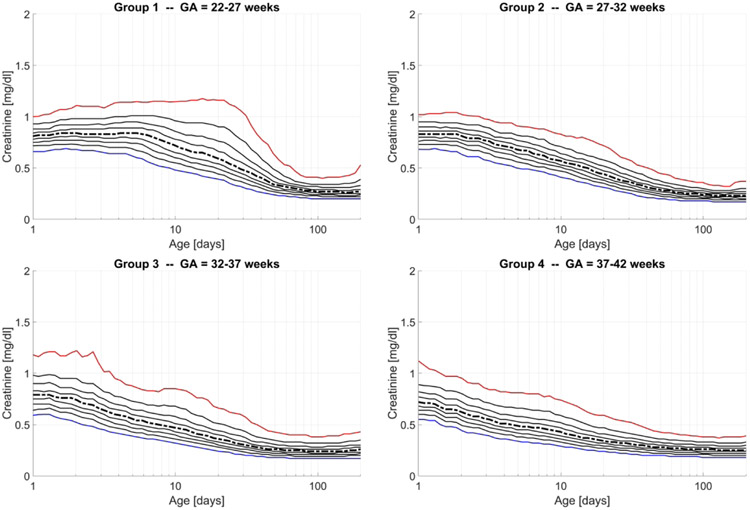
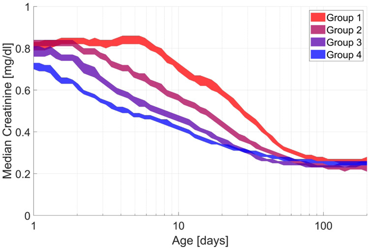
A total of 4,808 patients (55.7% male) met inclusion criteria providing 78,634 creatinine measurements for this study. Patient demographics are listed in Table 1. Using all the available labs, percentile curves as functions of age were developed for each of the GA groups as shown in Figure 1. The curves represent the moving 10th percentiles from 10 to 90. The 10th (blue line), 50th (black dashed line), and 90th (red line) percentiles are highlighted for easier reference. The 95% confidence region for the moving medians of the GA groups is displayed in Figure 2. These values are computed as functions of age in order to visualize the difference between these four GA groups in creatinine trend after birth.
Table 1.
Patient Characteristics
| Characteristic | Entire Cohort (N=4,808) |
Reduced Cohort (N=1,782) |
|---|---|---|
| Birth weight in grams, mean ± SD | 2340 ± 1070 | 2180 ± 1150 |
| Gestational age in weeks, mean ± SD | 34.41 ± 5 | 33.54 ± 5.7 |
| Male sex, n (%) | 2678 (55.7) | 1020 (57.2) |
| Race, n (%) | ||
| White | 3391 (70.5) | 1274 (71.5) |
| Black | 1019 (21.2) | 377 (21.2) |
| Other | 398 (8.3) | 131 (7.3) |
| Hispanic, n (%) | 1610 (33.5) | 599 (33.6) |
| Antenatal steroids, n (%) | 1915 (39.8) | 776 (43.6) |
| 5-minute APGAR, median [IQR] | 8 [7, 9] | 8 [7, 9] |
| Indomethacin, n (%) | 327 (6.8) | 252 (14.1) |
| Gentamicin, n (%) | 2219 (46.2) | 1094 (61.4) |
| Length of hospital stay in days, median [IQR] | 32 [12,67] | 60 [28,113] |
Figure 1.
10th to 90th Percentile curves by group number with highlighted 10th percentile (blue), 50th percentile (dashed black line), and 90th percentile (red line).
Figure 2.
95% Confidence region for moving median for each group
Results from advanced analysis
Quality of Model Fit
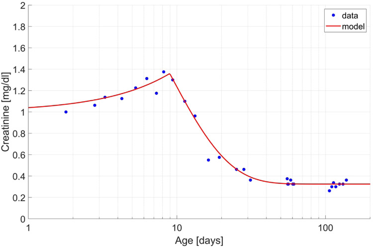
After implementing exclusion criteria, a total of 1,782 patients (57.2% male) were included in the advanced analysis. An example of the fitting result of the mathematical model is shown in Figure 3. The differential equation model fit the creatinine concentration data extremely well. The R2 value for goodness of fit was found to be >0.80 for more than 80% of the creatinine concentration profiles measured on study subjects. This indicates that 80% of the observed changes in creatinine in neonates in their early life can be explained by our model for over 80% of study subjects.
Figure 3.
Mathematical model fit to creatinine lab for one patient from Group 1
ANOVA
After the mathematical model was fit to the creatinine measurements, ANOVA was performed to test the effect of GA group, 5-minute APGAR score, receipt of antenatal steroids, and gentamicin or indomethacin administration on the filtration onset delay, filtration half-life, and steady-state creatinine concentration. Each of the parameters were log-transformed for this analysis.
Kidney Filtration Onset Delay
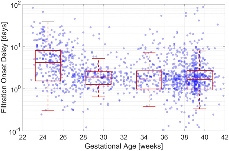
Figure 4 displays the filtration onset delay versus GA, and the boxplots (median, 25th and 75th percentiles, and robust extrema) for the kidney filtration onset delay from each of the four GA groups. Table 2 displays the median filtration onset delay and 95% confidence intervals. The median filtration onset delay for Group 1 is statistically greater than the medians for Groups 2, 3, and 4 (p< 0.001). There is an overall decreasing trend in filtration onset delay with increasing GA (Kruskal-Wallis Test, p<0.001). The ANOVA result for the filtration onset delay is shown in Table 3. The most significant factor is GA (p<0.0001), however, indomethacin administration also showed a significant effect (p<0.01).
Figure 4.
Filtration onset delay in days versus gestational age in weeks by Group
Table 2.
Median kidney filtration onset delay, filtration half-life, and steady state creatinine level with CI by gestational age group
| GA groups in weeks |
Median (95% CI) filtration onset delay in days |
Median (95% CI) filtration half-life in days |
Median (95% CI) steady state creatinine in mg/dl |
|---|---|---|---|
| 1 (22 < GA ≤ 27) | 4.26 (3.71, 4.89)* | 8.69 (7.99, 9.46)** | 0.28 (0.27, 0.29) |
| 2 (27 < GA ≤ 32) | 1.89 (1.76, 2.02) | 6.26 (5.69, 6.89)** | 0.29 (0.27, 0.31) |
| 3 (32 < GA ≤ 37) | 1.71 (1.57, 1.86) | 4.38 (3.91, 4.91)** | 0.26 (0.25, 0.27) |
| 4 (37 < GA ≤ 42) | 1.64 (1.53, 1.75) | 4.05 (3.74, 4.40)† | 0.27 (0.26, 0.28) |
Group 1 statistically different than Groups 2,3, and 4 (p<0.001)
Groups 1, 2, and 3 are significantly different from each other (p<0.001)
Group 4 is not significantly different from Group 3, but is from Groups 1 and 2 (p<0.001)
Table 3.
ANOVA
| Source | F | Prob>F |
|---|---|---|
| Filtration onset delay | ||
| Indomethacin | 4.7221 | 0.0091 |
| Antenatal steroid | 2.1813 | 0.1399 |
| APGAR 5min | 0.3627 | 0.5471 |
| Gentamicin | 0.0302 | 0.8620 |
| Gestational age | 15.1544 | <0.0001 |
| Filtration half-life | ||
| Indomethacin | 1.7518 | 0.1642 |
| Antenatal steroid | 2.0016 | 0.1144 |
| APGAR 5min | 0.2412 | 0.7446 |
| Gentamicin | 1.0178 | 0.3132 |
| Gestational age | 14.7475 | <0.0001 |
| Steady-state creatinine concentration | ||
| Indomethacin | 1.8278 | 0.1220 |
| Antenatal steroid | 0.0355 | 0.8505 |
| APGAR 5min | 1.8887 | 0.1696 |
| Gentamicin | 1.9829 | 0.1593 |
| Gestational age | 5.9230 | <0.001 |
Filtration Half-Life
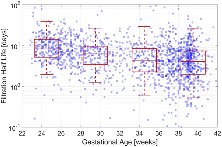
Figure 5 displays the half-life for creatinine filtration versus GA superimposed with the boxplots as previously described for each of the four GA groups. Table 2 displays the median filtration half-life and its 95% confidence interval for each group. The median creatinine clearance half-life for Groups 1, 2, and 3 are statistically different from each other (p<0.001) while there is no difference between Groups 3 and 4 (p=0.17). There is an overall decreasing trend in filtration half-life with increasing GA (Kruskal-Wallis Test, p<0.001). The ANOVA result for the filtration half-life is shown in Table 3. It shows that the only significant factor is GA.
Figure 5.
Half-life for creatinine filtration in days versus gestational age in weeks by Group
Steady-State Creatinine Concentration
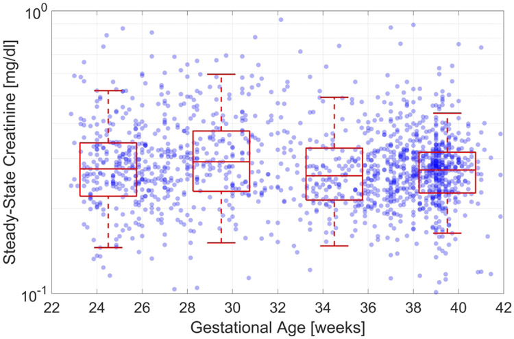
Figure 6 displays the steady-state creatinine concentration versus GA superimposed with the boxplots as previously described for each of the four GA groups. Table 2 displays the median steady-state creatinine concentration and its 95% confidence interval for each group. The median steady-state creatinine for the four GA groups are not statistically different from each other. The ANOVA result for the steady-state creatinine concentration is shown in Table 3. It shows that only GA has a statistically significant effect on the steady-state creatinine concentration.
Figure 6.
Steady-state creatinine concentration versus gestational age in weeks by Group
Discussion
Historically, the evaluation of GFR and creatinine clearance in neonates has been difficult. Assessments of kidney function in the neonatal population have been inconsistent or difficult to apply clinically at the bedside. Few studies include patients at the lower limits of viability and those that do are limited by low numbers of subjects. The current study examines kidney function in a novel way by using multiple creatinine measurements and mathematical modeling in neonates from 22 to 42 weeks’ gestation to describe the natural course of kidney function postnatally. We used the data to construct percentile curves based on GA that are representative of the expected renal function in neonates. Then we examined three key aspects of kidney function in this analysis: the filtration onset delay, the creatinine filtration rate, and the steady state creatinine concentration.
To our knowledge, this is the largest cohort of neonates studied to evaluate creatinine clearance across advancing GA that provides new perspective on the evolution of postnatal kidney development. In addition, by using multiple measurements of creatinine across the entire hospitalization for each subject, we could investigate the time it took for the kidney to begin clearance of creatinine, or the filtration onset delay. We then determined the creatinine filtration rate to evaluate the time it took for the subjects to reach steady state creatinine concentration. Using all these metrics combined, it was possible to get a more holistic understanding of the natural course of kidney development across the whole cohort.
Bateman et al. describes a phenomenon of serum creatinine going through phases in 218 appropriate for GA very low birth weight infants born between 25 and 33 weeks’ gestation with no history of congenital malformations or diagnosis of AKI.(7) They showed that creatinine will initially rise for 3-4 days, peak, and then decline over 7 weeks. Though informative, this study included a small number of subjects followed for only 60 days with none at the lower limit of viability (<25 weeks). They reported the mean and 95th percentile for the 3 GA groups evaluated.
In our study, we expanded on the evolution of kidney development. First, we were able to mathematically model the time at which the kidney began to function after birth and clear creatinine, known as the filtration onset delay. The filtration onset delay was inversely proportional to GA as the smallest infants had the longest filtration onset delays over the first 5 days of life. In addition, when examining factors associated with this delay, exposure to indomethacin and GA were found to be significant. Indomethacin is known to result in decreased GFR, and a relative contraindication to its use is renal dysfunction and it is therefore not surprising that early in life this impacted the filtration onset delay. Second, we were able to show that following this onset delay, the kidneys then started to clear the creatinine. This was demonstrated by showing the decay of creatinine using the filtration half-life as a metric for evaluating this function. It is notable that again, creatinine filtration half-life was also inversely proportional to GA. Finally, it appears that eventually, the kidneys reach a steady-state creatinine concentration regardless of GA. In this cohort, we showed median steady-state creatinine concentration was <0.3 mg/dl for all GA groups.
Contrary to the findings of Bateman et al., we show that in a majority of neonates, the creatinine does not rise a significant amount. Only in the lowest GA group (Group 1) was there a noticeable increase in creatinine level after birth. Our overall findings are also consistent with those of Bruel et al. who showed that creatinine values >1.6, 1.1, and 1.0 mg/dl for 24-27, 28-29, and 30-32 weeks GA groups, respectively, are critically elevated values and are associated with mortality or poor outcomes.(13) We showed that especially in the lowest GAs, the creatinine level at which there should be cause for concern is much less than 1.6 mg/dl. In fact, the 50th percentile curve for those infants at 22-27 weeks’ gestation is well below 1 mg/dl. These percentile curves may assist bedside clinicians in the NICU with interpretation of creatinine measurements for their patients and may lead to earlier recognition of poor renal function by allowing them to more easily recognize creatinine values that are >90th percentile for GA and postnatal age and/or crossing percentiles.
After determining the kidney filtration onset delay, the creatinine filtration rate, and the steady state creatinine concentration, we examined differences across GA. Specifically, we demonstrate the functions related to onset of creatinine filtration and overall clearance vary substantially between those infants at the lowest and highest ends of the GA spectrum. In fact, lowest GA infants (22 to 27 weeks) have both the longest filtration onset delay and filtration half-life compared to all other infants > 27 weeks’ gestation. The longer length of time for filtration onset and clearance is likely due to and representative of the on-going nephrogenesis in the smallest infants.
Importantly, the relative difference between creatinine measurements of the smallest and largest infants (GA groups 1 and 4) grows steadily over for the first 20 days of life, reaching more than 60% difference at its peak. At that point, the difference begins to reduce, and it returns to baseline at approximately 80 days of age (data not shown). This demonstrates the natural evolution of creatinine in the extremes of this cohort, but also shows that over time even the smallest infants eventually achieve similar kidney behavior compared to their term equivalent GA peers.
Limitations
There are several limitations to this study. It is a single center cohort study and a retrospective analysis of prospectively collected data. Despite these facts, we were still able to analyze a large number of patients in the population of interest. In addition, for advanced analysis, the smaller number of patients analyzed is still one of the largest cohorts evaluating creatinine clearance in neonates found in the literature. Since we were only able to include patients who had at least one creatinine measurement in this cohort, we are not able to analyze data from those patients with no creatinine measurements. In addition, standard practice at our institution is to check creatinine routinely, infants without measurements often represent short-stay or transitional nursery admissions and not the population of interest for this study. Finally, creatinine measurements were obtained at the discretion of the clinical team during this time period making uniformity of timing when these were made not possible. It would be ideal to have a regimented timing for this evaluation but was not possible in this retrospective study.
Conclusion
This study represents one of the largest cohorts of premature infants to monitor the evolution of kidney development and function over their entire hospitalization. We introduce the idea of the time it takes for the kidney to begin clearance of creatinine, or filtration onset delay, as an early indicator of kidney function. We also found that GA played a significant role in the filtration onset and creatinine clearance as the smallest cohort of infants 22 to 27 weeks’ gestation took the longest time to begin and complete maternal creatinine clearance. However, despite these developmental changes, all infants in this cohort eventually achieved a steady-state creatinine level of <0.3 mg/dL around 80 days of age regardless of GA.
Impact:
One of the largest cohorts of premature infants to describe the evolution of kidney development and function over their entire hospitalization.
New concept introduced of the kidney filtration onset delay, the time needed for the kidney to begin clearance of creatinine, and that it can be used as an early indicator of kidney function.
The smallest premature infants from 22 to 27 weeks’ gestation took the longest time to begin and complete maternal creatinine clearance.
Clinicians can easily compare the creatinine level of their patient to the normative curves to improve understanding of kidney function at the bedside
Acknowledgments
Statement of Financial Support: D.R.R. is supported by the National Institutes of Health (1K23HLI130522). C.J.R. is supported by the National Institutes of Health (1K23NS091382). C.G.R. is supported by National Institutes of Health (R01HL142994).
Footnotes
Disclosure Statement: Dr. Rusin has an SFI in Medical Informatics Corp. MIC has no financial interests in this work.
Bibliography:
- 1.Hinchliffe SA, Sargent PH, Howard CV, Chan YF, van Velzen D Human intrauterine renal growth expressed in absolute number of glomeruli assessed by the disector method and Cavalieri principle. Lab. Invest 64, 777–84 (1991). [PubMed] [Google Scholar]
- 2.Black MJ, et al. When birth comes early: Effects on nephrogenesis. Nephrology 18, 180–2 (2013). [DOI] [PubMed] [Google Scholar]
- 3.Go H, et al. Neonatal and maternal serum creatinine levels during the early postnatal period in preterm and term infants. PloS one 13, e0196721–e. (2018). [DOI] [PMC free article] [PubMed] [Google Scholar]
- 4.Lao TT, Loong EPL, Chin RKH, Lam YM Renal function in the newborn. Gynecol Obstet. Invest 28, 70–2 (1989). [DOI] [PubMed] [Google Scholar]
- 5.Guignard J-P., Drukker A Why do newborn infants have a high plasma creatinine? Pediatrics 103, e49–e (1999). [DOI] [PubMed] [Google Scholar]
- 6.Weintraub AS, Carey A, Connors J, Blanco V, Green RS Relationship of maternal creatinine to first neonatal creatinine in infants <30 weeks gestation. J. Perinatol 35, 401–4 (2015). [DOI] [PubMed] [Google Scholar]
- 7.Bateman DA, et al. Serum creatinine concentration in very low birth weight infants from birth to 34–36 weeks postmenstrual age. Pediatr. Res 77, 696–702 (2015). [DOI] [PMC free article] [PubMed] [Google Scholar]
- 8.Thayyil S, Sheik S, Kempley ST, Sinha A A gestation- and postnatal age-based reference chart for assessing renal function in extremely premature infants. J. Perinatol 28, 226–9 (2008). [DOI] [PubMed] [Google Scholar]
- 9.Gallini F, Maggio L, Romagnoli C, Marrocco G, Tortorolo G Progression of renal function in preterm neonates with gestational age ≤32 weeks. Pediatr. Nephrol 15, 119–24 (2000). [DOI] [PubMed] [Google Scholar]
- 10.Gubhaju L, et al. Assessment of renal functional maturation and injury in preterm neonates during the first month of life. Am. J. Physiol. Renal Physiol 307, F149–F58 (2014). [DOI] [PubMed] [Google Scholar]
- 11.Feldman H, Guignard JP Plasma creatinine in the first month of life. Arch. Dis. Child 57, 123–6 (1982). [DOI] [PMC free article] [PubMed] [Google Scholar]
- 12.Auron A, Mhanna MJ Serum creatinine in very low birth weight infants during their first days of life. J. Perinatol 26, 755 (2006). [DOI] [PubMed] [Google Scholar]
- 13.Bruel A, et al. Critical serum creatinine values in very preterm newborns. PloS one. 8, e84892–e (2013). [DOI] [PMC free article] [PubMed] [Google Scholar]
- 14.Miall LS, et al. Plasma creatinine rises dramatically in the first 48 hours of life in preterm infants. Pediatrics 104, e76–e (1999). [DOI] [PubMed] [Google Scholar]
- 15.Askenazi D, et al. Optimizing the AKI definition during first postnatal week using Assessment of Worldwide Acute Kidney Injury Epidemiology in Neonates (AWAKEN) cohort. Pediatr. Res 85(3), 329–38 (2019). [DOI] [PMC free article] [PubMed] [Google Scholar]
- 16.Walker MW, Clark RH, & Spitzer AR Elevation in plasma creatinine and renal failure in premature neonates without major anomalies: terminology, occurrence and factors associated with increased risk. J. Perinatol 31(3), 199–205 (2011). [DOI] [PubMed] [Google Scholar]
- 17.Weintraub AS, Connors J, Carey A, Blanco V, Green RS The spectrum of onset of acute kidney injury in premature infants less than 30 weeks gestation. J. Perinatol 36, 474–80 (2016). [DOI] [PubMed] [Google Scholar]
- 18.Kandasamy Y, Rudd D, Smith R The relationship between body weight, cystatin C and serum creatinine in neonates. J. Neonatal Perinatal Med 10, 419–23 (2017). [DOI] [PubMed] [Google Scholar]
- 19.Harer MW, Pope CF, Conaway MR, Charlton JR Follow-up of acute kidney injury in neonates during childhood years (FANCY): a prospective cohort study. Pediatr. Nephrol 32, 1067–76 (2017). [DOI] [PubMed] [Google Scholar]
- 20.Finney H, Newman DJ, Thakkar H, Fell JME, Price CP Reference ranges for plasma cystatin C and creatinine measurements in premature infants, neonates, and older children. Arch. Dis. Child 82, 71–5 (2000). [DOI] [PMC free article] [PubMed] [Google Scholar]
- 21.Garg P, Hidalgo G Glomerular filtration rate estimation by serum creatinine or serum cystatin C in preterm (<31 weeks) neonates. Indian Pediatr. 54, 508–9 (2017). [PubMed] [Google Scholar]


