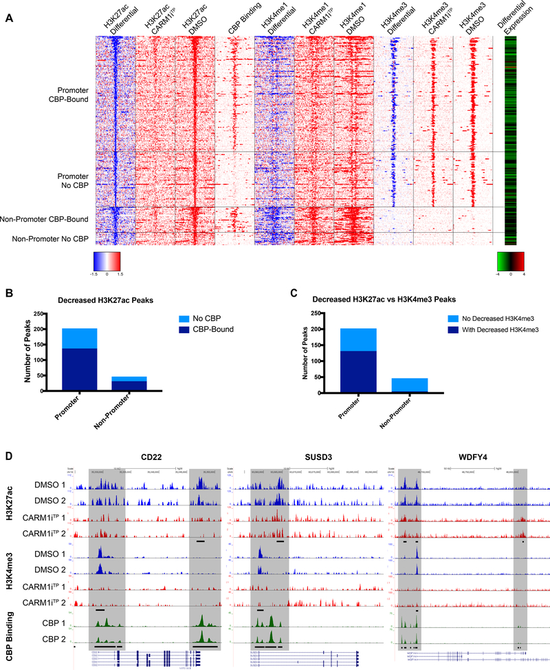
Figure 3. Deregulation of promoter and enhancer regions bound by CBP/p300 upon CARM1 inhibition.
A) Heatmap of Toledo ChIP-Seq signal around −10kb to 10kb of decreased H3K27ac peaks in CARM1iTP-treated cells. The decreased H3K27ac peaks were grouped by in-promoter and non-promoter, and also bound by CBP or not bound by CBP. CBP Binding column denotes whether CREBBP is bound at the locus in human GC B cells(41). The most right column in red-green color scale is the log2 expression ratio (log2 CARM1iTP-treated / DMSO) from RNA-Seq (Figure 2) for the genes that have the closest TSS to the H3K27ac peaks. For differential ChIP signal, blue color and red color indicate decreased signal and increased signal in CARM1iTP-treated cells, respectively. For log2 expression ratio, green color and red color indicate downregulated expression and upregulated expression in CARM1iTP-treated cells, respectively. B) Decreased H3K27ac peaks shown within promoter and outside promoter regions. Light blue bars represent no CBP binding in the region. Dark blue bars represent CBP binding within the region. P values of association with CBP binding by Fisher’s exact test were 3.3e-10 and 2.7e-10, respectively. C) Decreased H3K27ac peaks shown within promoter and outside promoter regions. Light blue bars represent no decrease in H3K4me3 in the region. Dark blue bars represent a concurrent decrease in both H3K4me3 and H3K27ac within the region. P values of association with CBP binding by Fisher’s exact test were 1.81e-4 and 9.22e-22, respectively. D) Representative ChIP-seq tracks of three CBP-target genes verified to have a significant decrease in expression in Figure 2E. Shaded areas indicate CBP-bound regions where peaks are significantly changed (FDR of 0.1 or lower) in CARM1iTP treated cells. Differential peaks are denoted by black bars in H3K27ac and H3K4me3 tracks (FDR of 0.1 or lower), CBP-bound peaks denoted by black bars in CBP track. Tracks represented include Input-subtracted H3K27ac peaks and H3K4me3 peaks measured in the Toledo B cell lymphoma line, and CBP binding measured in human germinal center B cells(41).

