Abstract
Background
Non-tuberculous mycobacterial pulmonary disease (NTM-PD) is becoming a significant health burden. Recent advances in analysis techniques have allowed the accurate identification of previously unknown NTM species. Here, we report a case of NTM-PD caused by a newly identified mycobacteria in an immunocompetent patient.
Case presentation
A 44-year-old woman was referred to our hospital due to the frequent aggravation of her chronic respiratory symptoms, with NTM-PD-compatible computed tomography findings. Unidentified mycobacterium was repeatedly isolated from respiratory specimens and we diagnosed her as NTM-PD of unidentified mycobacterium. Subsequent whole-genome analysis revealed that the unidentified mycobacterium was a novel mycobacterium genetically close to Mycolicibacterium mucogenicum. We started combination therapy with clarithromycin, moxifloxacin, amikacin, and imipenem/cilastatin, referring to drug sensitivity test results and observed its effect on M. mucogenicum infection. Her symptoms and radiological findings improved significantly.
Conclusion
We report a case of NTM-PD caused by a newly identified mycobacteria, Mycolicibacterium toneyamachuris, genetically close to M. mucogenicum. This pathogenic mycobacterium showed different characteristics from M. mucogenicum about clinical presentation and drug sensitivity. The clinical application of genomic sequencing will advance the identification and classification of pathogenic NTM species, and enhance our understanding of mycobacterial diseases.
Keywords: Non-tuberculous mycobacteria, Mycolicibacterium toneyamachuris, Mycolicibacterium mucogenicum, Rapid growing mycobacteria
Background
The prevalence of non-tuberculous mycobacterial pulmonary disease (NTM-PD) is increasing worldwide and is becoming a significant health burden [1]. Recent advances in analysis techniques have allowed the identification of previously unknown NTM species [2–5].
Here, we report a case of NTM-PD caused by a newly identified mycobacteria genetically close to Mycolicibacterium mucogenicum. This novel mycobacterium caused chronic and progressive pulmonary disease in an immunocompetent patient. Furthermore, drug susceptibility and clinical presentation were different from those reported for M. mucogenicum infections [6].
Case presentation
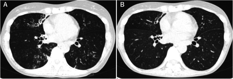
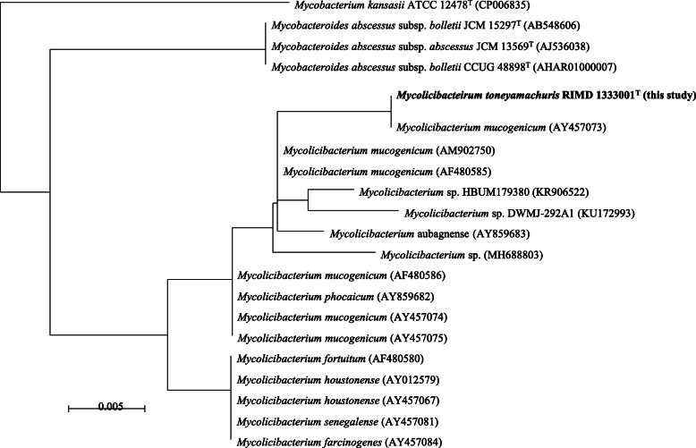
A 44-year-old woman, never smoker, was referred to our hospital 18 months ago due to chronic productive cough. She had asthma treated with inhalation therapy and allergic rhinitis. Chest computed tomography showed centrilobular nodules and bronchiectasis in the middle lobe and in the bilateral lower lobes (Fig. 1). Despite treatment with erythromycin and expectorants, her chronic respiratory symptoms worsened. Subsequently, a rapid growing mycobacterium (RGM), strain TY81, was cultured repeatedly from her sputum; however, its species/subspecies could not be identified by conventional methods such as AccuProbe (Gen-Probe Inc., San Diego, CA, USA), COBAS AMPLICOR (Roche Diagnostic, Tokyo, Japan), or DNA-DNA hybridization assay (Kyokuto Pharmaceutical Industrial, Tokyo, Japan). Therefore, we diagnosed her as NTM-PD of unidentifiable mycobacteria in accordance with the American Thoracic Society/Infectious Diseases Society of America (ATS/IDSA) criteria for the diagnosis of NTM-PD [7]. Multilocus sequence typing [8] revealed that the unidentified mycobacterium was genetically close to M. mucogenicum (Fig. 2).
Fig. 1.

Chest CT before treatment shows small centrilobular nodules in the middle and lower lobes and slight bronchiectasis with a consolidation in the middle lobe (a). After 2 months treatment, small centrilobular nodules almost vanished (b)
Fig. 2.
Approximately maximum likelihood phylogenetic tree using 16S rRNA sequences. Sequences were obtained from SILVA database release 138 as SSU Ref NR 99 sequences, which were showing larger than 98.7% of identity to the strain XXX T or derived from the type strains of Mycobacterium kansasii and Mycobacteroides abscessus
We performed a whole-genome analysis of TY81 and obtained the complete genome sequence (AP023362-AP023365). The DNA G + C content of the type strain was 67.18 mol%. The mean nucleotide identity (ANI) to M. mucogenicum was 93.3% and was the maximum value obtained among the type strains of 175 NTM species (Table 1). Phylogenetic analysis using the 16S rRNA sequence suggested that TY81 was closely related to M. mucogenicum and related strains. The TY81 strain satisfied three of four conserved signature indels of Mycolicibacterium [9]. The three conserved signatures were a 5 aa insertion of GDAQS at positions 197–201 in the LacI family transcriptional regulator gene, a 1 aa insertion of proline at position 60 in the CDP-diacylglycerol-glycerol-3-phosphate 3-phosphatidyl transferase gene, and 1 aa deletion at position 128 in the CDP-diacylglycerol-serine O-phosphatidyl transferase gene. The protein of Cyclase gene (Accession number; WP_066808156) was not detected by homology search using protein-protein Basic Local Alignment Search Tool (BLASTp) with the threshold of 90% of similarity.
Table 1.
ANI calculation
| Species name | Strain | Refseq accession number | Refseq category | ANI (%) |
|---|---|---|---|---|
| Mycolicibacterium mucogenicum | CSUR P2099 | GCF_001291445.1 | Representative genome | 93.3 |
| Mycolicibacterium phocaicum | JCM 15301T | GCF_010731115.1 | Representative genome | 92.7 |
| Mycolicibacterium aubagnense | JCM 15296T | GCF_010730955.1 | Representative genome | 88.4 |
| Mycolicibacterium houstonense | ATCC 49403T | GCF_900078665.2 | Representative genome | 80.5 |
| Mycolicibacterium senegalense | CK2 | GCF_001021425.1 | None | 80.4 |
| Mycobacterium fortuitum | CT6 | GCF_001307545.1 | Representative genome | 80.3 |
| Mycobacteroides abscessus | ATCC 19977T | GCF_000069185.1 | Reference genome | 78.2 |
Moreover, we performed additional examination, concerning 16S rRNA phylogeny may not distinguish between closely related species [10]. A comparison was made among TY81, the six strains of M. mucogenicum and the type strains of M. phocaicum and M. aubagnense by calculating the ANI values and constructing the phylogenetic tree based on core genomes consisting of 455 genes (Supplementary Fig. 1). The results from both analyses were consistent that all strains belonging to M. mucogenicum group are distinct from TY81. Note that the 4 of 6 strains of M. mucogenicum were closer to M. phocaicum as seen in Behra et al.10. Considering the genetic characteristics described above, we suggest that the TY81 strain is a novel species. The scientific name proposed for this species is Mycolicibacterium toneyamachuris sp. nov. with RIMD 1333001T as the type strain. The bacterium was named ‘Mycobacterium toneyamachuris’ after the place where it was discovered.
M. toneyamachuris is a gram-positive, acid-fast, aerobic, anaerobic, non-pigmented and non-motile bacillus. Colonies were grown on 2% Ogawa-medium, Tryptic Soy Agar (TSA), and 5% Sheep Blood Agar and appeared greyish without pigmentation (Supplementary Fig. 2). Growth was observed within 7 days at 25, 30, and 37 °C temperature with optimal growth at 37 °C.
Currently, the treatment of NTM-PD caused by rare species/subspecies is a process of trial and error. It often starts with the drug regimen clinically used for closely related species/subspecies, which is modified by in vitro drug sensitivity test results (Table 2). Because the closest species/subspecies to our strain was M. mucogenicum, an RGM for which macrolides, quinolones, and amikacin are the most commonly used drugs [6], we started combination therapy with clarithromycin (CAM) 800 mg/day, moxifloxacin (MFLX) 400 mg/day, amikacin (AMK) 400 mg/day, and imipenem/cilastatin (IPM/CS) 1500 mg/day. Her clinical symptoms and chest computed tomography 2 months after starting chemotherapy showed significant improvement (Fig. 1).
Table 2.
Antimicrobial drug susceptibility for Mycobacterium toneyamachuris
| Drug | Susceptibility | MIC (μg/ml) |
|---|---|---|
| CAM (3 days) | 0.5 | |
| CAM | S | 0.5 |
| AZM (3 days) | 2 | |
| AZM | 4 | |
| CFX | S | 16 |
| IPM | S | 1 |
| MEPM | S | 4 |
| FRPM | 8 | |
| AMK | S | 4 |
| TOB | R | 16 |
| MINO | R | > 16 |
| DOXY | R | > 16 |
| LZD | S | ≤ 4 |
| MFLX | I | 2 |
| CPFX | R | 16 |
| LVFX | 4 | |
| ST | S | ≤ 2/38 |
S Susceptible, I Intermediate, R Resistant
Discussion and conclusion
This novel mycobacterium has two different characteristics from the closest species [6]. First, M. mucogenicum tended to cause catheter-related bacteremia but little pulmonary disease. M. mucogenicum pulmonary disease are reported to be rare and mainly occurs in immunocompromised patients. The patient was immunocompetent. However, we could not exclude the possibilities that treatment with inhaled corticosteroid made her susceptible to the NTM-PD [11]. Second, our strain showed different drug susceptibility to tetracyclines and quinolones compared with M. mucogenicum, an RGM that is relatively susceptible to multidrug treatment. In general, M. mucogenicum is susceptible for amikacin, cefoxitin, clarithromycin, carbapenems, fluoroquinolones and tetracyclines, although, tetracycline resistant straines are detected in about 20 to 40% of patients. Our strain showed resistance to minocycline, doxycycline and ciprofloxacin.
NTM include mycobacteria other than M. tuberculosis and M. leprae and consist of approximately 200 NTM species that are potentially pathogenic [12, 13]. Because conventional methods have only identified a small number of NTM species, NTM cultured from respiratory samples are sometimes unidentifiable. However, we should strive to identify pathogenic NTM, since NTM have various pathogenicity and prognosis at species/subspecies level [1, 14, 15]. Actually, M. toneyamachuris have distinct characteristics from even the closest species; M. mucogenicum. Although one strain does not necessarily represent characteristic of its species, our case indicates importance of accurate identification and usefulness of genomic sequencing.
With the advancement of identification techniques, increasing numbers of novel bacterial species that are potentially pathogenic will be identified in human samples. The identification and classification of undiscovered pathogenic NTM species may enhance our understanding of mycobacterial diseases. In the future, data-sets of clinical phenotypes and bacterial DNA sequences might help elucidate the pathogenesis of NTM.
Supplementary Information
Additional file 1 Supplementary Figure 1. Whole genomic comparison of M. toneyamachurisand M. mucogenicumgroup. A) Mutual similarity using average nucleotide identity. B) Phylogenetic tree generated by core genome consisting of 455 genes. Supplementary Figure 2. The colony formation of TY81 on Tryptic Soy Agar at 30 °C at day 7. Scale bar indicates 2 mm.
Acknowledgments
We thank J. Ludovic Croxford, PhD, from Edanz Group (https://en-author-services.edanzgroup.com/ac) for editing a draft of this manuscript.
Abbreviations
- AMK
Amikacin
- ANI
Average nucleotide identity
- ATS/IDSA
American Thoracic Society/Infectious Diseases Society of America
- BLASTp
Protein-protein Basic Local Alignment Search Tool
- CAM
Clarithromycin
- CT
Computed tomography
- DDH
DNA − DNA hybridization assay
- IPM/CS
Imipenem/cilastatin
- MFLX
Moxifloxacin
- M. leprae
Mycobacterium lepraeTSA: Tryptic Soy AgarM. mucogenicum: Mycolicibacterium mucogenicum
- M. toneyamachuris
Mycobacterium toneyamachuris
- M. tuberculosis
Mycobacterium tuberculosis
- NTM
Non-tuberculous mycobacterial
- NTM-PD
Non-tuberculous mycobacterial pulmonary disease
- RGM
Rapid growing mycobacterium
Authors’ contributions
KF designed the project. KF and TK conducted clinical and laboratory data extraction and analysis. YA, EA, KH, HS, TN, AK, TK, TM, HK, KT, MM, KM, SK and HK assisted with data extraction and analysis. YO, KT, KY, KM, AH and YT assisted with data analysis. YM, DM and SN performed multi locus typing analysis and whole-genome analysis. TI performed phenotypic characterization of the culture isolate. KF wrote the manuscript. The authors have read and approved the manuscript.
Funding
This work was supported by AMED (Grant Number JP20fk0108129). The funder had no role in the design of the study; in the collection, analyses, or interpretation of data; in the writing of the manuscript, or in the decision to publish the results.
Availability of data and materials
The datasets supporting the conclusions of this article are included within the article.
Ethics approval and consent to participate
This study was approved by the institutional research ethics board, with a waived requirement for informed consent due to the retrospective nature of the analysis.
Consent for publication
Written permission for publication of patient information was obtained from the patient presented in this manuscript.
Competing interests
The authors declare no conflicts of interest to declare. Readers are welcome to comment on the online version of the paper. Correspondence and requests for materials should be addressed to KF. (fukushima@imed3.med.osaka-u.ac.jp).
Footnotes
Publisher’s Note
Springer Nature remains neutral with regard to jurisdictional claims in published maps and institutional affiliations.
Supplementary Information
The online version contains supplementary material available at 10.1186/s12879-020-05626-y.
References
- 1.Daley CL, Iaccarino JM, Lange C, Cambau E, Wallace RJ, Andrejak C, et al. Treatment of Nontuberculous mycobacterial pulmonary disease: an official ATS/ERS/ESCMID/IDSA clinical practice guideline: executive summary. Clin Infect Dis. 2020. 10.1093/cid/ciaa241.
- 2.Yoshida M, Fukano H, Ogura Y, Kazumi Y, Mitarai S, Hayashi T, et al. Complete genome sequence of mycobacterium shigaense. Genome Announc. 2018;6:e00552–e00518. doi: 10.1128/genomeA.00552-18. [DOI] [PMC free article] [PubMed] [Google Scholar]
- 3.Fukano H, Hiranuma O, Matsui Y, Tanaka S, Hoshino Y. The first case of chronic pulmonary mycobacterium shigaense infection in an immunocompetent patient. New Microbes New Infect. 2020;33:100630. doi: 10.1016/j.nmni.2019.100630. [DOI] [PMC free article] [PubMed] [Google Scholar]
- 4.Saito H, Iwamoto T, Ohkusu K, Otsuka Y, Akiyama Y, Sato S, et al. Mycobacterium shinjukuense sp. nov., a slowly growing, non-chromogenic species isolated from human clinical specimens. Int J Syst Evol Microbiol. 2011;61:1927–1932. doi: 10.1099/ijs.0.025478-0. [DOI] [PubMed] [Google Scholar]
- 5.Takeda K, Ohshima N, Nagai H, Sato R, Ando T, Kusaka K, et al. Six cases of pulmonary mycobacterium shinjukuense infection at a single hospital. Intern Med. 2016;55:787–791. doi: 10.2169/internalmedicine.55.5460. [DOI] [PubMed] [Google Scholar]
- 6.Adekambi T. MycobacteriuM. Mucogenicum group infections: a review. Clin Microbiol Infect. 2009;15:911–918. doi: 10.1111/j.1469-0691.2009.03028.x. [DOI] [PubMed] [Google Scholar]
- 7.Griffith DE, Aksamit T, Brown-Elliott BA, Catanzaro A, Daley C, Gordin F, et al. An official ATS/IDSA statement: diagnosis, treatment, and prevention of nontuberculous mycobacterial diseases. Am J Respir Crit Care Med. 2007;175:367–416. doi: 10.1164/rccm.200604-571ST. [DOI] [PubMed] [Google Scholar]
- 8.Matsumoto Y, Kinjo T, Motooka D, Nabeya D, Jung N, Uechi K, et al. Comprehensive subspecies identification of 175 nontuberculous mycobacteria species based on 7547 genomic profiles. Emerg Microbes Infect. 2019;8:1043–1053. doi: 10.1080/22221751.2019.1637702. [DOI] [PMC free article] [PubMed] [Google Scholar]
- 9.Gupta RS, Lo B, Son J. Phylogenomics and comparative genomic studies robustly support division of the genus mycobacterium into an emended genus mycobacterium and four novel genera. Front Microbiol. 2018;9:67. doi: 10.3389/fmicb.2018.00067. [DOI] [PMC free article] [PubMed] [Google Scholar]
- 10.Behra PRK, Pettersson BMF, Das S, Dasgupta S, Kirsebom LA. Comparative genomics of mycobacterium mucogenicum and mycobacterium neoaurum clade members emphasizing tRNA and non-coding RNA. BMC Evol Biol. 2019;19:124. doi: 10.1186/s12862-019-1447-7. [DOI] [PMC free article] [PubMed] [Google Scholar]
- 11.Brode SK, Campitelli MA, Kwong JC, Lu H, Marchand-Austin A, Gershon AS, et al. The risk of mycobacterial infections associated with inhaled corticosteroid use. Eur Respir J. 2017;50:1700037. doi: 10.1183/13993003.00037-2017. [DOI] [PubMed] [Google Scholar]
- 12.Fedrizzi T, Meehan CJ, Grottola A, Giacobazzi E, Fregni Serpini G, Tagliazucchi S, et al. Genomic characterization of Nontuberculous mycobacteria. Sci Rep. 2017;7:45258. doi: 10.1038/srep45258. [DOI] [PMC free article] [PubMed] [Google Scholar]
- 13.Philley JV, Griffith DE. Medical Management of Pulmonary Nontuberculous Mycobacterial Disease. Thorac Surg Clin. 2019;29:65–76. doi: 10.1016/j.thorsurg.2018.09.001. [DOI] [PubMed] [Google Scholar]
- 14.Boyle DP, Zembower TR, Reddy S, Qi C. Comparison of clinical features, virulence, and relapse among Mycobacterium avium complex species. Am J Respir Crit Care Med. 2015;191:1310–1317. doi: 10.1164/rccm.201501-0067OC. [DOI] [PubMed] [Google Scholar]
- 15.Kwak N, Dalcolmo MP, Daley CL, Eather G, Gayoso R, Hasegawa N, et al. Mycobacterium abscessus pulmonary disease: individual patient data meta-analysis. Eur Respir J. 2019;54:1801991. doi: 10.1183/13993003.01991-2018. [DOI] [PubMed] [Google Scholar]
Associated Data
This section collects any data citations, data availability statements, or supplementary materials included in this article.
Supplementary Materials
Additional file 1 Supplementary Figure 1. Whole genomic comparison of M. toneyamachurisand M. mucogenicumgroup. A) Mutual similarity using average nucleotide identity. B) Phylogenetic tree generated by core genome consisting of 455 genes. Supplementary Figure 2. The colony formation of TY81 on Tryptic Soy Agar at 30 °C at day 7. Scale bar indicates 2 mm.
Data Availability Statement
The datasets supporting the conclusions of this article are included within the article.


