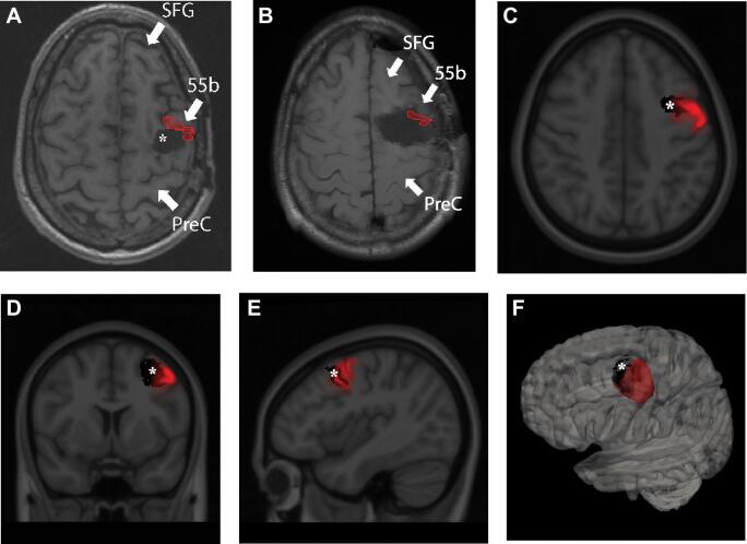
FIGURE 2.
A, Area 55b projected onto axial MRI of the patient prior to the resection causing his AOS. Asterix marks the first resection cavity sparing Area 55b (corresponding to the panel of Figure 1 labelled “initial resection”), which did not cause language deficits. B, Area 55b projected onto the cavity after the resection, which led to AOS deficits. C, Axial, D, coronal, and E, sagittal cuts of the MNI brain co-registered with probability maps of Area 55b (Red) and the defect prior to AOS-causing resection (black). F, 3D reconstruction with the probability map of Area 55b depicted in red and the defect prior to AOS-causing resection (black). SFG = Superior frontal gyrus; 55b = Area 55b; PreC = Pre-central gyrus; * = original resection cavity prior to AOS-causing resection.

