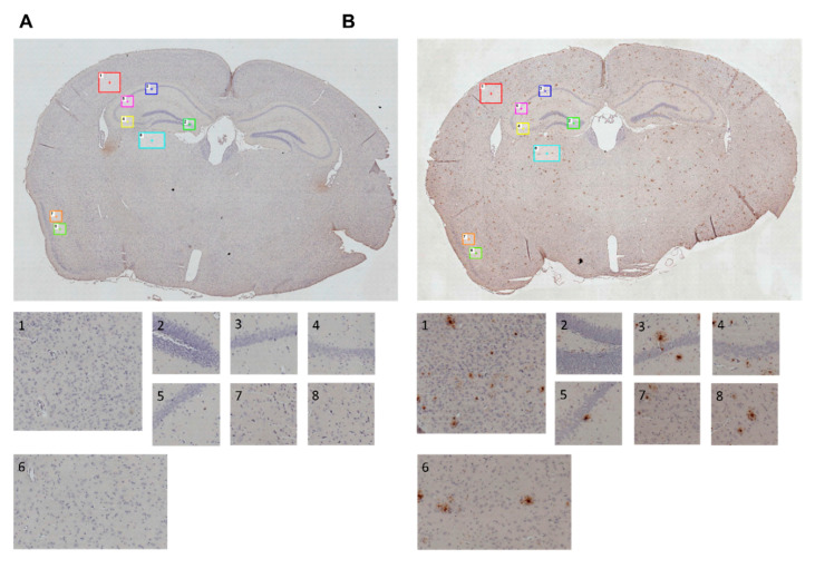
Figure 10.
Immunohistochemically stained sections of mouse brains; regions of interest (ROI) in cortical and hippocampal areas are marked in whole brain images (100× magnification) and depicted separately for further semi-quantification of amyloid plaques; 35 weeks of age, 30 weeks on experimental diet. (A) Exemplary section of a C57BL/6J wild type animal. (B) Exemplary section of an AppNL-G-F knock-in animal.

