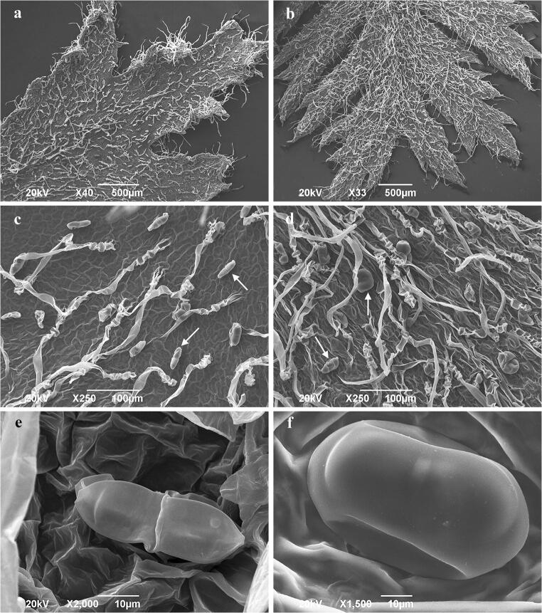
Fig. 2.
Scanning electron micrographs of in vitro grown tansy foliar surface. a Adaxial leaf surface; b abaxial leaf surface; c glandular (arrow) and non-glandular trichomes on the adaxial leaf surface; d glandular (arrow) and non-glandular trichomes on the abaxial leaf surface; e biseriate glandular trichome at the beginning of the secretory phase; f mature biseriate glandular trichome on the adaxial leaf surface in the full secretory phase

