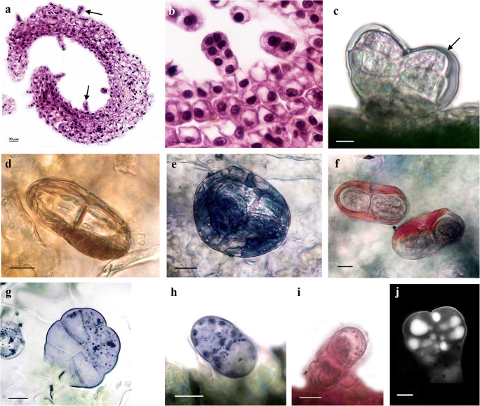
Fig. 3.
Structural and histochemical features of leaf glandular trichomes from in vitro grown tansy. a Cross-section of tansy leaf, note: trichomes (arrow) on adaxial and abaxial leaf surface; b young, immature leaf glandular trichomes, note: two basal cells, a short stalk, and secretory head of three pairs cells; c unstained biseriate trichome with subcuticular space (arrow); d orange-brown colored of secretory material in subcuticular space after stained with Sudan Red 7B/hematoxylin; e dark-blue color of lipophilic substance after staining with Sudan black B; f neutral lipids/essential oils stained red, while acid lipids stained blue with Nil blue A; g–h positive reaction with NADI reagent, violet-blue droplets indicate terpene secretion; i positive reaction with PAS; j UV-autofluorescence micrographs of leaf glandular trichomes. Bar = 10 μm

