Abstract
The symbiosis system comprising eukaryotic hosts, intracellular bacterium Wolbachia, and temperate bacteriophages WO is widely spread through nearly half the number of arthropod species. The relationships between the three components of the system are extremely intricate. Even though the bacteriophage WO can have diverse influences on the ecology and evolution of Wolbachia, little is known about the distribution and evolution of the phages. To the best of our knowledge, this study is the first to report that in infected fig wasps (Ceratosolen solmsi, Kradibia gibbosae, and Wiebesia pumilae), the genomes of all the Wolbachia strains had only one cryptic WO prophage, which contained defects in the genomic structural modules. This phenomenon was contrary to the widely accepted understanding that Wolbachia with cryptic prophages usually possesses at least one intact WO prophage consisting of gene sequences of the head, baseplate, and tail modules, through which the prophage could form intact virions. In addition to the genetic structure features, the phylogenetic relationships of WO and Wolbachia also revealed that bacteriophage WO can horizontally spread among a certain genus or a group of insect hosts, nearly free from the restriction of the affiliation of Wolbachia. Combined with the vertical transmission along with Wolbachia, the wide spread of WO phages can be explained. Furthermore, the gender preference and functional module preference for transcriptional activity of the genes in cryptic WOs implied the antagonized coevolutionary pattern between WO prophages and their Wolbachia hosts.
Keywords: bacteriophage WO, Wolbachia, genomic structure, distribution pattern, coevolutionary pattern
Introduction
Wolbachia is a genus of intracellular endosymbiotic bacteria belonging to the order Rickettsiale and is estimated to be distributed in more than 66% of arthropods and also shows mutualistic symbiosis in nematodes (Werren, 1997; Ferri et al., 2011). By phylogenetic analyses based on conserved coding genes used for multilocus sequence typing, such as 16S rRNAs and the Wolbachia surface protein (wsp) gene, Wolbachia has been classified into various strains under 18 supergroups (A–R) (Landmann, 2019). Wolbachia can manipulate the reproductive system of eukaryotic hosts using diverse methods, such as cytoplasmic incompatibility (CI), feminization of genetic male, parthenogenesis, and male killing (Werren et al., 2008), to avail the maternal transmission of its own population. It can also exert an influence on numerous processes in the host, including immune, behavioral, and metabolic processes (Landmann, 2019). Therefore, Wolbachia has great research potential as well as application value. For instance, it can be applied to control the populations of Aedes albopictus and Aedes aegypti, for inhibiting the widespread transmission of various arboviruses that are harmful to humans (Hoffmann et al., 2011; van den Hurk et al., 2012; Caragata et al., 2016).
WO, a bacteriophage that infects Wolbachia, is a λ phage-like temperate phage (Masui et al., 2001; Bordenstein et al., 2006). More than 80% of Wolbachia strains have WO-related gene fragments, and the infection proportion in supergroups A and B of Wolbachia is up to 90% (Gavotte et al., 2007). The CG contents and the codon preference of WO and Wolbachia bacteria are similar, so it has been speculated that WO and Wolbachia have coevolved for more than 100 million years (Masui et al., 2000). Therefore, there must be an intricate biological relationship between WO and Wolbachia.
As a temperate bacteriophage, WO has two different states. Viral DNA of the phage in the lysogenic state can be integrated into the genome of Wolbachia and duplicated with Wolbachia, which is usually called a prophage (Atanasoff, 1967). However, under certain conditions, the prophage can also convert into the lytic state, forming virions and causing the lysis of the bacteria (Echols, 1972). Prophages are under the selective pressure of their hosts, leading to various genomic defects of partial DNA, such as recombination, gene loss, or gradual degradation (Canchaya et al., 2003). The defective genomic prophages no longer have the ability to form virions and lyse host cells and are usually called defective prophages or cryptic prophages (Canchaya et al., 2003; Saridaki et al., 2011). Cryptic prophage WO is also found in the genome of Wolbachia. The most significant difference between intact and cryptic WO is that intact WO has a relatively complete gene module that codes for the proteins of the head, baseplate, and tail so that it can form active virions. Interestingly, Wolbachia with cryptic phages reported so far usually contain at least one intact prophage. However, there are exceptions. For example, in the Wolbachia strain of wRec that infects the Drosophila recens, it was reported for the first time that the host contained only a WO prophage with a fragmented structure (Metcalf et al., 2014). It has also been reported that only one cryptic prophage WOSol was found in Wolbachia infecting the fig wasp Ceratosolen solmsi, and interestingly, despite being a cryptic prophage, more than half of the WOSol genes were still transcriptionally active (Wang et al., 2014), which was far more than the previously reported numbers of the active genes in cryptic prophage WORiB of Drosophila simulans in which only two ank genes and one methyltransferase gene (met2) were shown to have transcriptional activity (Biliske et al., 2011), indicating that cryptic phages WO could have multiple gene functions.
It has been known that bacteriophages can have diverse influences on the ecology and evolution of bacterial hosts. For example, bacteriophages provide beneficial genes to the bacterial host (Abedon and Lejeune, 2005; Wang et al., 2010) or mediate the horizontal transfer of genes (Wommack and Colwell, 2000). Similarly, the phage can also mediate horizontal transfer of Wolbachia bacterial genes (Wang G. H. et al., 2016). Recent researches have further revealed that phage WO possibly plays an important role in the induction of CI in insect hosts by Wolbachia bacteria (LePage et al., 2017; Lindsey et al., 2018; Shropshire et al., 2018). Nevertheless, our understanding of WO is still incomplete, and the cause for the wide distribution of the phage in Wolbachia strains is unknown. Even though some evidences have validated that WO can be transmitted horizontally across different Wolbachia strains (even belonging to different supergroups) coinfecting the same cell of the insect host (Bordenstein and Wernegreen, 2004; Kent et al., 2011), and the phylogeny of phage WO found in fig wasps has revealed the strong specificity for insect hosts (Wang N. et al., 2016), they are still not sufficient to explain the widespread existence of WO. Therefore, this study was carried out to understand the distribution and evolution of the phage.
In this study, based on the well-assembled genomes of the three Wolbachia strains, infecting three species of pollinating fig wasps (C. solmsi, Kradibia gibbosae, and Wiebesia pumilae) (Hymenoptera, Chalcidoidea), we discovered that each Wolbachia strain contained only one cryptic prophage WO without the presence of any intact WO. To the best of our knowledge, this is the first study to report multiple Wolbachia strains containing only one cryptic phage WO in a certain group of insect hosts. By combining the genomes of the three cryptic WO phages and the WO-related genomic data of other strains of Wolbachia, our studies provided evidence that could prove the horizontal transmission of the WO phage among a genus or a certain group of insect hosts, which can elucidate the wide spread of phage WO.
Materials and Methods
The Collection of Fig Wasp Samples
We collected the fig wasp samples for the three pollinating fig wasps species, with the species of C. solmsi from Dongguan, Guangdong province, China (N22°39′, E113°31′); W. pumilae from Huangshan, Anhui province, China (E118°33′, N29°72′); and K. gibbosae from Danzhou, Hainan province, China (N19°30′, E109°29′). All fig wasps were collected at the stage of adults and then identified and classified under microscope Nikon SMZ80. Each gender of every species was conserved into RNA hold for RNA extraction.
Assembly of Wolbachia Genomes
The Wolbachia genomes were assembled based on the whole-genome sequencing data of the three fig wasp species in our laboratory (project accession number of PRJNA641212 for W. pumilae and K. gibbosae, PRJNA178998 for C. solmsi). We designed three different strategies to demonstrate the assembling procedures of each Wolbachia strain. (a) For the genome of Wolbachia infecting C. solmsi (wCsol), which was sequenced using second-generation Illumina HiSeq TM2000, the clean reads were aligned to the data set of the genomes of known Wolbachia using BLASTN (identity ≥ 70%, e-value ≤ 1e−5) to screen out homologous reads of Wolbachia for subsequent assembling using SOAPdenovo2 (Luo et al., 2012). During the assembling process, the parameters were adjusted constantly until an optimal result was obtained. (b) For the genome of Wolbachia infecting W. pumilae (wWpum), which was sequenced using third-generation PacBio Sequel, after assembling of corrected clean reads using smartdenovo-1.0, the assembly was polished using pilon-1.22. We then discovered an intact scaffold, which was highly homologous with Wolbachia (coverage ≥ 99.99%), and this scaffold was considered as the genome of wWpum. (c) For the genome of Wolbachia infecting K. gibbosae (wKgib), which was sequenced using third-generation PacBio Sequel, we used BLASTN against the data set of the genomes of known Wolbachia to find homologous reads of Wolbachia (identity ≥ 55%, coverage percent ≥ 55%, e-value ≤ 1e−5). The set of homologous reads was assembled using Canu-1.7.1 (Koren et al., 2017), and finally Purge Haplotigs (Roach et al., 2018) was used for filtering the redundancy with low coverage in the assembly to get more concise and consecutive genome sequences.
The genomic integrality test for all three genomes was conducted using the Benchmarking Universal Single-Copy Orthologs (BUSCO v4.0.6) (Simao et al., 2015) with a proteobacteria database, and the integrality of the four genomes of Wolbachia known to be intact (wHa, wMel, wAu, and wCauA) was also tested for contrast. The genomes were then submitted to GenBank which would be automatically annotated by the NCBI Prokaryotic Genome Annotation Pipeline (PGAP) (Tatusova et al., 2016), with the accession numbers of CP054557 for wWpum, CP054598 for wCsol and JABXYD000000000 for wKgib.
The Other Wolbachia Genomes Used in This Study
In addition to the above three Wolbachia in fig wasps, we also selected 49 known genomes of different strains of Wolbachia from GenBank for analysis. These strains were distributed in the supergroups A–F (Supplementary Table S1).
Protein Prediction and Reannotation of the Wolbachia Genomes
To ensure the consistency of our data, we repredicted the encoding proteins from all the 52 strains of Wolbachia. Each genome was predicted using Prodigal 2.6.3 (Hyatt et al., 2010). Then we reannotated all proteins of the Wolbachia strains in supergroup A with the following databases: NR database, Swiss-Prot, and InterProScan.
Construction of a Phylogenetic Tree Based on Single-Copy Orthologous Proteins
OrthoMCL (Li et al., 2003) (with default parameters) was used to search for orthologous proteins among the 52 strains of Wolbachia. The set of single-copy orthologous proteins was obtained by manually excluding the multicopy orthologous proteins.
All single-copy orthologous proteins in each strain were concatenated in a specific order into a super-protein sequence and aligned using MAFFT v7.313 (Katoh and Standley, 2013). Then we used Gblocks 0.91b (Talavera and Castresana, 2007) to select conserved blocks in the alignments. MrBayes (Altekar et al., 2004) was used to construct the phylogenetic tree of 52 super-proteins with the optimal amino acid substitution model predicted using ProtTest 3.4.2 (Darriba et al., 2011).
Search for Prophage WO in Wolbachia and Illustration of WO Structures
The location and number of prophage WO in each Wolbachia in supergroup A were ascertained through the combination of the annotation of genomes of Wolbachia (as obtained in this study) and the prediction from the platform PHASTER (Arndt et al., 2016). We uploaded the genomes of Wolbachia to PHASTER and compared them with the phage databases to obtain obscure positions of prophage WO. According to the functional annotation, the protein in the predicted position was confirmed to be the relative protein of prophage WO. If not, the protein was discarded, and if so, we continued to check the other proteins in its flanking region until the sequence consisted of non-WO genes at both ends. Then, the sequence contained in all these consecutive WO genes is designated as one prophage WO. The genomic structures of all prophages WO were illustrated using Illustrator for Biological Sequences (IBS) (Liu et al., 2015).
Construction of Phylogenetic Tree and Estimation of Divergence Time of WO
We extracted the conserved sequences including the head module and baseplate module (from phage terminase to tail protein I) from each WO prophage found in the Wolbachia strains of supergroup A. The sequences were aligned, and the conserved blocks were selected as described above, based on which the phylogenetic tree was constructed using MrBayes with the prediction of the nucleotide substitution model using ModelFinder. All software involved was integrated into the PhyloSuite v1.2.1 software (Zhang et al., 2020).
To estimate the divergence time of prophages, we used BEAST v1.10.4 (Suchard et al., 2018) to construct the time tree, and the chain length of MCMC was set as 100,000,000. We simulated six trees using different models (Bayesian Skyline, Constant Size and Exponential Growth model in Uncorrelated relaxed clock type and Strict clock type, respectively) and chose the optimal tree based on the result of the PS/SS MLE test (Supplementary Table S2). Given that there was little fossil evidence to calibrate the divergence time of prophage WO, we referred to the method in the essay of William R. Corner, setting a relative time of the node between WOCsol and WOKgib as 1 to construct an arbitrary scaling time tree (Conner et al., 2017), to analyze the relative divergence order of prophages WO.
Synteny Analyses of the Sequences
The synteny analyses were mainly performed using MCscanX (Wang et al., 2012) with the parameter of e-value ≤ 1e−10. In a few conditions, we employed Mauve 2.4.0 (Darling et al., 2004) using its default parameters.
RNA Extraction and Polymerase Chain Reaction Verification of Transcriptional Activity of Prophage WO Genes
For each species of fig wasp, we set eight samples (four of females and four of males). The individual numbers in each sample ranged from 30 to 50 according to the body size of the fig wasp (for C. solmsi and W. pumilae, there were 30 individuals per sample, whereas for K. gibbosae, there were 50 individuals per sample). We used TransZol Up Plus RNA Kit (TransGenBiotech, Beijing, China) to extract the RNA for each sample and finally dissolved them in 35 μL RNase-free water. The concentration and purity of RNA were tested according to the OD values using Thermo Scientific NanoDrop One. All RNA samples were used to performed PCR test with Wolbachia wsp81F/691R universal primers (Zhou et al., 1998) as the negative controls to confirm that they did not contain massive genomic DNA contamination. Then the cDNA was synthesized, and at the same time, the remaining genomic DNA contamination was removed using EasyScript® II One-Step gDNA Removal and cDNA Synthesis SuperMix (TransGenBiotech, Beijing, China). The reaction mixture was incubated at 42°C for 30 min. We designed primers for PCR verification on the conserved functional genes of the prophages WO. To reduce the probability of false positives, we did not choose unspecific genes, such as Ankyrin repeats and transposase, which were probably also present in the insect and bacterial hosts. We verified 23 genes in WOCsol and WOKgib, and 15 genes in WOWpum (primers are listed in Supplementary Table S3). The expression of wsp gene in each cDNA sample was tested by PCR as the positive control to demonstrate the cDNA in all samples was well synthesized. The PCR products with bands of target sizes were sequenced to confirm the correct amplification of the target genes.
Results
Genome Assemblies and Supergroup Typing of Wolbachia Strains Infecting the Three Fig Wasp Species
We obtained the genome assemblies of three Wolbachia strains that infected the three fig wasp species. The genome of Wolbachia strain wWpum was an intact sequence that could be circularized with the size of 1.28 Mb. The genome of wCsol consisted of a single scaffold with the size of 1.21 Mb, but it could not be circularized. The genome of wKgib contained three scaffolds, with the total size of 1.45 Mb and N50 length of the assembly 948,431 bp. The integrality of each Wolbachia genome tested using BUSCO was wWpum = 84.5%, wCsol = 83.6%, and wKgib = 84.9%. Compared with the integrality of the other four intact Wolbachia strains (wHa = 84%, wMel = 84.5%, wAu = 84.9%, and wCauA = 84.5%), the genomes of Wolbachia infecting fig wasps did not present significant differences (p value of wWpum = 1.000, wCsol = 0.147, and wKgib = 0.264), indicating that our assemblies of genomes were sufficiently complete.
The assembling statistics of each Wolbachia infecting the fig wasps were annotated using PGAP and was compared with 49 other Wolbachia strains (Supplementary Table S4). The results showed few significant differences, whether compared with all strains or with the 24 strains in supergroup A, except in the case of wKgib, where the numbers of rRNA (n = 6, p = 0.026 < 0.05 to all strains) and ncRNA (n = 5, p = 0.002 < 0.01 to all strains, and p = 0.001 < 0.01 to supergroup A) increased significantly (Table 1).
TABLE 1.
Mann-Whitney U tests for assembling statistics of each Wolbachia in fig wasp against other strains.
| wWpum vs. all strains | wCsol vs. all strains | wKgib vs. all strains | wWpum vs. supergroup A | wCsol vs. supergroup A | wKgib vs. supergroup A | |
| Size | 0.917 | 0.488 | 0.282 | 0.889 | 0.366 | 0.126 |
| GC% | 0.327 | 0.972 | 0.727 | 0.711 | 0.165 | 0.379 |
| Protein | 0.51 | 0.425 | 0.177 | 0.488 | 0.267 | 0.127 |
| rRNA | 0.828 | 0.828 | 0.026* | 0.853 | 0.853 | 0.115 |
| tRNA | 0.108 | 0.846 | 0.072 | 0.14 | 1 | 0.104 |
| ncRNA | 0.758 | 0.758 | 0.002** | 0.827 | 0.827 | 0.001** |
| Gene | 0.652 | 0.386 | 0.225 | 0.782 | 0.267 | 0.127 |
| Pseudogene | 0.912 | 0.883 | 0.658 | 0.693 | 0.693 | 0.478 |
The numbers in the cells represent the p-values. *p < 0.05, **p < 0.01.
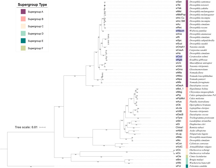
Based on 253 groups of single-copy orthologous proteins, we constructed a Bayesian phylogenetic tree with 52 strains of Wolbachia (Figure 1). It revealed that all the three Wolbachia strains in fig wasps were classified into supergroup A, with the strains of wCsol and wKgib clustered together to form a single branch, whereas the strain of wWpum showing a closer relationship with several Wolbachia strains that infect fruit flies on another branch.
FIGURE 1.
Phylogeny of 52 strains of Wolbachia. The tree was constructed based on 253 groups of single-copy orthologous proteins using MrBayes and the amino acid substitution model CpRev + I + G + F, and wFol was set as the outgroup. The three Wolbachia strains infecting fig wasps were marked with a violet background. Each supergroup was labeled using different color stripes, and after the stripes, the eukaryotic host that each Wolbachia strain belongs to was listed.
Structural Diversity of WO Prophages in the Wolbachia Strains in Supergroup A
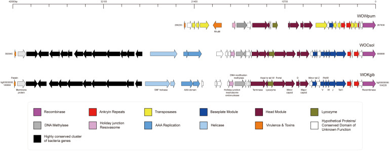
Based on the three well-assembled genomic sequences of Wolbachia strains in the fig wasps, we searched for and annotated the sequences of prophages WO and noticed that each Wolbachia strain possessed merely one prophage WO, named as WOCsol, WOKgib, and WOWpum, respectively, corresponding to the Wolbachia strains of wCsol, wKgib, and wWpum (Figure 2). The length of WOCsol was 42,744 bp, the length of WOKgib was 42,757 bp, and WOWpum was 22,596 bp. Interestingly, no gene member of the tail module was found in any of the three prophages. The defect in the genomic structure indicated that these WO were cryptic phages with no capacity to form virions. To the best of our knowledge, it is the first time that cryptic prophages WO existing independently without any intact prophage WO in the Wolbachia strains among a group of insect hosts is being reported. WOCsol and WOKgib were very similar in terms of structure and sequence length, and the similarity of nucleotide sequences was 99.82%. However, compared with WOCsol or WOkgib, the sequence of WOWpum was almost half the length, which was owing to the lack of insertion of a highly conserved cluster of bacteria genes and the absence of genes of other modules, including that of the head, baseplate, helicase, and AAA replication. These results suggested a relatively distant relationship between WOWpum and the other two WOs infecting fig wasps, which seemed to be consistent with the phylogeny of their bacterial host, Wolbachia.
FIGURE 2.
Genomic structures of the three cryptic WO prophages in Wolbachia found in fig wasps. The structures of the core regions of the prophages were illustrated by IBS, and the functional annotation of genes was labeled. In WOWpum, the highly conserved cluster of bacterial genes was absent, and quite a few genes of the head module, baseplate module, AAA replication, and helicase were missing or degraded.
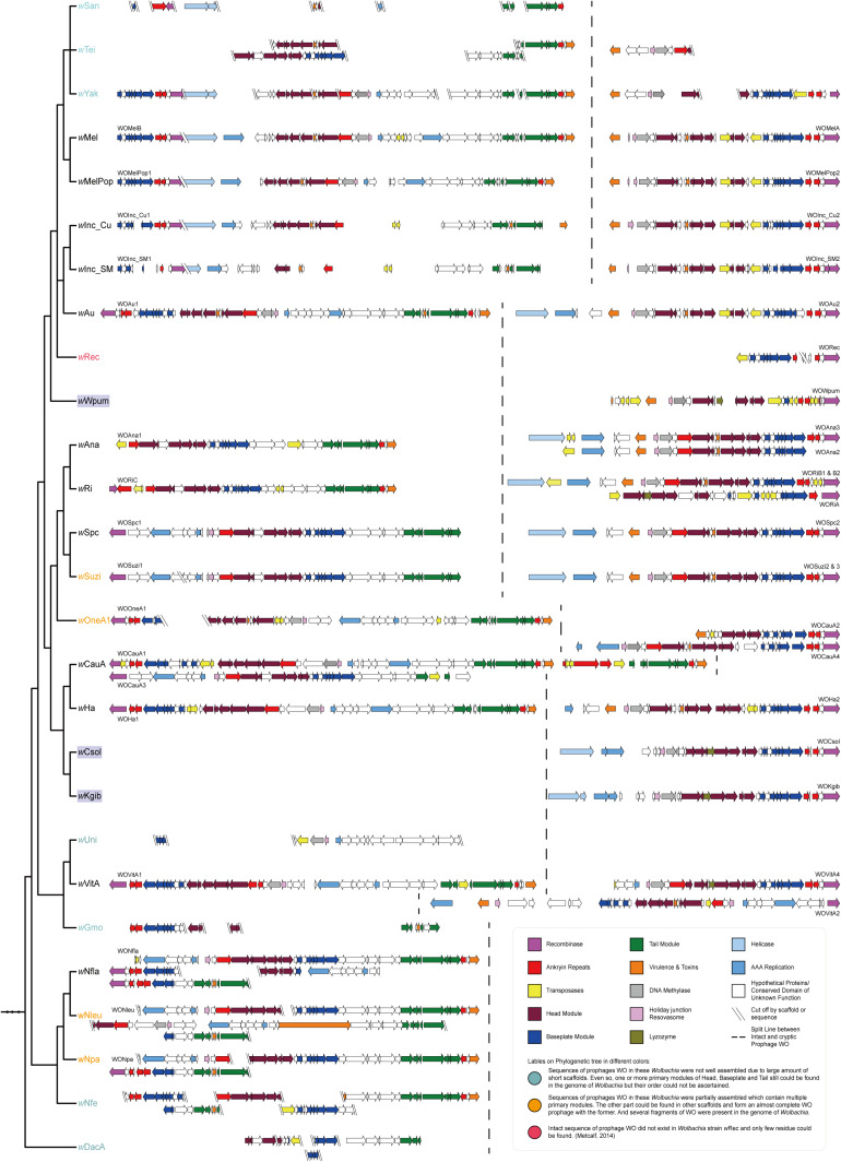
To determine whether it is a special phenomenon that Wolbachia that infect fig wasps contain only cryptic prophages without the existence of any intact WO, as well as to understand the tendency of structural change and the evolutionary relationship of the WO prophages, we searched for and annotated the prophages WO that infect the other 24 Wolbachia strains in supergroup A. We then mapped the structures of the WO onto the phylogeny of Wolbachia (Figure 3). The results showed that except the three Wolbachia that infected the three fig wasps studied and the previously reported wRec, each Wolbachia in supergroup A possessed at least one intact WO. Even though some prophages were not well assembled because of the severe fragile assemblies of their bacterial host, the sequence fragments of the head, baseplate, and tail modules (wSan, wTei, wYak, wGmo, wNfe, wDacA) or the homologous fragments of other intact WO (wUni) could still be found, implying the presence of intact prophages in these Wolbachia strains. Furthermore, simply in terms of structure, within a Wolbachia strain, the intact WO is usually quite different from the cryptic WO, while the intact prophages WO or cryptic prophages WO among Wolbachia strains with close relationships were much more similar. Therefore, we reasonably speculated that the cryptic WO of each Wolbachia strain was not derived directly from the intact WO of the same Wolbachia strain.
FIGURE 3.
Mapping of the genomic structures of WO in 27 Wolbachia strains in supergroup A to the phylogenetic tree of their bacterial hosts. In each of the Wolbachia strains, there were different numbers of cryptic WO prophages with at least one intact prophage WO, but exceptionally, each of the three strains infecting fig wasps possessed only one cryptic prophage WO without the existence of any intact prophage WO. The dashed line was used for separating intact WO (on the left) and cryptic WO (on the right) sequences. Phylogenetic tree of Wolbachia was the same as the section of supergroup A in Figure 1, ignoring branch length.
Phylogeny and Estimated Divergence Time of the WO Prophages
To verify the above speculation, we constructed an arbitrary scaling divergence time tree and phylogenetic tree for all prophages referred to in the previous section (the prophages without consecutive sequences were excluded), based on their conserved nucleotide sequences of the head and baseplate modules.
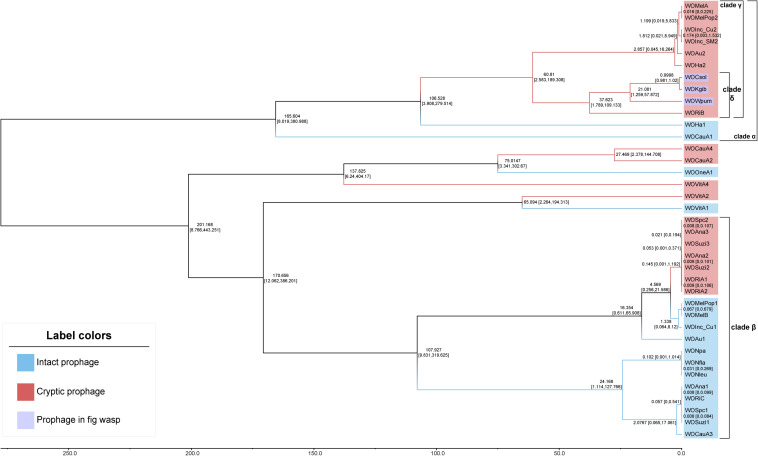
The divergence time tree (Figure 4) showed that intact WO was unsurprisingly more ancient than cryptic WO. For most of the phages, such as those in clade α and clade β, all cryptic WOs were newly produced after intact WOs. Furthermore, the estimated divergence time of the WO prophages of Wolbachia strains infecting Drosophila (WOSpc, WOAna, WOSuzi, and WORi) and Nomada (WONpa, WONfla, and WONleu) in clade β was very late, even later than the time set as 1 for the node between WOCsol and WOKgib. In the Wolbachia strains of Drosophila, the divergence time of both cryptic and intact WO was extremely close, and even the estimated divergence time was exactly the same (in 0.008 or 0.009 relative time), such as in the WO of wSpc, wAna, wSuzi, and wRi, revealing a clear pattern of coevolution and codivergence. Surprisingly, WOWpum was clustered with WOCsol/WOKgib, showing a very close phylogenetic relationship, rather than the greater distance between their bacterial hosts. However, the divergence time of WOWpum was indeed earlier than the other two WOs, indicating that more selection was carried out in its case, which was consistent with its more fragmented genome structure as mentioned above.
FIGURE 4.
Chronograms for WO prophages. The chronogram depicted a relative divergence time of each WO prophage to the node of WOCsol–WOKgib, where we set an arbitrary scaling of relative age at 1. The estimated divergence time of each node is shown with 95% confidence intervals. Branches and labels of this unrooted tree are shown in two diverse colors to distinguish between the intact and cryptic prophages. In both of clade α and clade β, intact WOs usually appeared earlier than cryptic WOs. Clade γ was a typical clade in which all prophages were cryptic. All four WOs in clade δ were found to have genes with transcriptional activities.
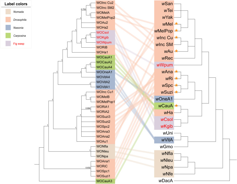
When we integrated the phylogeny of the WO prophages, Wolbachia, and their insect hosts together to form an intuitive relationship among them (Figure 5), the results showed that (a) the phenomenon that prophages WO among distant Wolbachia strains presented close relationships not only appeared in the group of fig wasps, but also in the insect hosts of the Hymenoptera genus of Nasonia. For example, the Wolbachia strain wOneA1 infecting Nasonia oneida was apparently evolutionarily distant from the strain infecting Nasonia vitripennis, wVitA, but their prophages WOVitA4 and WOOneA1 were relatively close and were even clustered into one clade. (b) However, in the genus of Drosophila, the topological structure of the phylogeny of Wolbachia (except for wHa and wAu) was pretty similar to that of their prophages, such as the clade of wMel: wAu and the clade of wAna: wSuzi. This relationship was also seen in the Hymenoptera genus of Nomada. (c) Interestingly, consistent with our speculation that in the same Wolbachia strain, the cryptic WO is not similar to the intact WO, except for in the case of WOVitA and WOHa, a few cryptic WO (in 9/20 of Wolbachia strains, which are labeled using orange asterisks) were evolutionarily dissimilar to the intact WO in the same strain. Moreover, if we counted only the Wolbachia strains possessing both intact and cryptic WO, the percentage would rise to 82% (9/11), indicating a substantial probability that cryptic prophages WO were not derived directly from the intact ones in the same bacterial host.
FIGURE 5.
Phylogenetic relationships of the WO prophages, Wolbachia strains, and insect hosts. The tree of WO on the left was constructed using MrBayes in the nucleotide substitution model GTR + F + I + G4, and WOCauA3 is set as the outgroup. The tree on the right is the same as the section depicting supergroup A of Wolbachia as shown in Figure 1, in which the branch length is ignored. We connected each prophage to their bacterial host with lines, the colors of which labeled the insect host group. The WO/Wolbachia in the same color belonged to one specific genus or a biological group of insects. Orange asterisks represent the Wolbachia strains with the intact and cryptic WO relatively far apart.
High Proportion of Genes of the WO Prophages Were Actively Transcribed in the Fig Wasps
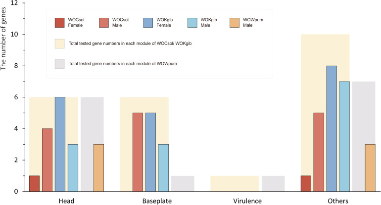
We verified the transcriptional activity of each characteristic functional gene in the core region of the WO prophages of the respective fig wasps using reverse transcription PCR (RT-PCR). Twenty-three genes were verified in WOCsol and WOKgib, respectively, and 15 genes were verified in WOWpum. In WOCsol, only two genes showed transcription in females, whereas 14 genes were active in males, with the percentage of actively transcribed genes at 61% (14/23). In WOKgib, 19 genes were transcribed in females, and 13 were transcribed in males, with the percentage of actively transcribed genes at 83% (19/23). In WOWpum, the mRNA of these genes was not detected in females, whereas in males, six genes showed transcriptional activity, with the percentage of active genes at 40% (6/15) (Table 2). Thus, these actively transcribed WO genes showed a certain gender preference in the fig wasp hosts (Figure 6), with WOCsol and WOWpum having significantly more actively transcribed genes in wasp males than in females, while WOKgib having more actively transcribed genes in the female host. Furthermore, the functional module preference could also be discovered, although the genes of the baseplate and head modules were both conserved, genes of the head module showed more activity than that of the baseplate module. For the genes of the baseplate module, only the genes of WOCsol were transcribed actively in male hosts, and the genes of WOKgib were transcribed in both genders of the host, whereas in other samples, they showed no activity (in WOCsol-F, WOWpum-F, and WOWpum-M). In contrast, genes of the head module showed high transcriptional ratios in four samples, except for the samples of WOCsol-F (one active gene) and WOWpum-F (no active gene).
TABLE 2.
RNA transcription of the three cryptic WO prophage in fig wasps.
| Module | Gene | C-F | C-M | K-F | K-M | W-F | W-M |
| Others | Site-specific recombinase | − | + | + | + | − | + |
| Others | Putative phage−related protein | − | + | + | + | − | + |
| Baseplate | Tail I | − | + | + | − | ||
| Baseplate | Baseplate assembly protein J | − | − | + | − | ||
| Baseplate | Prophage lambda W1, baseplate assembly protein W | − | + | − | − | ||
| Baseplate | PAAR motif | − | + | + | + | ||
| Baseplate | Baseplate assembly protein GpV | − | + | + | + | ||
| Others | Hypothetical protein So0009 | − | + | + | − | ||
| Baseplate | Prophage minor tail protein Z (GPZ) | − | + | + | + | − | − |
| Others | Hypothetical protein So0011 | + | + | + | + | − | + |
| Head | Putative major capsid protein | − | + | + | − | − | + |
| Head | Bacteriophage lambda head decoration protein D | + | + | + | + | − | − |
| Head | Putative minor capsid protein C | − | − | + | − | − | + |
| Head | Phage portal protein | − | + | + | + | − | − |
| Others | Lyzozyme M1 | − | − | + | + | − | + |
| Head | Head-to-tail joining protein W | − | + | + | + | − | − |
| Head | Phage terminase large subunit family protein | − | − | + | − | − | − |
| Others | Hypothetical protein So0019 | − | + | + | + | − | − |
| Others | DNA modification methylase | − | − | − | − | − | − |
| Others | Putative Holliday junction resolvasome endonuclease | − | − | + | + | − | − |
| Others | DEAD/DEAH box helicase | − | − | − | − | ||
| Others | Putative membrane protein | − | − | + | + | ||
| Virulence | Patatin-like phospholipase | − | − | − | − | ||
| Virulence | Virulence RhuM family protein | − | − |
The abbreviation of each sample’s name: C, WOCsol; K, WOkgib; W, WOWpum; M, male; F, female.The plus sign (+) represents the gene expression is positive in at least two of four samples. The minus sign (−) represents no gene expression is characterized in all four samples.
FIGURE 6.
Sexual-specific and module-specific transcription activities of genes in the three WO prophages. The numbers of genes of the prophages verified by RT-PCR were counted. The thick bars in the background represent the total numbers of genes for which we designed primers for amplification, and the narrow bars represent the actual numbers of genes possessing transcriptional activities in each WO sample. Results of six WO samples were split into four groups according to the different functional modules. WOCsol and WOWpum showed a higher transcription activity in males than that in females, and in WOKgib, the pattern was observed to be the opposite. Meanwhile, the genes of head modules of the three WOs were transcribed to a greater extent than those of the baseplate modules, which showed functional module preference on conserved modules.
Discussion
In this study, based on the three well-assembled genome sequences of the Wolbachia strains of wWpum, wKgib, and wCsol that infect three fig wasp species, we ascertained the numbers and structural features of the temperate WO prophages. Each of these three strains contained only one typical cryptic prophage WO, which contained the head and baseplate modules, but no gene of the tail module. However, we found no intact prophages capable of forming active virions in these Wolbachia strains, which was consistent with previous results that the Wolbachia strain infecting fig wasp C. solmsi merely had one cryptic prophage ascertained using the method of q-PCR (Wang et al., 2013, 2014). Furthermore, we explored the distribution of the WO prophages of the previously reported 24 Wolbachia strains in supergroup A and found that the phenomenon of only one cryptic prophage existing without an intact prophage WO was unique to the Wolbachia strains of fig wasps. This result was contrary to the widely accepted understanding that Wolbachia with cryptic prophages usually possesses at least one intact WO prophage (Kent et al., 2011). It is worth mentioning that although the Wolbachia strain of wRec possessed only fragmented sequences of WO (Metcalf et al., 2014), the assembly of the wRec genome was not consecutive. Therefore, the detection and analysis of WO may be biased, and many of the WO sequences might have been discarded during the assembly process. In contrast, in the present study, the assemblies of the Wolbachia genomes in fig wasps are much more complete, which would be more convincing to indicate the absence of an intact WO and the existence of a single cryptic WO in the bacterial hosts. To the best of our knowledge, this is the first report describing the phenomenon of a cryptic WO prophage existing solely in the absence of an intact prophage in the Wolbachia strains of a specific group of insect hosts.
We reannotated and illustrated the genomic structures of the core regions of all the WO prophages of Wolbachia in supergroup A involved in this study. Simultaneously, we constructed a phylogenetic tree and an arbitrary scaling divergence time tree based on the relatively conserved sequences ranging from the head to the baseplate modules commonly found in WO, and these phylogenetic trees were then mapped to the phylogenetic tree of their bacterial hosts. Both the structural features and phylogenetic results indicated that most of the cryptic prophages WO were not derived from the intact ones in the same Wolbachia strain; instead, their relationship was relatively distant. In addition, as the phages of Wolbachia, WOs showed a close relationship when the bacterial hosts were relatively distant, and interestingly, the insect hosts of the prophages with this feature were usually from the same genus or the same group of organisms. These results indicate that WO could be actively transmitted horizontally among different Wolbachia strains. WO has been reported to be able to transmit horizontally between different Wolbachia strains (even belonging to different supergroups) coinfected in the same host cell (Bordenstein and Wernegreen, 2004; Kent et al., 2011). Our results were expanded on this basis, and we believed that WO could be transmitted horizontally among a certain genus or a group of organisms and even spread across species over long distances driven by certain unknown factors (such as the spread of WOs between fruit fly and fig wasps in clade δ in Figure 4). Of course, the evidence of the codivergence of WO in the Drosophila genus suggested that WO could also be transmitted vertically in the Wolbachia host. Combined with the evidence that the estimated divergence time between wSpc and wSuzi was between 1,000 and 10,000 years ago (Conner et al., 2017), it can be inferred that the vertical transmission of WO might be a recent event compared with the event of horizontal transmission. In summary, we speculate that the rapid horizontal transmission of WO among a wide range of eukaryotic hosts and vertical transmission along with newly divergent Wolbachia strains could explain the wide distribution of WO phages in Wolbachia, and the horizontal (Conner et al., 2017) or vertical transmission (Werren et al., 2008) of Wolbachia could further promote the spread of WO among eukaryotic hosts.
Besides, we could draw more inferences or speculations based on the above results to elucidate the selection undergone by the WO phages and their evolutionary path after insertion into their Wolbachia hosts. (a) The genes of the four prophages WO in clade δ (in Figure 4), including the three WOs in fig wasps and WORiB, were all detected to have different levels of transcriptional activities, and the more recently WO diverged, the more genes with transcriptional activities it preserved. These discoveries verified that the prophages could be inactivated gradually by their Wolbachia hosts. Therefore, we reasonably inferred that the ancestor of clade γ was possibly an intact prophage WO with horizontal transmission capability in its virion form when it diverged from its Most Recent Common Ancestor, along with WOHa1 at the relative time of 106.528. After horizontal transmission across multiple Wolbachia strains, all the WOs derived from the ancestor WO phages became cryptic, explaining why several cryptic prophages WO were in the clade γ. (b) Given that Wolbachia was able to affect the WO phages negatively by causing the degradation and inactivation of their genes, WOs may also resist the selective pressures of their Wolbachia hosts. We noticed that the genes of both WOCsol and WOWpum presented higher transcriptional activities in male hosts than in females, which showed a certain gender preference. As Wolbachia is an endosymbiont with maternal transmission, it meant that the bacteria infecting males cannot be inherited by descendants. Considering the “non-compliance” of WOs to their Wolbachia hosts and the congruence between the phylogeny of WOs and the groups of insect hosts, we suggested two possible explanations for the higher transcription of WO phage genes in male hosts than in females. First, in the male insect host, WOs may increase the gene transcription to activate its horizontal transmission as quickly as possible, in order to escape the dead end of vertical transmission in the male insect host. Second, in the tripartite symbiont of insect-Wolbachia-WO, male insect hosts might tend to align with the prophages to antagonize the Wolbachia strain, so prophages WO could show higher activity in male insects where the Wolbachia strain is relatively “weak.” In the coevolution between the Wolbachia hosts and prophages, we could also observe the antagonism between them. In the analysis of the cryptic WOs, genes of the head module showed higher transcriptional activities than those of the baseplate module, which revealed that the genes of head module might be more crucial for WO. Combined with the absence of the tail modules in the cryptic prophages, we could speculate that under the selective pressure exerted by Wolbachia, WOs would be degraded in the order of the functional modules of the tail, baseplate, and head, which may be to maximize the retention time of its activity.
Data Availability Statement
The datasets presented in this study can be found in online repositories. The names of the repository/repositories and accession number(s) can be found in the article/Supplementary Material.
Author Contributions
Y-hM, J-hX, and D-wH designed the study. Y-hM performed the experiments. Y-hM and J-hX analyzed the data and wrote the manuscript. All authors have read and approved the submitted manuscript.
Conflict of Interest
The authors declare that the research was conducted in the absence of any commercial or financial relationships that could be construed as a potential conflict of interest.
Acknowledgments
We thank Dr. Li-Ming Niu and Guang-Chang Ma for help in collecting fig wasps.
Footnotes
Funding. This study was supported by the National Natural Science Foundation of China (Nos. 31830084, 31970440, and 31672336), and also supported by the construction funds for the “Double First-Class” initiative for Nankai University (Nos. 96172158, 96173250, and 91822294).
Supplementary Material
The Supplementary Material for this article can be found online at: https://www.frontiersin.org/articles/10.3389/fmicb.2020.595629/full#supplementary-material
References
- Abedon S. T., Lejeune J. T. (2005). Why bacteriophage encode exotoxins and other virulence factors. Evol. Bioinform. Online 1 97–110. [PMC free article] [PubMed] [Google Scholar]
- Altekar G., Dwarkadas S., Huelsenbeck J. P., Ronquist F. (2004). Parallel metropolis coupled markov chain monte carlo for bayesian phylogenetic inference. Bioinformatics 20 407–415. 10.1093/bioinformatics/btg427 [DOI] [PubMed] [Google Scholar]
- Arndt D., Grant J. R., Marcu A., Sajed T., Pon A., Liang Y., et al. (2016). PHASTER: a better, faster version of the PHAST phage search tool. Nucleic Acids Res. 44 W16–W21. 10.1093/nar/gkw387 [DOI] [PMC free article] [PubMed] [Google Scholar]
- Atanasoff D. (1967). Interaction among virus. Cell and organism. J. Phytopathol. 59 267–276. 10.1111/j.1439-0434.1967.tb02969.x [DOI] [Google Scholar]
- Biliske J. A., Batista P. D., Grant C. L., Harris H. L. (2011). The bacteriophage WORiC is the active phage element in wRi of Drosophila simulans and represents a conserved class of WO phages. BMC Microbiol. 11:251. 10.1186/1471-2180-11-251 [DOI] [PMC free article] [PubMed] [Google Scholar]
- Bordenstein S. R., Marshall M. L., Fry A. J., Kim U., Wernegreen J. J. (2006). The tripartite associations between bacteriophage. Wolbachia, and Arthropods. PLoS Pathog 2:e106 10.1371/journal.ppat.0020106 [DOI] [PMC free article] [PubMed] [Google Scholar]
- Bordenstein S. R., Wernegreen J. J. (2004). Bacteriophage flux in endosymbionts (Wolbachia): infection frequency, lateral transfer, and recombination rates. Mol. Biol. Evol. 21 1981–1991. 10.1093/molbev/msh211 [DOI] [PubMed] [Google Scholar]
- Canchaya C., Proux C., Fournous G., Bruttin A., Brussow H. (2003). Prophage genomics. Microbiol. Mol. Biol. Rev. 67 238–276. 10.1128/mmbr.67.2.238-276.2003 [DOI] [PMC free article] [PubMed] [Google Scholar]
- Caragata E. P., Dutra H. L., Moreira L. A. (2016). Inhibition of Zika virus by Wolbachia in Aedes aegypti. Microb. Cell 3 293–295. 10.15698/mic2016.07.513 [DOI] [PMC free article] [PubMed] [Google Scholar]
- Conner W. R., Blaxter M. L., Anfora G., Ometto L., Rota-Stabelli O., Turelli M. (2017). Genome comparisons indicate recent transfer of wRi-like Wolbachia between sister species Drosophila suzukii and D. subpulchrella. Ecol. Evol. 7 9391–9404. 10.1002/ece3.3449 [DOI] [PMC free article] [PubMed] [Google Scholar]
- Darling A. C., Mau B., Blattner F. R., Perna N. T. (2004). Mauve: multiple alignment of conserved genomic sequence with rearrangements. Genome Res. 14 1394–1403. 10.1101/gr.2289704 [DOI] [PMC free article] [PubMed] [Google Scholar]
- Darriba D., Taboada G. L., Doallo R., Posada D. (2011). ProtTest 3: fast selection of best-fit models of protein evolution. Bioinformatics 27 1164–1165. 10.1093/bioinformatics/btr088 [DOI] [PMC free article] [PubMed] [Google Scholar]
- Echols H. (1972). Developmental pathways for the temperate phage: lysis vs lysogeny. Annu. Rev. Genet. 6 157–190. 10.1146/annurev.ge.06.120172.001105 [DOI] [PubMed] [Google Scholar]
- Ferri E., Bain O., Barbuto M., Martin C., Lo N., Uni S., et al. (2011). New insights into the evolution of Wolbachia infections in filarial nematodes inferred from a large range of screened species. PLoS One 6:e20843. 10.1371/journal.pone.0020843 [DOI] [PMC free article] [PubMed] [Google Scholar]
- Gavotte L., Henri H., Stouthamer R., Charif D., Charlat S., Bouletreau M., et al. (2007). A Survey of the bacteriophage WO in the endosymbiotic bacteria Wolbachia. Mol. Biol. Evol. 24 427–435. 10.1093/molbev/msl171 [DOI] [PubMed] [Google Scholar]
- Hoffmann A. A., Montgomery B. L., Popovici J., Iturbe-Ormaetxe I., Johnson P. H., Muzzi F., et al. (2011). Successful establishment of Wolbachia in Aedes populations to suppress dengue transmission. Nature 476 454–457. 10.1038/nature10356 [DOI] [PubMed] [Google Scholar]
- Hyatt D., Chen G. L., Locascio P. F., Land M. L., Larimer F. W., Hauser L. J. (2010). Prodigal: prokaryotic gene recognition and translation initiation site identification. BMC Bioinformatics 11:119. 10.1186/1471-2105-11-119 [DOI] [PMC free article] [PubMed] [Google Scholar]
- Katoh K., Standley D. M. (2013). MAFFT multiple sequence alignment software version 7: improvements in performance and usability. Mol. Biol. Evol. 30 772–780. 10.1093/molbev/mst010 [DOI] [PMC free article] [PubMed] [Google Scholar]
- Kent B. N., Funkhouser L. J., Setia S., Bordenstein S. R. (2011). Evolutionary genomics of a temperate bacteriophage in an obligate intracellular bacteria (Wolbachia). PLoS One 6:e24984. 10.1371/journal.pone.0024984 [DOI] [PMC free article] [PubMed] [Google Scholar]
- Koren S., Walenz B. P., Berlin K., Miller J. R., Bergman N. H., Phillippy A. M. (2017). Canu: scalable and accurate long-read assembly via adaptive k-mer weighting and repeat separation. Genome Res. 27 722–736. 10.1101/gr.215087.116 [DOI] [PMC free article] [PubMed] [Google Scholar]
- Landmann F. (2019). The Wolbachia endosymbionts. Microbiol. Spectr. 7:BAI-0018-2019. 10.1128/microbiolspec.BAI-0018-2019 [DOI] [PMC free article] [PubMed] [Google Scholar]
- LePage D. P., Metcalf J. A., Bordenstein S. R., On J., Perlmutter J. I., Shropshire J. D., et al. (2017). Prophage WO genes recapitulate and enhance Wolbachia-induced cytoplasmic incompatibility. Nature 543 243–247. 10.1038/nature21391 [DOI] [PMC free article] [PubMed] [Google Scholar]
- Li L., Stoeckert C. J., Jr., Roos D. S. (2003). OrthoMCL: identification of ortholog groups for eukaryotic genomes. Genome Res. 13 2178–2189. 10.1101/gr.1224503 [DOI] [PMC free article] [PubMed] [Google Scholar]
- Lindsey A. R. I., Rice D. W., Bordenstein S. R., Brooks A. W., Bordenstein S. R., Newton I. L. G. (2018). Evolutionary genetics of cytoplasmic incompatibility Genes cifA and cifB in prophage WO of Wolbachia. Genome Biol. Evol. 10 434–451. 10.1093/gbe/evy012 [DOI] [PMC free article] [PubMed] [Google Scholar]
- Liu W., Xie Y., Ma J., Luo X., Nie P., Zuo Z., et al. (2015). IBS: an illustrator for the presentation and visualization of biological sequences. Bioinformatics 31 3359–3361. 10.1093/bioinformatics/btv362 [DOI] [PMC free article] [PubMed] [Google Scholar]
- Luo R., Liu B., Xie Y., Li Z., Huang W., Yuan J., et al. (2012). SOAPdenovo2: an empirically improved memory-efficient short-read de novo assembler. Gigascience 1:18. 10.1186/2047-217X-1-18 [DOI] [PMC free article] [PubMed] [Google Scholar]
- Masui S., Kamoda S., Sasaki T., Ishikawa H. (2000). Distribution and evolution of bacteriophage WO in Wolbachia, the endosymbiont causing sexual alterations in arthropods. J. Mol. Evol. 51 491–497. [DOI] [PubMed] [Google Scholar]
- Masui S., Kuroiwa H., Sasaki T., Inui M., Kuroiwa T., Ishikawa H. (2001). Bacteriophage WO and virus-like particles in Wolbachia, an endosymbiont of arthropods. Biochem. Biophys. Res. Commun. 283 1099–1104. 10.1006/bbrc.2001.4906 [DOI] [PubMed] [Google Scholar]
- Metcalf J. A., Jo M., Bordenstein S. R., Jaenike J., Bordenstein S. R. (2014). Recent genome reduction of Wolbachia in Drosophila recens targets phage WO and narrows candidates for reproductive parasitism. PeerJ 2:e529. 10.7717/peerj.529 [DOI] [PMC free article] [PubMed] [Google Scholar]
- Roach M. J., Schmidt S. A., Borneman A. R. (2018). Purge Haplotigs: allelic contig reassignment for third-gen diploid genome assemblies. BMC Bioinformatics 19:460. 10.1186/s12859-018-2485-7 [DOI] [PMC free article] [PubMed] [Google Scholar]
- Saridaki A., Sapountzis P., Harris H. L., Batista P. D., Biliske J. A., Pavlikaki H., et al. (2011). Wolbachia prophage DNA adenine methyltransferase genes in different Drosophila-Wolbachia associations. PLoS One 6:e19708. 10.1371/journal.pone.0019708 [DOI] [PMC free article] [PubMed] [Google Scholar]
- Shropshire J. D., On J., Layton E. M., Zhou H., Bordenstein S. R. (2018). One prophage WO gene rescues cytoplasmic incompatibility in Drosophila melanogaster. Proc. Natl. Acad. Sci. U.S.A. 115 4987–4991. 10.1073/pnas.1800650115 [DOI] [PMC free article] [PubMed] [Google Scholar]
- Simao F. A., Waterhouse R. M., Ioannidis P., Kriventseva E. V., Zdobnov E. M. (2015). BUSCO: assessing genome assembly and annotation completeness with single-copy orthologs. Bioinformatics 31 3210–3212. 10.1093/bioinformatics/btv351 [DOI] [PubMed] [Google Scholar]
- Suchard M. A., Lemey P., Baele G., Ayres D. L., Drummond A. J., Rambaut A. (2018). Bayesian phylogenetic and phylodynamic data integration using BEAST 1.10. Virus Evol. 4:vey016. 10.1093/ve/vey016 [DOI] [PMC free article] [PubMed] [Google Scholar]
- Talavera G., Castresana J. (2007). Improvement of phylogenies after removing divergent and ambiguously aligned blocks from protein sequence alignments. Syst. Biol. 56 564–577. 10.1080/10635150701472164 [DOI] [PubMed] [Google Scholar]
- Tatusova T., DiCuccio M., Badretdin A., Chetvernin V., Nawrocki E. P., Zaslavsky L., et al. (2016). NCBI prokaryotic genome annotation pipeline. Nucleic Acids Res. 44 6614–6624. 10.1093/nar/gkw569 [DOI] [PMC free article] [PubMed] [Google Scholar]
- van den Hurk A. F., Hall-Mendelin S., Pyke A. T., Frentiu F. D., McElroy K., Day A., et al. (2012). Impact of Wolbachia on infection with chikungunya and yellow fever viruses in the mosquito vector Aedes aegypti. PLoS Negl. Trop. Dis. 6:e1892. 10.1371/journal.pntd.0001892 [DOI] [PMC free article] [PubMed] [Google Scholar]
- Wang G. H., Niu L. M., Ma G. C., Xiao J. H., Huang D. W. (2014). Large proportion of genes in one cryptic WO prophage genome are actively and sex-specifically transcribed in a fig wasp species. BMC Genomics 15:893. 10.1186/1471-2164-15-893 [DOI] [PMC free article] [PubMed] [Google Scholar]
- Wang G. H., Sun B. F., Xiong T. L., Wang Y. K., Murfin K. E., Xiao J. H., et al. (2016). Bacteriophage WO can mediate horizontal gene transfer in endosymbiotic Wolbachia genomes. Front. Microbiol. 7:1867. 10.3389/fmicb.2016.01867 [DOI] [PMC free article] [PubMed] [Google Scholar]
- Wang N., Jia S., Xu H., Liu Y., Huang D. (2016). Multiple horizontal transfers of bacteriophage WO and host Wolbachia in fig wasps in a closed community. Front. Microbiol. 7:136. 10.3389/fmicb.2016.00136 [DOI] [PMC free article] [PubMed] [Google Scholar]
- Wang G. H., Xiao J. H., Xiong T. L., Li Z., Murphy R. W., Huang D. W. (2013). High-efficiency thermal asymmetric interlaced PCR (hiTAIL-PCR) for determination of a highly degenerated prophage WO genome in a Wolbachia strain infecting a fig wasp species. Appl. Environ. Microbiol. 79 7476–7481. 10.1128/AEM.02261-13 [DOI] [PMC free article] [PubMed] [Google Scholar]
- Wang X., Kim Y., Ma Q., Hong S. H., Pokusaeva K., Sturino J. M., et al. (2010). Cryptic prophages help bacteria cope with adverse environments. Nat. Commun. 1:147. 10.1038/ncomms1146 [DOI] [PMC free article] [PubMed] [Google Scholar]
- Wang Y., Tang H., Debarry J. D., Tan X., Li J., Wang X., et al. (2012). MCScanX: a toolkit for detection and evolutionary analysis of gene synteny and collinearity. Nucleic Acids Res. 40:e49. 10.1093/nar/gkr1293 [DOI] [PMC free article] [PubMed] [Google Scholar]
- Werren J. H. (1997). Biology of Wolbachia. Annu. Rev. Entomol. 42 587–609. 10.1146/annurev.ento.42.1.587 [DOI] [PubMed] [Google Scholar]
- Werren J. H., Baldo L., Clark M. E. (2008). Wolbachia: master manipulators of invertebrate biology. Nat. Rev. Microbiol. 6 741–751. 10.1038/nrmicro1969 [DOI] [PubMed] [Google Scholar]
- Wommack K. E., Colwell R. R. (2000). Virioplankton: viruses in aquatic ecosystems. Microbiol. Mol. Biol. Rev. 64 69–114. 10.1128/mmbr.64.1.69-114.2000 [DOI] [PMC free article] [PubMed] [Google Scholar]
- Zhang D., Gao F., Jakovlic I., Zou H., Zhang J., Li W. X., et al. (2020). PhyloSuite: an integrated and scalable desktop platform for streamlined molecular sequence data management and evolutionary phylogenetics studies. Mol. Ecol. Resour. 20 348–355. 10.1111/1755-0998.13096 [DOI] [PubMed] [Google Scholar]
- Zhou W. G., Rousset F., O’Neill S. (1998). Phylogeny and PCR-based classification of Wolbachia strains using wsp gene sequences. Proc. R. Soc. B Biol. Sci. 265 509–515. 10.1098/rspb.1998.0324 [DOI] [PMC free article] [PubMed] [Google Scholar]
Associated Data
This section collects any data citations, data availability statements, or supplementary materials included in this article.
Supplementary Materials
Data Availability Statement
The datasets presented in this study can be found in online repositories. The names of the repository/repositories and accession number(s) can be found in the article/Supplementary Material.


