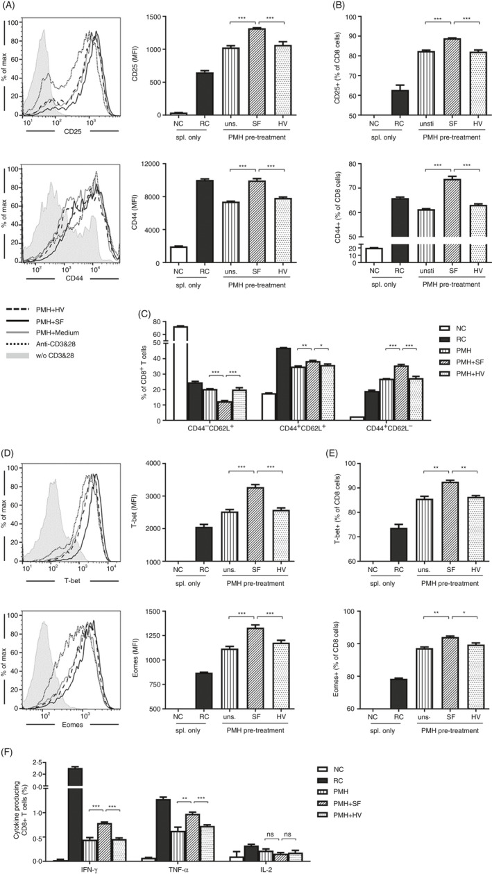
FIGURE 1.

Stimulation of PMHs by SF improved the activation and function of CD8+ T cells. PMHs from WT C57BL/6 mice were pretreated with SF or HV protein (2.5 µg/ml) for 24 h and then co‐cultured with TLR5−/− splenocytes in the presence of anti‐CD3 and anti‐CD28 for an additional 24 h. (A, B) Expression of the activation markers CD25 and CD44 on CD8+ T cells. Grey shadow, without (w/o) CD3 and CD28; dark dotted line, anti‐CD3 and anti‐CD28; grey solid line, PMH + medium; dark solid line, PMH + SF; dark dashed line, PMH + HV. (C) Differentiation of CD8+ T cells was characterized as naïve cells (CD62L+CD44−), effector cells (CD62L−CD44+) and memory cells (CD62L+CD44+). (D, E) Expression of the transcription factors T‐bet and Eomes and (F) production of IFN‐γ, TNF‐α and IL‐2 in CD8+ T cells were analysed by flow cytometry. Data are representative of at least three independent experiments. Bars: mean ± SD. *: p < 0.05; **: p < 0.01; ***: p < 0.001, statistical relevance was determined by one‐way ANOVA.
