Figure 5.
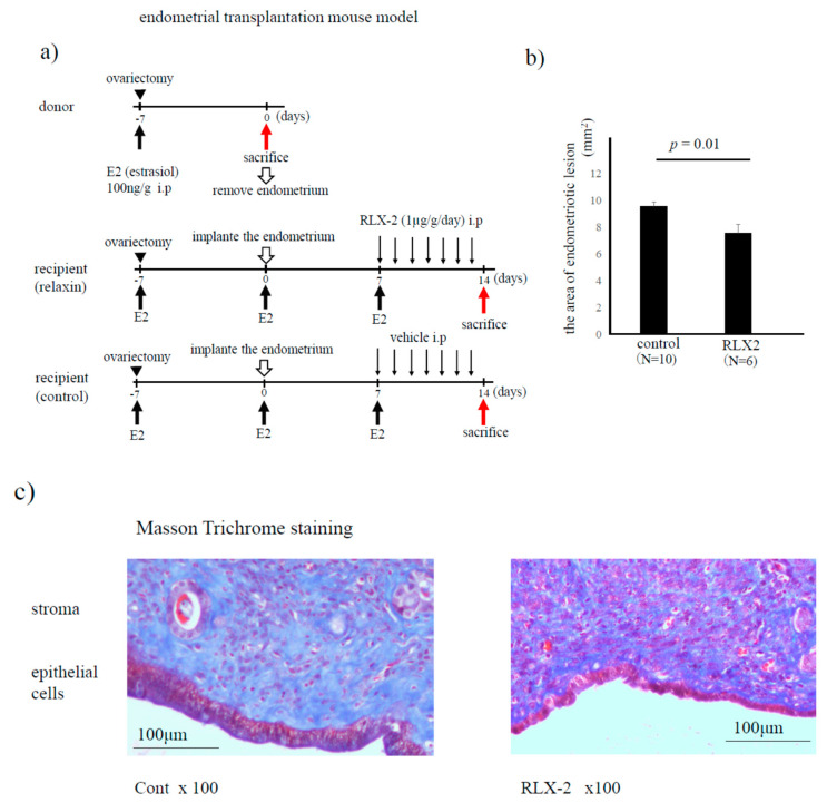
Endometrial transplantation mouse model. Both donor and recipient mice were treated with estradiol (E2) once a week from the day of ovariectomy. Seven days after the ovariectomy, endometrial fragments from donor mice were transplanted into the peritoneum of recipient mice. Seven days after the transplantation, we sacrificed a few mice to confirm the formation of endometriotic lesions. Then, the recipient mice were treated with RLX-2 (1 µg/g/day) or 0.1% BSA/PBS (control) every day. Fourteen days after transplantation, we sacrificed recipient mice, and tissue samples were used following experiments (a). The area of collected endometriotic-like lesions (control group N = 10, and RLX-2 group N = 6) was compared (b), and the extent of fibrosis (stained with blue color) was evaluated by the Masson trichrome staining method in control and RLX-2 groups (c).

