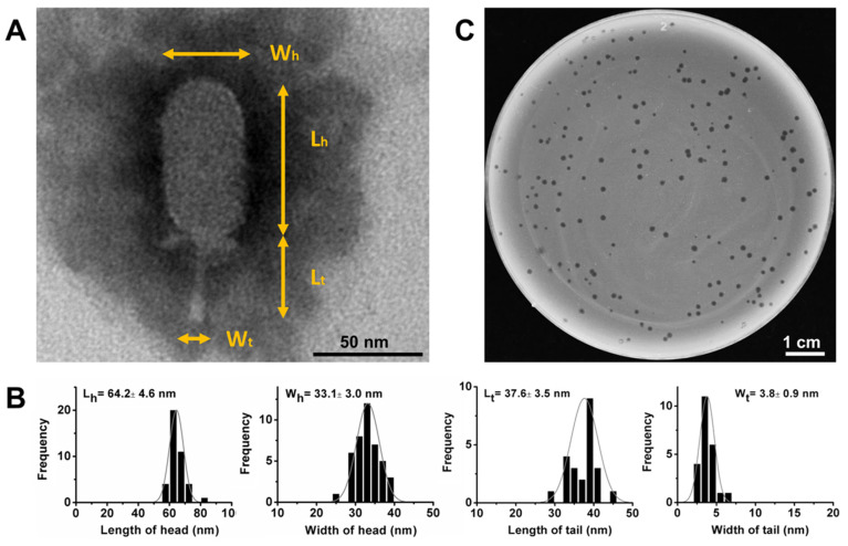
Figure 1.
The morphology and size of phage DLc1 and phage plaques. (A) TEM of DLc1, indicating each measured part of virions (Wh, the width of the head; Lh, the length of the head; Wt, the width of the tail; Lt, the length of the tail); (B) Average size and statistical histogram of each part measured in at least 20 individual virions; (C) Phage plaques formed on a double-layer agar plate (0.4% agar in top layer) using B. cereus 1582-3B as the host.

