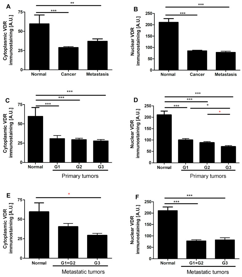
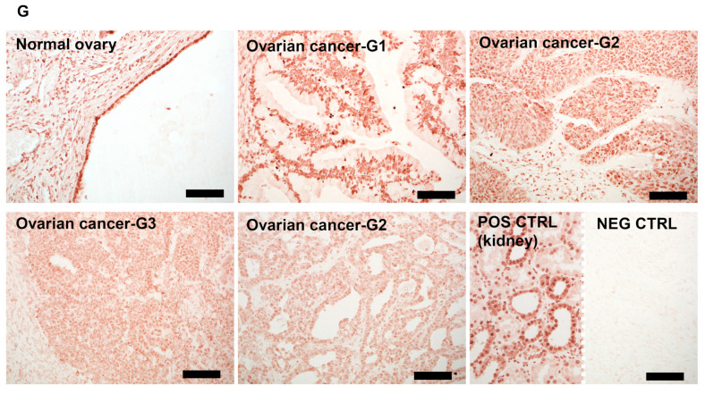
Figure 4.
Cytoplasmic and nuclear vitamin D receptor (VDR) immunostaining in normal ovary and ovarian tumors (n = 20) (A,B), in relation to tumors grade in primary tumors (n = 57) (C,D) and in metastatic tumors (n = 13) (E,F). Statistically significant differences are denoted with asterisks as determined by ANOVA (black asterisks) or Student’s t-test (red asterisks, * p < 0.05, ** p < 0.01, *** p < 0.001). (G) Representative VDR immunostaining in normal epithelia, ovarian cancers, and positive (POS CTRL) and negative (NEG CTRL) controls. Scale bars = 50 µm.


