Abstract
Background
Retinoblastoma (RB) is a common malignancy in children eyes. Aberrant microRNA (miR) expression is observed in many cancer cases. miR-515-5p is reported to be concerned with the course of many cancers. This study explores the role of miR-515-5p in proliferation and drug sensitivity of RB cells.
Methods
Human RB cell lines (WERI-RB1, SO-RB50 and Y79) and human retinal pigment epithelial cell line ARPE-19 were utilized in this study. Drug-resistant cells SO-RB50/VCR and SO-RB50/CBP were constructed for the following experiments. The expressions of miR-515-5p and Notch1 in RB cells were detected. Notch1 was significantly upregulated in RB cells while miR-515-5p was notably downregulated. Then, the binding relationship between miR-515-5p and Notch1 was predicted and verified.
Results
miR-515-5p negatively regulated Notch1 expression. In vitro and in vivo experiments revealed that overexpressed miR-515-5p inhibited RB cell proliferation and enhanced drug sensitivity. Functional rescue experiment suggested that miR-515-5p regulated RB cell proliferation and drug sensitivity via inhibiting Notch1 expression.
Conclusion
It could be concluded that overexpressed miR-515-5p suppressed proliferation and drug resistance of RB cells by targeting Notch1 expression, indicating that miR-515-5p might constitute a promising therapy target for RB.
Keywords: retinoblastoma, microRNA-515-5p, drug sensitivity, Notch1, proliferation
Introduction
Retinoblastoma (RB) belongs to a kind of malignant tumor caused by immature cells in the retina of one or both eyes, which usually occurs in children under 5 years old.1 RB is a highly malignant ocular tumor with a low survival rate and a high metastasis rate.2 The clinical symptoms of RB are marked by pupil abnormality, vision decline, leukocoria, red and irritated eyes, growth retardation or retardation.3 The knowledge of chemotherapy pharmacokinetics may be helpful to guide the safety and efficacy of RB treatment.4 Currently, the major treatments for RB include enucleation, radiotherapy, chemotherapy and focal therapies.5 Delivery of chemotherapeutic drugs to ophthalmic artery for the therapy of RB has greatly improved the efficacy of this fatal but curable ophthalmic cancer.6 However, the clinical application of RB first-line drugs is severely limited because of acquired drug resistance in long-term treatment.7 Multidrug resistance is a primary obstacle limiting the overall survival of patients, particularly for children, since solid tumors account for approximately 30% of all childhood cancers.8 Cellular drug resistance represents a multi-causes phenomenon, concerning many biological molecules and interrelated pathways.9 In short, RB still poses a considerable threat to children’ health and quality of life. Therefore, further elucidating the mechanism of drug resistance of RB cells remains the urgent issue to be solved in oncology to improve the therapy response of RB patients.
Notch1 plays a vital part in cell proliferation, differentiation and apoptosis, and Notch1 dysregulation can bring about tumorigenesis.10 Chen et al show that colorectal cancer cell growth and aggressiveness are promoted by activation of Notch1.11 More importantly, it is reported that inhibition of Notch1 can suppress the malignant development of RB.12 Emerging evidences has demonstrated that the effect of Notch1 on human diseases is realized by cooperating with microRNAs (miRs).13–15 For example, miR‑34a inhibits the proliferation and promotes the chemosensitivity of RB cells by downregulating Notch1 expression.16 miRs are endogenous non-coding RNAs that regulate protein-coding genes post-transcriptionally.17 Much information has obtained about the key role of miRs in tumor development and drug resistance.18 For instance, Wan et al reveal that miR-25-3p promotes malignant transformation of RB cells by inhibiting PTEN.19 Studies have reported the anti-cancer effect of miR-515-5p.20,21 miR-515 expression is decreased significantly in RB cells and miR-515 overexpression inhibits the activity of RB cells.22 Based on the previous findings, we investigated the role of miR-515-5p and Notch1 in the malignant transformation and drug resistance of RB cells, which shall shed lights on the targeted gene therapy for RB.
Materials and Methods
Cell Line and Culture23,24
Human RB cell line (WERI-RB1, SO-Rb50 and Y79) and human normal retinal pigment epithelium vascular endothelial cell line ACBRI-181 were obtained from American Type Culture Collection (Manassas, Virginia, USA). Cells were cultured in Roswell Park Memorial Institute-1640 medium (HyClone, Logan, UT, USA) added with 10% heat-inactivated fetal bovine serum (FBS) (Life Technologies, Gaithersburg, MD, USA), 100 U/mL penicillin and 100 μg/mL streptomycin (Beyotime Biotechnology Co. Ltd., Shanghai, China) in humidified air at 37°C with 5% CO2. SO-RB50 cells were exposed to increased doses of vincristine (VCR) (Solarbio, Beijing, China) and carboplatin (CBP) (Sigma-Aldrich, Merck KGaA, Darmstadt, Germany), respectively, to generate anti-VCR SO-RB50 (SO-RB50/VCR) and anti-CBP SO-RB50 (SO-RB50/CBP). SO-RB50/VCR and SO-RB50/CBP cell culture media were added with 200 ng/mL VCR and 1.0 mg/mL to maintain drug resistance.
Cell Transfection23,25
miR-515-5p mimic and miR negative control (miR-NC) were synthesized by GenePharma (Shanghai, China). Notch1 was cloned into pcDNA3.1 vector (Invitrogen, Carlsbad, CA, USA; Thermo Fisher Scientific Inc., Waltham, MA, USA) to generate overexpressed vector of pcDNA-Notch 1. The wild type (WT) vectors (Notch1-WT) and the mutant type (MUT) vectors (Notch1-MUT) containing miR-515-5p binding site were constructed and then cloned to the downstream of pmirGLO promoter vector (Promega, Madison, WI, USA). The cells in logarithmic growth phase were seeded into 6-well plates at the density of 2 × 10⁵ cells/well. The culture medium was free of antibiotics and cultured in a cell incubator with 5% CO2 and 37°C. When cell confluence reached 70%-80%, RNA oligonucleotides were transfected into cells using the Lipofectamine 2000™ (Invitrogen). The subsequent experiments were performed after 48 hours of transfection.
Dual-Luciferase Reporter Gene Assay23
The targeting relationship between miR-515-5p and Notch1 was predicted by Targetscan (http://www.targetscan.org/vert71/) and miRNAmap (http://mirnamap.mbc.nctu.edu.tw/). The Notch1 fragment containing miR-515-5p binding site was cloned to the pmirGLO vector (Promega). Subsequently, the Notch1-WT vectors and the Notch1-MUT vectors were co-transfected with miR-515-5p mimic or miR-NC into HEK-293T cells using the Lipofectamine 2000™ (Invitrogen). Luciferase activity was detected after 48 hours of transfection by dual-luciferase reporter gene assay system (Promega), and the relative activity was calculated as the ratio of luciferase activity of firefly to that of renilla. The experiment was conducted 3 times.
Reverse Transcription Quantitative Polymerase Chain Reaction (RT-qPCR)26,27
Total RNA of cells was extracted using the TRIzol reagent (15596026, Invitrogen). Next, miR and mRNA were reversely transcribed into cDNA using the TaqMan™ microRNA reverse transcription kit (Applied Biosystems, Foster City, CA, USA) and the PrimeScript-RT reagent kit (Takara, Dalian, China). The qPCR was performed using ABI 7500 quantitative PCR instrument (Applied Biosystems) on the instructions of SYBR® Premix Ex TapTM Ⅱ (Perfect Real Time) kit (DRR081, Takara, Japan). The reaction system included 10 μL SYBR® Premix Ex TaqTM II, 0.8 μL forward primers, 0.8 μL reverse primers, 0.4 μL ROX Reference Dye II, 2 μL cDNA and 6 μL ddH2O. PCR was performed on the following conditions: pre-denaturation at 95°C for 10 minutes; 40 cycles of denaturing at 95°C for 15 seconds and annealing at 60°C for 1 minute. Three duplicated wells were set in each sample and the Ct value of each well was recorded. The relative expression of miR and mRNA was calculated by 2−ΔΔCt method, with U6 and glyceraldehyde-3-phosphate dehydrogenase (GAPDH) acting as the internal reference. Primers (Table 1) were synthesized by Sangon Biotech Co., Ltd, (Shanghai, China).
Table 1.
Primer Sequence for RT-qPCR
| Gene | Sequence |
|---|---|
| miR-515-5p | F: 5ʹ-CGGGTTCTCCAAAAGAAAGCA-3’ |
| R: 5ʹ-CAGCCACAAAAGAGCACAAT-3’ | |
| U6 | F: 5ʹ-CTCGCTTCGGCAGCACA-3’ |
| R: 5ʹ-AACGCTTCACGAATTTGCGT-3’ | |
| Notch1 | F: 5ʹ-AGGCATCATGCATGTCAAAC-3’ |
| R: 5ʹ-TGTGTTGCTGGAGCATCTTC-3’ | |
| β-actin | F: 5ʹ-CTTAGTTGCGTTACACCCTTTCTTG-3’ |
| R: 5ʹ-CTGTCACCTTCACCGTTCCAGTTT-3’ |
Western Blot27
At 48 h post transfection, the cells were seeded into a 6-well plate and treated under the specified conditions. Then, cells were lysed in the radio-immunoprecipitation assay buffer (P0013B, Beyotime). Equal amount of protein was separated on sodium dodecyl sulfate-polyacrylamide gel electrophoresis and transferred onto polyvinylidene difluoride membranes (Bio-Rad Laboratories, Hercules, CA, USA). The membranes were blocked with 5% skim milk, washed by tris buffered saline tween and incubated with primary antibodies at 4°C overnight: Notch1 (#3608, 1:1000, Cell Signaling Technology, Beverly, MA, USA) and β-actin (#4970, 1:1000, Cell Signaling Technology). Afterwards, the membranes were cultured with secondary antibody horseradish peroxidase (HRP)-conjugated goat anti-rabbit immunoglobulin G (IgG) H&L (1:2000, ab205718, Abcam) for 1 hour. Subsequently, the membranes were developed and visualized using the enhanced chemiluminescence reagent (Thermo Scientific Pierce, Rockford, IL, USA). The protein band was observed with β-actin acting as the internal reference.
In vitro Chemosensitivity Assay23,24
At 48 h post transfection, the cells were seeded into a 96-well plate (5×10³ cell/well) and then supplemented with different concentrations of VCR (200 ng/mL, Solarbio), CBP (1.0 mg/mL, Sigma-Aldrich), etoposide (500 ng/mL, VP-16, Sigma-Aldrich), cisplatin (1.0 mg/mL, DDP, Sigma-Aldrich) and 5-fluorouracil (10.0 μg/mL, 5-FU, Sigma-Aldrich). Cell counting kit-8 (CCK-8) assay was employed to test cell viability after 48 hours. The half maximal inhibitory concentration (IC50) of each drug was evaluated and the drug resistance index was calculated: drug resistance index = IC50 (RB cell/drug-resistant cell)/IC50 (RB cell/parent cell).
CCK-8 Assay24,28
At 48 h post transfection, the cells were seeded into a 96-well plate (5×10³ cells/well) and then cultured overnight. Then, the cells were cultured with 10 μL CCK-8 solution (C0039, Beyotime) for 2 hours at 37°C. The optical density (OD) at a wavelength of 450 nm of each well was evaluated using the microplate reader (BioTek Instruments Inc., Winooski, VT, USA). Cell survival/proliferation was evaluated by subtracting the absorbance of the blank well from the OD of the test well.
Colony Formation Assay24,28
At 48 h post transfection, cells were seeded into a 6-well plate at 1×10³ cells/well at 37°C for 2 weeks. The supernatant was discarded and washed with PBS for 3 times. Then, the cells were fixed with methanol for 15 min and stained with 0.1% crystal violet solution (1 mg/mL, Sigma-Aldrich) for 20 min. The residual dyes were washed by PBS and dried naturally. The cell colonies were counted under the inverted microscope (Shanghai Optical Instrument Co., Ltd, Shanghai, China). The experiment was independently performed three times.
Xenograft Tumor in Nude Mice23,29
Human miR-515-5p precursor (GenePharma) was synthesized and cloned into pSuper-green fluorescent protein (GFP)-Luciferase (Luc) (pSuper-GFP-Luc). SO-RB50 cell line stably transfected with pSuper-GFP-Luc (pSuper) or pSuper-GFP-Luc-miR-515-5p (pSuper-miR-515-5p) was constructed by G418 (Sigma-Aldrich) selection.23 Then, 5 × 10⁶ cells were subcutaneously seeded into 5-week-old female BALB/C nude mice (Chinese Academy of Sciences, Shanghai, China). The nude mice were assigned into 7 groups (6 mice in each group): Control group (injected with SO-RB50 cells), pSuper group (injected with SO-RB50 cells transfected with pSuper-GFP-Luc), pSuper-miR-515-5p group (injected with SO-RB50 cells transfected with pSuper-GFP-Luc-miR-515-5p), pSuper+VCR group (injected with SO-RB50 cells transfected with pSuper-GFP-Luc, and treated with VCR), pSuper-miR-515-5p+VCR group (injected with SO-RB50 cells transfected with pSuper-GFP-Luc-miR-515-5p, and treated with VCR), pSuper+CBP group (injected with SO-RB50 cells transfected with pSuper-GFP-Luc, and treated with CBP), and pSuper-miR-515-5p+CBP group (injected with SO-RB50 cells transfected with pSuper-GFP-Luc-miR-515-5p, and treated with CBP). One week after injection, VCR (1.0 mg/kg) or CBP (50 mg/kg) was injected intraperitoneally into each mouse twice a week. Four weeks later, the nude mice were euthanized by an intraperitoneal injection of excessive pentobarbital sodium. Thereafter, the tumors were dissected and weighed for immunohistochemistry.
Immunohistochemistry
The tumor specimens were fixed in 4% paraformaldehyde solution and embedded in paraffin and sliced. The sections were routinely dewaxed and washed twice with PBS for 2 min each time. The citrate (pH=6.0) and high pressure steam were used to repair the antigen. The sections were incubated in 3% H2O2 for 10–15 rain to reduce the specific background staining caused by endogenous peroxidase, following three PBS washes for 2 min each time. Ki67 (ab16667, 1:200, Abcam) and Notch1 (ab52627, 1:150, Abcam) were added to the sections, incubated at 37°C for 3 h, and washed 5 times with PBS, 2 min each time. Next, HRP-labeled goat anti-rabbit IgG secondary antibody (ab224426, 1:50, Abcam) was added into the sections and incubated at 37°C for 10–15 min, and then washed 5 times with PBS, 2 min each time. Sections were stained with 2,4-diaminobutyric acid for 3–5 min, rinsed with tap water for 3 min, washed with distilled water once, counterstained with hematoxylin, washed with running water, dehydrated, cleared and sealed. In the negative control group, PBS was used instead of the primary antibody. Two independent pathologists read the film blindly. Ki67 and Notch1 staining scores were calculated as the product of staining intensity and percentage, and the staining intensity was divided into four grades (no staining 0, weak staining 1, moderate staining 2, strong staining 3). The percentage of positive cells was divided into four grades: 0 (0%), 1 (1%-25%), 2 (26%-50%), 3 (51%-75%) and 4 (76%-100%).30
Statistical Analysis
Data analysis was introduced using the SPSS 21.0 (IBM Corp., Armonk, NY, USA). Data are expressed as mean ± standard deviation. The t test was adopted for analysis of comparisons between two groups. One-way analysis of variance (ANOVA) or two-way ANOVA was employed for the comparisons among multiple groups, and Tukey’s multiple comparison test was applied for the post hoc test after ANOVA. The p value was obtained from a two-tailed test, and p < 0.05 meant statistically significant.
Results
Notch1 Was Upregulated in RB Cells and miR-515-5p Inhibited Notch 1 Expression
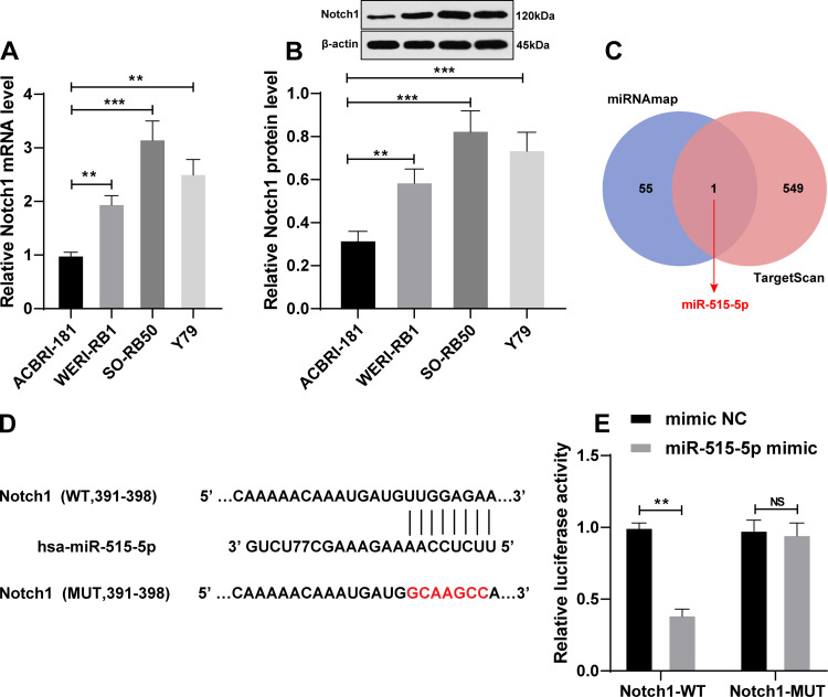
Notch1 was reported to involve in the malignant phenotype of RB12,31 and chemosensitivity of multiple tumors.32–36 The results of RT-qPCR and Western blot showed that Notch1 was significantly upregulated in RB cells (p < 0.05) (Figure 1A and B). However, the upstream miR regulating Notch1 expression remained unclear. We predicted the 56 upstream miRs of Notch1 using miRNAmap and 550 miRs using TargetScan. Venn map was drawn on the online website (http://bioinformatics.psb.ugent.be/webtools/Venn/). After taking the intersection, it was found that only miR-515-5p targeted Notch1 (Figure 1C). The target binding site of miR-515-5p and Notch1 was shown in Figure 1D. The dual-luciferase reporter gene assay further showed that miR-515-5p mimic significantly reduced the luciferase activity of the Notch1-WT group (p < 0.05), while the luciferase activity of the Notch1-MUT group did not change (Figure 1E). It was suggested that there might be a direct interaction between miR-515-5p and Notch1 in RB.
Figure 1.
miR-515-5p was the upstream miR of Notch1. (A) Notch1 mRNA expression in RB cells was detected using RT-qPCR; (B) Notch1 protein level in RB cells was detected using Western blot; (C) Venn diagram of the upstream miRs of Notch1 predicted by miRNAmap and TargetScan; (D) The binding site of miR-515-5p and Notch1 3ʹUTR was predicted by TargetScan; (E) The target binding relationship between miR-515-5p and Notch1 was verified using dual-luciferase reporter gene assay. Each experiment was repeated for three times independently. Data are presented as mean ± standard deviation. The t test was adopted for analysis of comparisons between two groups. One-way ANOVA was employed for the comparisons among multiple groups and Tukey’s multiple comparison test was applied for the post hoc test, **p < 0.01, ***p < 0.001.
Abbreviations: NS, no significant; NC, negative control; RB, retinoblastoma; RT-qPCR, reverse transcription quantitative polymerase chain reaction; ANOVA, analysis of variance.
miR-515-5p Was Downregulated in RB Cells and Overexpression of miR-515-5p Inhibited RB Cell Proliferation
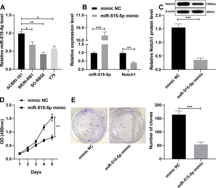
miR-515-5p expression was downregulated in RB cell lines, especially in SO-RB50 cells (all p < 0.05) (Figure 2A). To determine the function of miR-515-5p in RB, we transfected miR-515-5p mimic into SO-RB50 cells. After overexpression of miR-515-5p, miR-515-5p expression in SO-RB50 cells was significantly upregulated, and Notch1 expression was downregulated (all p < 0.05) (Figure 2B and C). The results of CCK-8 assay (Figure 2D) and colony formation assay (Figure 2E) revealed that the proliferation ability of SO-RB50 cells in the miR-515-5p mimic group was dramatically lower than that in the mimic NC group (p < 0.05). These results indicated that overexpressed miR-515-5p notably inhibited RB cell proliferation.
Figure 2.
miR-515-5p inhibited SO-RB50 cell proliferation. (A) Expression of miR-515-5p in RB cells was detected using RT-qPCR; (B) Expressions of miR-515-5p and Notch1 mRNA in SO-RB50 cells after overexpression of miR-515-5p were detected using RT-qPCR; (C) Notch1 protein level in SO-RB50 cells after overexpression of miR-515-5p was detected using Western blot; (D and E) The proliferation ability of SO-RB50 cells after overexpression of miR-515-5p was detected using CCK-8 assay and colony formation assay. Each experiment was repeated for three times independently. Data are presented as mean ± standard deviation. The t test was adopted for analysis of comparisons between two groups. One-way ANOVA was employed for the comparisons among multiple groups and Tukey’s multiple comparison test was applied for the post hoc test, *p < 0.05, **p < 0.01, ***p < 0.001.
Abbreviations: NC, negative control; RB, retinoblastoma; OD, optical density; RT-qPCR, reverse transcription quantitative polymerase chain reaction; ANOVA, analysis of variance.
miR-515-5p Enhanced Drug Sensitivity in vivo
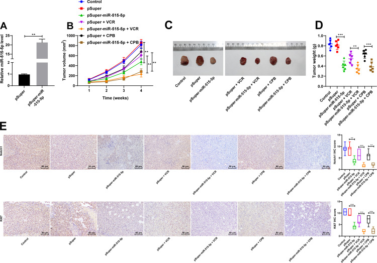
To determine the function of miR-515-5p in drug sensitivity in vivo, we injected SO-RB50 cells with stable and miR-515-5p (pSuper-miR-515-5p) into mice subcutaneously, followed by VCR or CBP treatment. RT-PCR confirmed that miR-515-5p was upregulated by 21.5 folds (p < 0.05) in cells stably expressing miR-515-5p (Figure 3A). Under the VCR and CBP treatment, the tumor growth rate of mice in the pSuper group was notably faster than that in the pSuper-miR-515-5p group (p < 0.05) (Figure 3B). Four weeks later, the tumors of nude mice after injection of SO-RB50 cells stably transfected with overexpressing miR-515-5p were shown in Figure 3C. The average weight of tumors of mice in the pSuper-miR-515-5p group was dramatically lower than that in the pSuper group (all p < 0.05) (Figure 3D). Notch l was positively expressed in the cytoplasm and nucleus, and Ki67 was located in the nucleus. The immunohistochemical scores of Notch L and Ki67 in pSuper-miR-515-5p group were significantly lower than those in pSuper group. After VCR and CBP treatment, the effect of pSuper-miR-515-5p on Notch1 and Ki67 was enhanced (Figure 3E). These results indicated that overexpression of miR-515-5p significantly inhibited the growth of RB and enhanced multidrug resistance in vivo.
Figure 3.
miR-515-5p enhanced drug sensitivity in vivo. (A) Relative expression of miR-515-5p in SO-RB50 cells stably expressing miR-515-5p was detected using RT-qPCR; (B) Effect of VCR or CBP on tumor volume in nude mice; (C) A representative image of tumors in nude mice 4 weeks after injection of SO-RB50 cells stably overexpressing miR-515-5p; (D) Effect of VCR or CBP on tumor weight in nude mice; (E) Immunohistochemistry was used to detect the expression of Notch1 and Ki67 in tumor tissues of nude mice. N = 6. Data are presented as mean ± standard deviation. The t test was adopted for analysis of comparisons between two groups. One-way ANOVA was employed for the comparisons among multiple groups and Tukey’s multiple comparison test was applied for the post hoc test, **p < 0.01, ***p < 0.001.
Abbreviations: VCR, vincristine; CBP, carboplatin; RT-qPCR, reverse transcription quantitative polymerase chain reaction.
miR-515-5p Enhanced Drug Sensitivity in vitro
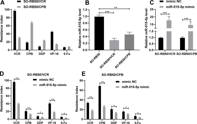
To define the function of miR-515-5p in chemotherapeutic drug sensitivity of RB cells, we exposed SO-RB50 cells to increased doses of VCR or CBP to construct two drug-resistant cell lines, SO-RB50/VCR and SO-RB50/CBP (Figure 4A, attached Figure 1). miR-515-5p expression in SO-RB50/VCR cells and SO-RB50/CBP cells was 70% and 53% lower than that in SO-RB50 cells, respectively (Figure 4B). Then, the drug sensitivity was evaluated by transfection of miR-515-5p mimic into SO-RB50/VCR and SO-RB50/CBP cells. RT-qPCR verified that miR-515-5p expression was significantly promoted (both p < 0.05) (Figure 4C). The drug resistance index of the miR-515-5p mimic group was notably lower than that of the mimic NC group (all p < 0.05), which indicated that the drug sensitivity of the miR-515-5p mimic group increased (Figure 4D and E, attached Table 1). Overexpressed miR-515-5p significantly enhanced the drug sensitivity of RB cells.
Figure 4.
miR-515-5p enhanced drug sensitivity in vitro. (A) The drug resistance index of VCR, CBP, DDP, VP-16 and 5-Fu in SO-RB50/VCR and SO-RB50/CBP cells; (B) Expression of miR-515-5p in SO-RB50/VCR, SO-RB50/CBP and SO-RB50 cells was detected using RT-qPCR; (C) Expression of miR-515-5p in SO-RB50/VCR and SO-RB50/CBP cells after overexpression of miR-515-5p was detected using RT-qPCR; (D and E) SO-RB50 cell proliferation after overexpression of miR-515-5p was detected using CCK-8 assay and colony formation assay. Each experiment was repeated for three times independently. Data are presented as mean ± standard deviation. The t test was adopted for analysis of comparisons between two groups. One-way ANOVA was employed for the comparisons among multiple groups and Tukey’s multiple comparison test was applied for the post hoc test, *p < 0.05, **p < 0.01, ***p < 0.001.
Abbreviations: VCR, vincristine; CBP, carboplatin; DDP, cisplatin; VP-16, etoposide; 5-Fu, 5-fluorouracil; NC, negative control; RT-qPCR, reverse transcription quantitative polymerase chain reaction; ANOVA, analysis of variance.
miR-515-5p Inhibited RB Cell Proliferation via Downregulating Notch1
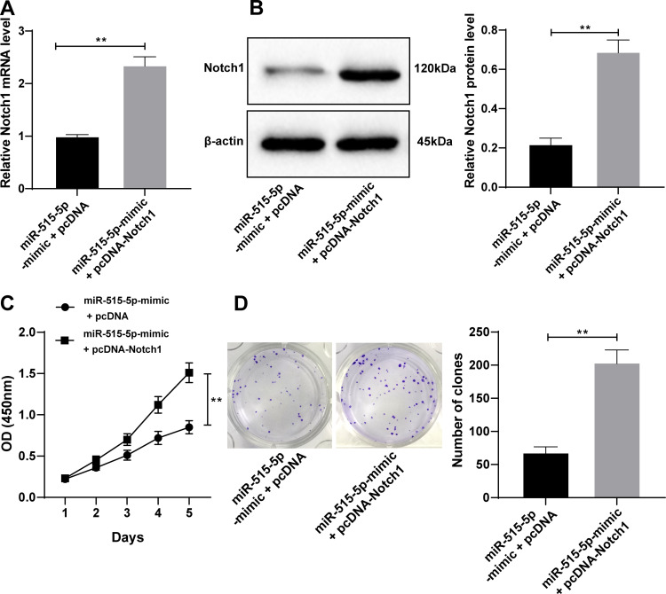
The pcDNA-Notch1 with miR-515-5p mimic or miR-NC into SO-RB50 cells to verify the role of Notch1 in RB cell proliferation. Notch1 expression in miR-515-5p mimic + pcDNA-Notch1 cells was significantly higher than that in miR-515-5p mimic + pcDNA cells (all p < 0.05) (Figure 5A and B). Overexpressed Notch1 attenuated the inhibitory effect of miR-515-5p on SO-RB50 cell proliferation (all p < 0.05) (Figure 5C and D).
Figure 5.
miR-515-5p inhibited RB cell proliferation via downregulating Notch1. (A) Expression of Notch1 mRNA in SO-RB50 cells of each transfection group was detected using RT-qPCR; (B) Notch1 protein level in SO-RB50 cells of each transfection group was detected using Western blot; (C and D) SO-RB50 cell proliferation of each transfection group was detected using CCK-8 assay and colony formation assay. Each experiment was repeated for three times independently. Data are presented as mean ± standard deviation. The t test was adopted for analysis of comparisons between two groups, **p < 0.01.
Abbreviations: RB, retinoblastoma; OD, optical density; RT-qPCR, reverse transcription quantitative polymerase chain reaction; ANOVA, analysis of variance.
miR-515-5p Promoted Drug Sensitivity of RB Cells via Downregulating Notch1
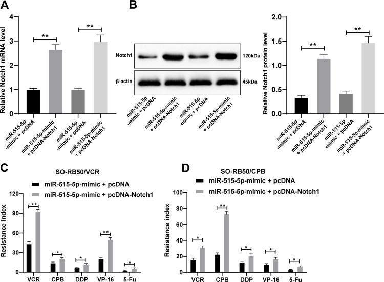
SO-RB50/VCR and SO-RB50/CBP cells were cotransfected with pcDNA-Notch1 and miR-515-5p mimic or miR-NC. Notch1 expression was significantly increased in cells transfected with pcDNA-Notch1 (all p < 0.05) (Figure 6A and B). In vitro experiment further showed that overexpressed Notch1 weakened the promoting effect of miR-515-5p on drug sensitivity (all p < 0.05) (Figure 6C and D, attached Table 1).
Figure 6.
miR-515-5p promoted drug sensitivity of RB cells via downregulating Notch1. (A) Expression of Notch1 mRNA in SO-RB50/VCR and SO-RB50/CBP cells of each transfection group was detected using RT-qPCR; (B) Notch1 protein level in SO-RB50/VCR and SO-RB50/CBP cells of each transfection group was detected using Western blot; (C and D) The drug resistance index of VCR, CBP, DDP, VP-16 and 5-Fu in SO-RB50/VCR and SO-RB50/CBP cells. Each experiment was repeated for three times independently. Data are presented as mean ± standard deviation. The t test was adopted for analysis of comparisons between two groups, *p < 0.05, **p < 0.01.
Abbreviations: VCR, vincristine; CBP, carboplatin; DDP, cisplatin; VP-16, etoposide; 5-Fu, 5-fluorouracil.
Discussion
RB was a kind of intraocular malignant tumor in children.3 If left untreated, RB might deteriorate into sporadic or hereditary forms, or even be fatal.37 Chemotherapy had been conducted to ameliorate the prognosis of patients with RB to a large extent, but multidrug resistance was still the major obstacle to improving the overall survival of patients.8 Hence, the researches on the improvement of drug sensitivity were in urgency to meliorate the clinical survival and prognosis of RB patients. We focused on Notch1 and specifically explored its role in RB cell proliferation and drug sensitivity. We found that miR-515-5p suppressed RB cell proliferation and enhanced drug sensitivity by targeting Notch1.
It was reported that Notch1 contributed to the malignant development of RB.38 Consistently, we found that Notch1 expression was notably upregulated in RB cells. Emerging evidence had showed that miRs extensively modulated RB pathological process.39 For instance, Jin et al showed that miR-101-3p could inhibit the proliferation of RB cells.40 Zhou et al also demonstrated that miR-338-5p served as an oncogenic role in the RB progression.41 Hence, we then explored the upstream miR of Notch1 in the regulation of proliferation and drug sensitivity of RB cells. Notch1 was predicted to be the target gene of miR-515-5p by miRNAmap and TargetScan. The target binding relationship between miR-515-5p and Notch1 was further verified using dual-luciferase reporter gene assay. miR-515-5p had been manifested to be a tumor suppressor.42 A previous study showed that miR-515-5p had a critical influence on the progression of human cancers, such as prostate cancer, lung cancer and breast cancer.43 Lyv et al had also revealed that miR-515 participated in the progression of RB.22 However, the exact role of miR-515-5p in the course of RB had not been clarified. Our study was the first to reveal that miR-515-5p expression was downregulated in RB cell line and overexpressed miR-515-5p could suppress the proliferation of RB cells. We might unveil an inhibitory role of miR-515-5p in RB progression.
Then, we constructed two drug-resistant cell lines SO-RB50/VCR and SO-RB50/CBP to determine the role of miR-515-5p mimic in drug sensitivity of RB cells. In vivo experiments, SO-RB50 cells stably transfected with pSuper GFP Luc (pSuper) or pSuper-GFP-Luc-miR-515-5p (pSuper-miR-515-5p) were subcutaneously injected into 5-week-old female BALB/c nude mice. One week later, VCR (1.0 mg/kg) or CBP (50 mg/kg) was injected intraperitoneally. Both in vitro and in vivo experiments showed that the enhanced expression of miR-515-5p could improve the drug sensitivity of RB cells by targeting Notch. Intriguingly, previous literature had also implied that miR-515-5p was involved in the bladder cancer cell proliferation and chemo-resistance.44 miR-515-3p was reported to promote the chemosensitivity of mucinous ovarian cancer cell to oxaliplatin.45 Furthermore, Notch1 was reported to exert an effect on chemo-resistance and poor outcome.32 Bai et al showed that inhibition of Notch1 could promote drug sensitivity of ovarian cancer cells.35 Suppression of Notch1 inhibited cell growth and improved drug sensitivity of breast cancer.33 The combined effect of miR and Notch1 in RB had been published, but the interaction between miR-515-5p and Notch1 in RB was studied for the first time. Thereafter, functional rescue experiments were conducted, which indicated that overexpressing Notch1 attenuated the inhibitory effect of miR-515-5p on RB cell proliferation and the promotion of drug sensitivity. Li et al also reported that restoration of Notch1 expression could partially reverse the inhibitory effect of overexpressing miR-433 on RB cell proliferation.12
Conclusion
To summarize, our study demonstrated that overexpressing miR-515-5p reduced proliferation and drug resistance of RB cells via inhibiting Notch1 expression. The research on the drug resistance of RB cells was of great significance to adjust the clinical practice of chemotherapy. This study might hint the possibility of miR-515-5p as a potential target for RB patients with chemoresistance. Considering the limitation of our experimental technology and funding, we constructed a subcutaneous retinoblastoma model.23,29,46,47 The microenvironment and its immune response greatly affect proliferation and survival of tumor. Subcutaneous environment vary greatly with intraocular space and retina tissue. When the following technology is mature and the conditions are feasible, we will establish a xenograft tumor model of vitreous RB in nude mice to verify the effect of miR-515-5p on RB in vivo. In the future, we shall conduct more researches on the feasibility and safety of miR-515-5p in the treatment of RB, with the aim to transform miR-515-5p from a gene tool into a clinical means.
Ethics Statement
The study got the approval of the Clinical Ethical Committee of The Affiliated Hospital of Qingdao University (SYXK-2019-0010). All experimental procedures were implemented on the Ethical Guidelines for the Study of Experimental Pain in Conscious Animals.
Disclosure
All authors declare that they have no conflicts of interest for this work.
References
- 1.Lohmann D. Retinoblastoma. Adv Exp Med Biol. 2010;685:220–227. doi: 10.1007/978-1-4419-6448-9_21 [DOI] [PubMed] [Google Scholar]
- 2.Zheng Q, Zhu Q, Li C, et al. microRNA-144 functions as a diagnostic and prognostic marker for retinoblastoma. Clinics (Sao Paulo). 2020;75:e1804. doi: 10.6061/clinics/2020/e1804 [DOI] [PMC free article] [PubMed] [Google Scholar]
- 3.Yun J, Li Y, Xu CT, Pan BR. Epidemiology and Rb1 gene of retinoblastoma. Int J Ophthalmol. 2011;4(1):103–109. doi: 10.3980/j.issn.2222-3959.2011.01.24 [DOI] [PMC free article] [PubMed] [Google Scholar]
- 4.Schaiquevich P, Fabius AW, Francis JH, Chantada GL, Abramson DH. Ocular pharmacology of chemotherapy for retinoblastoma. Retina. 2017;37(1):1–10. doi: 10.1097/IAE.0000000000001275 [DOI] [PubMed] [Google Scholar]
- 5.Sun X, Shen H, Liu S, Gao J, Zhang S. Long noncoding RNA SNHG14 promotes the aggressiveness of retinoblastoma by sponging microRNA124 and thereby upregulating STAT3. Int J Mol Med. 2020;45(6):1685–1696. doi: 10.3892/ijmm.2020.4547 [DOI] [PMC free article] [PubMed] [Google Scholar] [Retracted]
- 6.Manjandavida FP, Stathopoulos C, Zhang J, Honavar SG, Shields CL. Intra-arterial chemotherapy in retinoblastoma - a paradigm change. Indian J Ophthalmol. 2019;67(6):740–754. doi: 10.4103/ijo.IJO_866_19 [DOI] [PMC free article] [PubMed] [Google Scholar]
- 7.Zhu X, Xue L, Yao Y, et al. The FoxM1-ABCC4 axis mediates carboplatin resistance in human retinoblastoma Y-79 cells. Acta Biochim Biophys Sin (Shanghai). 2018;50(9):914–920. doi: 10.1093/abbs/gmy080 [DOI] [PubMed] [Google Scholar]
- 8.Fruci D, Cho WC, Nobili V, Locatelli F, Alisi A. Drug transporters and multiple drug resistance in pediatric solid tumors. Curr Drug Metab. 2016;17(4):308–316. doi: 10.2174/1567205010666131212110948 [DOI] [PubMed] [Google Scholar]
- 9.Griguer CE, Oliva CR. Bioenergetics pathways and therapeutic resistance in gliomas: emerging role of mitochondria. Curr Pharm Des. 2011;17(23):2421–2427. doi: 10.2174/138161211797249251 [DOI] [PubMed] [Google Scholar]
- 10.Qi R, An H, Yu Y, et al. Notch1 signaling inhibits growth of human hepatocellular carcinoma through induction of cell cycle arrest and apoptosis. Cancer Res. 2003;63(23):8323–8329. [PubMed] [Google Scholar]
- 11.Chen J, Yuan W, Wu L, et al. PDGF-D promotes cell growth, aggressiveness, angiogenesis and EMT transformation of colorectal cancer by activation of Notch1/Twist1 pathway. Oncotarget. 2017;8(6):9961–9973. doi: 10.18632/oncotarget.14283 [DOI] [PMC free article] [PubMed] [Google Scholar]
- 12.Li X, Yang L, Shuai T, Piao T, Wang R. MiR-433 inhibits retinoblastoma malignancy by suppressing Notch1 and PAX6 expression. Biomed Pharmacother. 2016;82:247–255. doi: 10.1016/j.biopha.2016.05.003 [DOI] [PubMed] [Google Scholar]
- 13.Guan YJ, Li J, Yang X, et al. Evidence that miR-146a attenuates aging- and trauma-induced osteoarthritis by inhibiting Notch1, IL-6, and IL-1 mediated catabolism. Aging Cell. 2018;17(3):e12752. doi: 10.1111/acel.12752 [DOI] [PMC free article] [PubMed] [Google Scholar]
- 14.Li J, Li Q, Lin L, et al. Targeting the Notch1 oncogene by miR-139-5p inhibits glioma metastasis and epithelial-mesenchymal transition (EMT). BMC Neurol. 2018;18(1):133. doi: 10.1186/s12883-018-1139-8 [DOI] [PMC free article] [PubMed] [Google Scholar]
- 15.Xu Z, Huang B, Zhang Q, He X, Wei H, Zhang D. NOTCH1 regulates the proliferation and migration of bladder cancer cells by cooperating with long non-coding RNA HCG18 and microRNA-34c-5p. J Cell Biochem. 2019;120(4):6596–6604. doi: 10.1002/jcb.27954 [DOI] [PubMed] [Google Scholar]
- 16.Yin W, Gao F, Zhang S. MicroRNA34a inhibits the proliferation and promotes the chemosensitivity of retinoblastoma cells by downregulating Notch1 expression. Mol Med Rep. 2020;22(2):1613–1620. doi: 10.3892/mmr.2020.11238 [DOI] [PubMed] [Google Scholar]
- 17.Shioya M, Obayashi S, Tabunoki H, et al. Aberrant microRNA expression in the brains of neurodegenerative diseases: miR-29a decreased in Alzheimer disease brains targets neurone navigator 3. Neuropathol Appl Neurobiol. 2010;36(4):320–330. doi: 10.1111/j.1365-2990.2010.01076.x [DOI] [PubMed] [Google Scholar]
- 18.Hijazi ZM, Suradi H. Intracardiac echocardiography-guided interventions: do we need trials to prove equivalency/superiority to transesophageal echocardiography? JACC Cardiovasc Interv. 2014;7(9):1045–1047. doi: 10.1016/j.jcin.2014.07.001 [DOI] [PubMed] [Google Scholar]
- 19.Wan W, Wan W, Long Y, et al. MiR-25-3p promotes malignant phenotypes of retinoblastoma by regulating PTEN/Akt pathway. Biomed Pharmacother. 2019;118:109111. doi: 10.1016/j.biopha.2019.109111 [DOI] [PubMed] [Google Scholar]
- 20.Guo F, Li S, Guo C, et al. Circular RNA circMAGI3 accelerates the glycolysis of non-small cell lung cancer through miR-515-5p/HDGF. Am J Transl Res. 2020;12(7):3953–3963. [PMC free article] [PubMed] [Google Scholar]
- 21.Ni JS, Zheng H, Ou YL, et al. miR-515-5p suppresses HCC migration and invasion via targeting IL6/JAK/STAT3 pathway. Surg Oncol. 2020;34:113–120. doi: 10.1016/j.suronc.2020.03.003 [DOI] [PubMed] [Google Scholar]
- 22.Lyv X, Wu F, Zhang H, Lu J, Wang L, Ma Y. Long noncoding RNA ZFPM2-AS1 knockdown restrains the development of retinoblastoma by modulating the MicroRNA-515/HOXA1/Wnt/beta-catenin axis. Invest Ophthalmol Vis Sci. 2020;61(6):41. doi: 10.1167/iovs.61.6.41 [DOI] [PMC free article] [PubMed] [Google Scholar]
- 23.Yang L, Zhang L, Lu L, Wang Y. miR-214-3p regulates multi-drug resistance and apoptosis in retinoblastoma cells by targeting ABCB1 and XIAP. Onco Targets Ther. 2020;13:803–811. doi: 10.2147/OTT.S235862 [DOI] [PMC free article] [PubMed] [Google Scholar]
- 24.Yang L, Zhang L, Lu L, Wang Y. lncRNA UCA1 increases proliferation and multidrug resistance of retinoblastoma cells through downregulating miR-513a-5p. DNA Cell Biol. 2020;39(1):69–77. doi: 10.1089/dna.2019.5063 [DOI] [PubMed] [Google Scholar]
- 25.Yang G, Fu Y, Lu X, Wang M, Dong H, Li Q. miR34a regulates the chemosensitivity of retinoblastoma cells via modulation of MAGEA/p53 signaling. Int J Oncol. 2019;54(1):177–187. doi: 10.3892/ijo.2018.4613 [DOI] [PubMed] [Google Scholar]
- 26.Qiao K, Ning S, Wan L, et al. LINC00673 is activated by YY1 and promotes the proliferation of breast cancer cells via the miR-515-5p/MARK4/Hippo signaling pathway. J Exp Clin Cancer Res. 2019;38(1):418. doi: 10.1186/s13046-019-1421-7 [DOI] [PMC free article] [PubMed] [Google Scholar]
- 27.He TG, Xiao ZY, Xing YQ, Yang HJ, Qiu H, Chen JB. Tumor suppressor miR-184 enhances chemosensitivity by directly inhibiting SLC7A5 in retinoblastoma. Front Oncol. 2019;9:1163. doi: 10.3389/fonc.2019.01163 [DOI] [PMC free article] [PubMed] [Google Scholar]
- 28.Tian T, Fu X, Lu J, et al. MicroRNA-760 inhibits doxorubicin resistance in hepatocellular carcinoma through regulating Notch1/Hes1-PTEN/Akt signaling pathway. J Biochem Mol Toxicol. 2018;32(8):e22167. doi: 10.1002/jbt.22167 [DOI] [PubMed] [Google Scholar]
- 29.Park EY, Chang E, Lee EJ, et al. Targeting of miR34a-NOTCH1 axis reduced breast cancer stemness and chemoresistance. Cancer Res. 2014;74(24):7573–7582. doi: 10.1158/0008-5472.CAN-14-1140 [DOI] [PubMed] [Google Scholar]
- 30.Liu H, Wu Z, Zhou H, et al. The SOX4/miR-17-92/RB1 axis promotes prostate cancer progression. Neoplasia. 2019;21(8):765–776. doi: 10.1016/j.neo.2019.05.007 [DOI] [PMC free article] [PubMed] [Google Scholar]
- 31.Gao Y, Luo X, Zhang J. LincRNA-ROR is activated by H3K27 acetylation and induces EMT in retinoblastoma by acting as a sponge of miR-32 to activate the Notch signaling pathway. Cancer Gene Ther. 2020. doi: 10.1038/s41417-020-0181-z [DOI] [PubMed] [Google Scholar]
- 32.Zhang Z, Zhou Z, Zhang M, et al. High Notch1 expression affects chemosensitivity of head and neck squamous cell carcinoma to paclitaxel and cisplatin treatment. Biomed Pharmacother. 2019;118:109306. doi: 10.1016/j.biopha.2019.109306 [DOI] [PubMed] [Google Scholar]
- 33.Zang S, Chen F, Dai J, et al. RNAi-mediated knockdown of Notch-1 leads to cell growth inhibition and enhanced chemosensitivity in human breast cancer. Oncol Rep. 2010;23(4):893–899. doi: 10.3892/or_00000712 [DOI] [PubMed] [Google Scholar]
- 34.Takam Kamga P, Dal Collo G, Midolo M, et al. Inhibition of notch signaling enhances chemosensitivity in B-cell precursor acute lymphoblastic leukemia. Cancer Res. 2019;79(3):639–649. doi: 10.1158/0008-5472.CAN-18-1617 [DOI] [PubMed] [Google Scholar]
- 35.Bai L, Wang A, Zhang Y, Xu X, Zhang X. Knockdown of MALAT1 enhances chemosensitivity of ovarian cancer cells to cisplatin through inhibiting the Notch1 signaling pathway. Exp Cell Res. 2018;366(2):161–171. doi: 10.1016/j.yexcr.2018.03.014 [DOI] [PubMed] [Google Scholar]
- 36.Alshaer W, Alqudah DA, Wehaibi S, et al. Downregulation of STAT3, beta-Catenin, and Notch-1 by single and combinations of siRNA treatment enhance chemosensitivity of wild type and doxorubicin resistant MCF7 breast cancer cells to doxorubicin. Int J Mol Sci. 2019;20:15. doi: 10.3390/ijms20153696 [DOI] [PMC free article] [PubMed] [Google Scholar]
- 37.Zhang J, Benavente CA, McEvoy J, et al. A novel retinoblastoma therapy from genomic and epigenetic analyses. Nature. 2012;481(7381):329–334. doi: 10.1038/nature10733 [DOI] [PMC free article] [PubMed] [Google Scholar]
- 38.Asnaghi L, Tripathy A, Yang Q, et al. Targeting Notch signaling as a novel therapy for retinoblastoma. Oncotarget. 2016;7(43):70028–70044. doi: 10.18632/oncotarget.12142 [DOI] [PMC free article] [PubMed] [Google Scholar]
- 39.Zhang Y, Zhu X, Zhu X, et al. MiR-613 suppresses retinoblastoma cell proliferation, invasion, and tumor formation by targeting E2F5. Tumour Biol. 2017;39(3):1010428317691674. doi: 10.1177/1010428317691674 [DOI] [PubMed] [Google Scholar]
- 40.Jin Q, He W, Chen L, Yang Y, Shi K, You Z. MicroRNA-101-3p inhibits proliferation in retinoblastoma cells by targeting EZH2 and HDAC9. Exp Ther Med. 2018;16(3):1663–1670. doi: 10.3892/etm.2018.6405 [DOI] [PMC free article] [PubMed] [Google Scholar]
- 41.Zhou P, Li X. Serum miR-338-5p has potential for use as a tumor marker for retinoblastoma. Oncol Lett. 2019;18(1):307–313. doi: 10.3892/ol.2019.10331 [DOI] [PMC free article] [PubMed] [Google Scholar] [Retracted]
- 42.Li J, Tang Z, Wang H, et al. CXCL6 promotes non-small cell lung cancer cell survival and metastasis via down-regulation of miR-515-5p. Biomed Pharmacother. 2018;97:1182–1188. doi: 10.1016/j.biopha.2017.11.004 [DOI] [PubMed] [Google Scholar]
- 43.Wang D, Liu K, Chen E. LINC00511 promotes proliferation and invasion by sponging miR-515-5p in gastric cancer. Cell Mol Biol Lett. 2020;25:4. doi: 10.1186/s11658-020-0201-x [DOI] [PMC free article] [PubMed] [Google Scholar]
- 44.Gong P, Xu R, Zhuang Q, He X. A novel circular RNA (hsa_circRNA_102336), a plausible biomarker, promotes the tumorigenesis by sponging miR-515-5p in human bladder cancer. Biomed Pharmacother. 2020;126:110059. doi: 10.1016/j.biopha.2020.110059 [DOI] [PubMed] [Google Scholar]
- 45.Hisamatsu T, McGuire M, Wu SY, et al. PRKRA/PACT expression promotes chemoresistance of mucinous ovarian cancer. Mol Cancer Ther. 2019;18(1):162–172. doi: 10.1158/1535-7163.MCT-17-1050 [DOI] [PMC free article] [PubMed] [Google Scholar]
- 46.Zhong W, Yang J, Li M, Li L, Li A. Long noncoding RNA NEAT1 promotes the growth of human retinoblastoma cells via regulation of miR-204/CXCR4 axis. J Cell Physiol. 2019;234(7):11567–11576. doi: 10.1002/jcp.27812 [DOI] [PubMed] [Google Scholar]
- 47.Kim DY, Choi JA, Koh JY, Yoon YH. Efficacy and safety of aflibercept in in vitro and in vivo models of retinoblastoma. J Exp Clin Cancer Res. 2016;35(1):171. doi: 10.1186/s13046-016-0451-7 [DOI] [PMC free article] [PubMed] [Google Scholar]


