Abstract
Purpose
Dissection after plain balloon angioplasty is required to achieve adequate luminal area; however, it is associated with a high risk of vascular events. This study aimed to examine the relationship between non-flow limiting coronary dissections and subsequent lumen loss and long-term clinical outcomes following successful drug-coated balloon (DCB) treatment of de novo coronary lesions.
Materials and Methods
A total of 227 patients with good distal flow (Thrombolysis in Myocardial Infarction flow grade 3) following DCB treatment were retrospectively enrolled and stratified according to the presence or absence of a non-flow limiting dissection. The primary endpoint was late lumen loss (LLL) at 6-month angiography, and the secondary endpoint was target vessel failure (TVF, a composite of cardiac death, target vessel myocardial infarction, target vessel revascularization, and target vessel thrombosis).
Results
The cohort consisted of 95 patients with and 132 patients without a dissection. There were no between-group differences in LLL (90.8%) returning for angiography at 6 months (0.05±0.19 mm in non-dissection and 0.05±0.30 mm in dissection group, p=0.886) or in TVF (6.8% in non-dissection and 8.4% in dissection group, p=0.799) at a median follow-up of 3.4 years. In a multivariate analysis, the presence of dissection and its severity were not associated with LLL or TVF. Almost dissections (93.9%) were completely healed, and there was no newly developed dissection at 6-month angiography.
Conclusion
The presence of a dissection following successful DCB treatment of a de novo coronary lesion may not be associated with an increased risk of LLL or TVF (Impact of Drug-coated Balloon Treatment in de Novo Coronary Lesion; NCT04619277).
Keywords: Balloon angioplasty, dissection, coronary heart disease, outcome research
INTRODUCTION
Restenosis after balloon angioplasty (BA) is strongly associated with incomplete lesion dilatation in de novo coronary lesions.1,2,3 Oversizing predilation balloons lower the risk of restenosis compared to undersized balloons; however, their use is associated with increased complications, and particularly, a higher risk of coronary dissection.4,5 Reassuringly, previous studies demonstrated that uncomplicated dissections after BA were not associated with higher restenosis rates or adverse outcomes.6,7,8 Disappointingly, the overall restenosis rate in patients who had undergone successful BA using a plain balloon remained approximately 30% through a combination of acute recoil, chronic constrictive remodeling, and neointimal hyperplasia,3,9,10 prompting the introduction of metal stents with local antiproliferative drug delivery. Unfortunately, late stent thrombosis and restenosis, with a hazard rate of nearly 2% per year after drug-eluting stent (DES) implantation, remained a concern11 driving the development of drug-coated balloons (DCB).
The rationale behind DCB technology was a combination therapy of balloon and drug to treat coronary lesions, eliminating stent thrombosis and achieving lower rates of restenosis by leaving no metal behind.12 Furthermore, DCBs have been shown to have comparable efficacy to DES for the treatment of small vessel disease.13 However, concerns over acute vessel closure and restenosis have hampered their use for de novo coronary lesions. In contemporary practice, more advanced technology and medications are available for lowering the risk of vascular events after DCB treatment. Nevertheless, it remains unclear whether untreated dissections after DCB application affect the risk of restenosis or future vascular events. Accordingly, this study was conducted to determine if dissections after DCB treatment would affect angiographic results and long-term clinical outcomes.
MATERIALS AND METHODS
Study population
This retrospective registry enrolled patients from the Peking University Shougang Hospital and Ulsan Medical Center between July 2014 and August 2018, who had had successful percutaneous coronary intervention (PCI) performed with a DCB in a de novo coronary lesion, including those in vessels >3.0 mm in diameter (Impact of Drug-coated Balloon Treatment in de Novo Coronary Lesion; NCT04619277). All patients with successful treatment using a DCB, who had either no dissection or a Type A–C dissection following predilation, were included in the study. Patients were excluded if there was any use of a DCB for in-stent restenosis; they presented with ST-segment elevation myocardial infarction or were hemodynamically unstable at presentation; or had a life expectancy <1 year. The study protocol was approved by the Institutional Review Board or Ethics Committee (USH-20-004), and was in accordance with the Declaration of Helsinki (2013).
Procedure
All patients were pretreated with aspirin 200 mg and clopidogrel 300–600 mg loading dose, and 100 U/kg of unfractionated heparin was injected intravenously to maintain an activated clotting time of ≥250 s during the procedure. Intracoronary nitroglycerin (200 µg) was administered routinely before diagnostic coronary angiography. The intervention was performed according to international guidelines and the Asian consensus paper on DCB treatment.14,15 Specifically, predilation with a plain balloon, including a scoring balloon, was mandatory (the recommended balloon to vessel ratio was 0.8:1.0). In cases of flow-limiting dissection after predilation, PCI using a DES was recommended without using a DCB, and the patients were excluded. The practice at both institutions was not to stent Type A to C coronary dissections [National Heart, Lung, and Blood Institute (NHBLI) classification system for intimal tears by the Coronary Angioplasty Registry] in the absence of symptoms, electrocardiogram changes, hemodynamic disturbance or the persistence of a Thrombolysis In Myocardial Infarction (TIMI) flow grade 3. Stenting was performed for Type D or higher coronary dissections and/or impaired distal flow after predilation. The DCB was inflated for 30 to 60 s at nominal pressure. After using a DCB, a final assessment was undertaken at least 5 minutes after administering a bolus of intracoronary vasodilator, in order to catch acute vessel recoil. In the event of this, bailout stent implantation was considered. The use of glycoprotein IIb/IIIa receptor inhibitors were allowed in cases of high thrombus burden. The duration of prescribed dual antiplatelet treatment was 1 to 3 months, after which patients were prescribed aspirin monotherapy.
Definitions
Angiographic success was defined as final residual stenosis by visual estimate ≤30%, with TIMI flow grade 3. Procedural success was defined as angiographic success without the occurrence of in-hospital adverse cardiac events [defined as any occurrence of cardiac death, myocardial infarction, target vessel revascularization (TVR) or target vessel thrombosis].
Follow-up
All patients underwent clinical follow-up after the index procedure, with 90.8% having an angiographic follow-up with quantitative coronary assessment after 6 months. All measurements were performed on angiograms recorded after 200 mg of intracoronary nitroglycerin administration. Identical projections were used for each comparison. Quantitative analysis of angiographic data was analyzed off-line by a single independent expert, using the CAAS system (5.10, Pie Medical Imaging B.V., Maastricht, The Netherlands). The following parameters were analyzed: reference vessel diameter (RVD), minimal lumen diameter (MLD), percent diameter stenosis, acute lumen gain (defined as the difference between MLD after index PCI and MLD at baseline), net lumen gain (defined as the difference between MLD at follow up and MLD at baseline), late lumen loss (LLL, defined as the difference between MLD after index PCI and MLD at follow up), lesion length, binary restenosis, and persistence of dissection (NHBLI classification). Measurements included the whole segment treated plus 5 mm proximally and distally. Binary restenosis was defined as stenosis of at least 50% of the luminal diameter at angiographic follow-up.
Endpoints
The primary endpoint was LLL, and the secondary endpoint was target vessel failure (TVF, composed of cardiac death, target vessel myocardial infarction, TVR, and target vessel thrombosis).
Statistical analysis
Categorical variables are presented as counts and percentages, and were compared by either Pearson's chi-square test or Fisher's exact test. Continuous variables are presented as mean±standard deviations (SD) or median [interquartile ranges (IQR)] according to normal distribution by the Kolmogorov-Smirnov test. Correlations between parameters were tested with Spearman correlation coefficient. The cumulative incidence of clinical events was compared by the log-rank test. Hazard ratio (HR) with 95% confidence interval (CI) was analyzed using the Cox proportional hazard model. For multivariable-adjusted analysis, adjustments for age, sex, hypertension, diabetes mellitus, current smoking, clinical presentation, prior PCI, multivessel disease, scoring balloon use, DCB-to-reference vessel ratio, dissection presence, RVD, lesion length, and MLD were performed. Linear regression analysis was used to estimate the correlation coefficient between quantitative variables. All probability values were two-sided, and p values <0.05 were considered statistically significant. Statistical analyses were performed using the R version 3.6.3 (R Foundation for Statistical Computing, Vienna, Austria).
RESULTS
The study population consisted of 227 patients who were all treated with a SeQuent Please paclitaxel-coated balloon (B. Braun Melsungen AG, Berlin, Germany) and stratified according to the presence or absence of a flow-limiting dissection after DCB treatment. There was no bailout stenting during the hospitalization period. Procedural success was achieved in all patients. Baseline clinical characteristics are shown in Table 1. Thirty-nine percent of patients had diabetes mellitus, and the clinical indications were stable angina in 85.0%, unstable angina in 7.5%, and non-ST-segment elevation myocardial infarction in 7.5%. Stable angina and a lesion in the left anterior descending artery were more frequent in the dissection group.
Table 1. Patient Clinical Characteristics.
| Total (n=227) | Non-dissection (n=132) | Dissection (n=95) | p value | |
|---|---|---|---|---|
| Age (yr) | 59.4±9.7 | 59.5±10.2 | 59.2±9.0 | 0.876 |
| Men | 167 (73.6) | 106 (80.3) | 61 (64.2) | <0.001 |
| Hypertension | 146 (64.3) | 89 (67.4) | 57 (60.0) | 0.249 |
| Hypercholesterolemia | 161 (70.9) | 104 (78.8) | 57 (60.0) | 0.002 |
| Diabetes | 89 (39.2) | 54 (40.9) | 35 (36.8) | 0.536 |
| Current smoker | 80 (35.2) | 49 (37.1) | 31 (32.6) | 0.485 |
| Prior MI | 18 (7.9) | 11 (8.3) | 7 (7.4) | 0.791 |
| Prior PCI | 52 (22.9) | 29 (22.0) | 23 (24.2) | 0.692 |
| Prior stroke | 37 (16.3) | 24 (18.2) | 13 (13.7) | 0.365 |
| Clinical presentation | ||||
| Stable CAD | 193 (85.0) | 104 (78.8) | 89 (93.7) | 0.002 |
| Acute coronary syndrome | 34 (15.0) | 28 (21.2) | 6 (6.3) | 0.131 |
| Culprit vessel | ||||
| Left anterior descending artery | 81 (35.7) | 38 (28.8) | 43 (45.3) | 0.011 |
| Left circumflex artery | 90 (39.6) | 57 (43.2) | 33 (34.7) | 0.199 |
| Right coronary artery | 56 (24.7) | 37 (28.0) | 19 (20.0) | 0.166 |
| Multivessel disease | 143 (63.0) | 95 (72.0) | 48 (50.5) | 0.001 |
| Peak troponin I, ng/mL | 0.04 (0.02–0.10) | 0.05 (0.02–0.11) | 0.04 (0.02–0.10) | 0.193 |
MI, myocardial infarction; PCI, percutaneous coronary intervention; CAD, coronary artery disease.
Values are mean±SD, median (interquartile ranges, 25th–75th), or n (%).
Baseline angiographic and procedural characteristics are shown in Table 2. The RVD was comparable in both groups, being 2.5 mm (IQR: 2.1–2.8) in the non-dissection group vs. 2.4 mm (IQR: 2.1–2.7) in the dissection population (p=0.126). Predilation balloon diameter, predilation balloon-to-reference vessel ratio, DCB diameter, and DCB-to-reference vessel ratio were larger in the dissection group. None of the patients had a stent implanted during the hospitalization period. The angiographic outcomes from the 206 patients (90.8%) who returned for scheduled angiographic follow-up with quantitative coronary assessment after 6 months (IQR: 5 months to 9 months) are presented in Table 2. Lesion length was longer in the dissection group. The pre-procedure, post-procedure, and follow-up MLD and DS were all worse in the dissection group. Notably, acute lumen gain and net lumen gain were larger in the non-dissection group.
Table 2. Angiographic and Procedural Characteristics.
| Total (n=227) | Non-dissection (n=132) | Dissection (n=95) | p value | |
|---|---|---|---|---|
| Scoring balloon for predilation | 22 (9.7) | 12 (9.1) | 10 (10.5) | 0.443 |
| Predilation balloon diameter, mm | 2.5 (2.0–3.0) | 2.5 (2.0–2.8) | 2.8 (2.5–3.0) | 0.012 |
| Predilation balloon to reference vessel ratio | 1.09±0.18 | 1.04±0.14 | 1.15±0.20 | <0.001 |
| DCB diameter, mm | 2.5 (2.5–3.0) | 2.5 (2.1–2.8) | 2.8 (2.5–3.0) | 0.010 |
| DCB diameter ≥3 mm | 70 (30.8) | 31 (23.5) | 39 (41.1) | 0.006 |
| DCB to reference vessel ratio | 1.10±0.17 | 1.05±0.13 | 1.18±0.19 | <0.001 |
| DCB to predilation balloon ratio | 1.03±0.15 | 1.02±0.15 | 1.04±0.15 | 0.365 |
| DCB length, mm | 20 (17–20) | 20 (15–20) | 20 (20–26) | <0.001 |
| DCB maximal pressure, atm | 8 (7–9) | 8 (7–9) | 8 (7–10) | 0.032 |
| DCB inflation duration, second | 60 (45–60) | 50 (40–60) | 60 (55–60) | <0.001 |
| Quantitative coronary angiography | ||||
| Pre-procedure | ||||
| Reference vessel diameter, mm | 2.5 (2.1–2.8) | 2.5 (2.1–2.8) | 2.4 (2.1–2.7) | 0.126 |
| lesion length, mm | 15.2 (9.8–20.0) | 11.3 (9.2–15.8) | 17.2 (13.0–21.6) | <0.001 |
| Minimal lumen diameter, mm | 0.8 (0.4–1.1) | 1.0 (0.8–1.1) | 0.7 (0.4–1.1) | 0.006 |
| Diameter stenosis, % | 66.2±16.5 | 63.72±15.57 | 69.52±17.29 | 0.009 |
| Post-procedure | ||||
| Minimal lumen diameter, mm | 2.3 (1.9–2.7) | 2.5 (2.0–2.7) | 2.1 (1.8–2.5) | <0.001 |
| Diameter stenosis, % | 9.6±11.2 | 4.5±7.3 | 16.3±12.3 | <0.001 |
| Acute lumen gain, mm | 1.48±0.50 | 1.54±0.45 | 1.39±0.55 | 0.020 |
| Follow-up | n=206 | n=124 | n=82 | |
| Minimal lumen diameter, mm | 2.3 (2.0–2.6) | 2.5 (2.0–2.7) | 2.1 (1.8–2.5) | <0.001 |
| Diameter stenosis, % | 9.8±12.0 | 5.1±7.4 | 16.5±14.2 | <0.001 |
| Net lumen gain, mm | 1.42±0.52 | 1.48±0.48 | 1.32±0.57 | 0.028 |
| Late lumen loss, mm | 0.05±0.24 | 0.05±0.19 | 0.05±0.30 | 0.886 |
| Binary restenosis | 2 (1.0) | 0 | 2 (2.4) | 0.081 |
| Dissection right after predilation balloon | <0.001 | |||
| None | 133 (58.6) | 132 (100.0) | 1 (1.1) | |
| A | 59 (26.0) | 0 | 59 (62.1) | |
| B | 22 (9.7) | 0 | 22 (23.2) | |
| C | 13 (5.7) | 0 | 13 (13.7) | |
| Dissection right after DCB | <0.001 | |||
| None | 132 (58.1) | 132 (100.0) | 0 | |
| A | 58 (25.6) | 0 | 58 (61.0) | |
| B | 24 (10.6) | 0 | 24 (25.3) | |
| C | 13 (5.7) | 0 | 13 (13.7) | |
| Dissection at follow-up | n=206 | n=124 | n=82 | 0.021 |
| None | 201 (97.6) | 124 (100.0) | 77 (93.9) | |
| A | 4 (1.9) | 0 | 4 (4.9) | |
| B | 1 (0.5) | 0 | 1 (1.2) | |
| C | 0 | 0 | 0 |
DCB, drug-coated balloon.
Values are mean±SD, median (interquartile ranges, 25th–75th), or n (%).
The primary endpoint of LLL was low and comparable in both groups: (0.05±0.19 mm in the non-dissection and 0.05±0.30 mm in the dissection group, p=0.886).
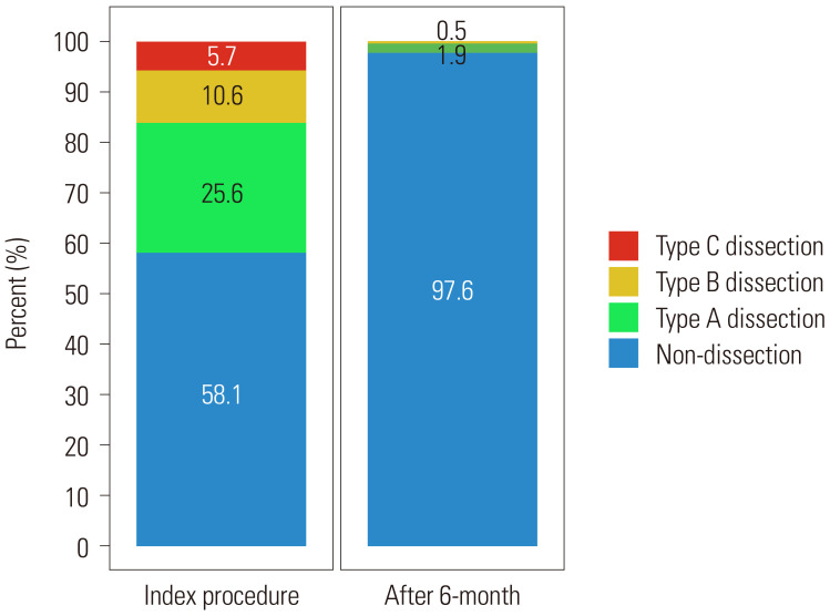
During follow-up angiography, there were no new or worse dissections; and whilst complete vascular healing occurred in 93.9%, five had persistent non-progressive uncomplicated dissections (4 Type A, 1 Type B) (Fig. 1). Over a median follow-up of 3.4 years (IQR: 25 months to 53 months), TVF occurred in 17 patients (7.5%) with similar rates in both groups: 9 patients (6.8%) in the non-dissection group and 8 patients (8.4%) in dissection group (p=0.799); and driven primarily by TVR (Table 3). There was one target vessel myocardial infarction at 22 months in the dissection group, which was related to a target lesion revascularization.
Fig. 1. The fate of dissections after drug-coated balloon treatment.
Table 3. Comparison of Clinical Outcomes According to Dissection.
| Total (n=227) | Non-dissection (n=132) | Dissection (n=95) | p value* | |
|---|---|---|---|---|
| Cardiac death | 0 | 0 | 0 | |
| Target vessel myocardial infarction | 1 (0.4) | 0 | 1 (1.1) | 0.419 |
| Target lesion revascularization | 6 (2.6) | 2 (1.5) | 4 (4.2) | 0.24 |
| Target vessel revascularization | 17 (7.5) | 9 (6.8) | 8 (8.4) | 0.799 |
| Target vessel thrombosis | 0 | 0 | 0 | |
| Target vessel failure | 17 (7.5) | 9 (6.8) | 8 (8.4) | 0.799 |
Values are n (%). Target vessel failure consisted of cardiac death, target vessel myocardial infarction, target vessel revascularization, and target vessel thrombosis.
*p value is from the log-rank test.
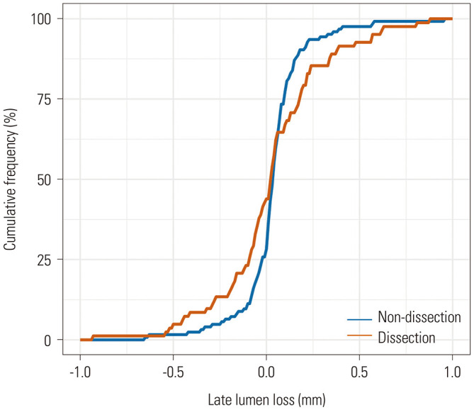
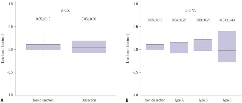
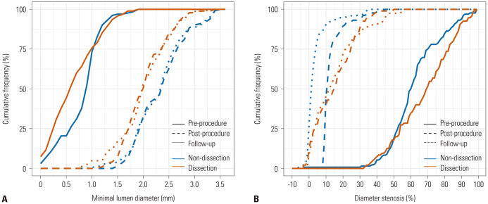
In the multivariable analysis, women, stable angina, higher DCB to reference vessel ratio and longer lesion length were independently associated with the presence of dissection. Women had more dissections after DCB; and dissections had a significant association with sex [odds ratio (OR)=2.69, 95% CI: 1.29–5.73, p=0.009], stable coronary artery disease (CAD, OR=5.17, 95% CI: 1.82–17.34, p=0.004), DCB-to-reference vessel ratio per 0.1 (OR=1.36, 95% CI: 1.06–1.79, p=0.020), and lesion length (OR=1.11, 95% CI: 1.04–1.18, p=0.001), even after adjusting for clinical and procedural characteristics (Table 4). Women were the only independent risk factor for LLL, while sex was not associated with TVF. Diabetes, acute coronary syndrome, and lesion length were independent predictors of TVF (Table 5). The presence of dissection and its severity were not associated with LLL (Fig. 2) or TVF. Cumulative frequency of MLD and diameter stenosis are shown in Fig. 3 and Fig. 4 shows the cumulative frequency distribution curves of LLL according to dissection.
Table 4. Predictors of Dissection after DCB Treatment.
| OR | 95% CI | p value | |
|---|---|---|---|
| Women | 2.69 | 1.29–5.73 | 0.009 |
| Age (per yr) | 0.99 | 0.96–1.02 | 0.526 |
| Stable coronary artery disease | 5.17 | 1.82–17.34 | 0.004 |
| DCB-to-reference vessel ratio (per 0.1) | 1.36 | 1.06–1.79 | 0.020 |
| DCB diameter (per 1 mm) | 2.12 | 1.00–4.59 | 0.053 |
| Lesion length (per 1 mm) | 1.11 | 1.04–1.18 | 0.001 |
| Minimal lumen diameter (per 1 mm) | 0.46 | 0.20–1.06 | 0.074 |
OR, odds ratio; CI, confidence interval; DCB, drug-coated balloon.
OR for continuous variables is per increase by 1 unit.
Table 5. Predictors of Target Vessel Failure.
| HR | 95% CI | p value | |
|---|---|---|---|
| Women | 1.44 | 0.36–5.68 | 0.764 |
| Age | 1.03 | 0.98–1.08 | 0.371 |
| Hypertension | 1.06 | 0.36–3.13 | 0.797 |
| Diabetes mellitus | 2.79 | 1.02–7.62 | 0.035 |
| Smoking | 3.05 | 0.95–9.80 | 0.149 |
| Prior PCI | 2.17 | 0.76–6.22 | 0.141 |
| Acute coronary syndrome | 4.59 | 1.54–13.67 | 0.012 |
| Dissection | 1.29 | 0.45–3.64 | 0.194 |
| Lesion length | 0.90 | 0.08–1.00 | 0.045 |
HR, hazard ratio; CI, confidence interval; PCI, percutaneous coronary intervention.
Fig. 2. Comparison of late lumen loss according to the presence of dissection (A) and the severity of dissection (B).
Fig. 3. Cumulative frequency distribution curves of minimal lumen diameter (A) and percent diameter stenosis (B) before procedure, after procedure, and at follow-up.
Fig. 4. Cumulative frequency distribution curves of late lumen loss according to dissection.

DISCUSSION
We investigated the angiographic and clinical outcomes of DCB treatment on de novo coronary lesions according to the presence of dissection, and our main findings were as follows: 1) there was no correlation between the presence or absence of dissection after DCB treatment and LLL at follow-up angiography; 2) TVF was not driven by the presence of dissection or its severity; 3) most dissections were completely healed, and there was no newly developed dissection at 6-month follow-up angiography.
Restenosis after BA is strongly associated with incomplete initial dilatation. This was overcome during the BA era by using oversized balloons with a balloon-to-artery diameter ratio of 1.1–1.3; however, this was associated with an increased incidence of arterial dissection and acute complications.16 Increased experience, improved instrumentation, and advanced medications eventually lead to less residual stenosis and better outcomes after PCI. The introduction of stenting alleviated the limitations of BA, which were related to elastic recoil and flow-limiting dissections. DESs, which elute an antiproliferative drug into the vessel wall, were also developed to further improve outcomes; however, late stent thrombosis and restenosis, with a hazard of nearly 2% per year after implantation, remained a concern11 and contributed to the development of DCB.
DCB continues to demonstrate some of the inherent limitations of BA, such as flow-limiting dissections and acute vessel closure, as the basic treatment mechanism is to dilatate the lesion with balloon-like behavior. However, if successful, the clinical outcomes following DCB treatment have been reported to be as good as those of DES.13,17,18,19 Successful DCB treatment should be preceded by successful BA, which is described in the International & Asian DCB consensus group as the absence of a flow-limiting dissection after BA, and a residual diameter stenosis of ≤30% or a post-balloon fractional flow reserve value of ≥0.75.14,15 In this study, we analyzed patients who were successfully treated with a DCB to determine the angiographic outcomes when a dissection persisted after DCB treatment and to assess the effect of these dissections on clinical outcomes.
In the BA era, the term “therapeutic dissection” was accepted, since dissection was considered essential to reduce restenosis. However, dissection was a double-edged sword, which was necessary to secure the lumen area; however, severe dissections resulted in complications such as myocardial infarction and emergency coronary artery bypass graft surgery.4,5 However, uncomplicated moderate dissections (Types A–C) after BA had good outcomes without stenting, and additional stenting did not actually improve the outcomes.20 These results were from studies conducted in the absence of current medications that improve prognosis, such as dual antiplatelets and high-intensity statin therapy. Therefore, the impact of residual dissection needs to be reevaluated after DCB treatment in contemporary practice. In our study, unlike with plain BA, dissection or its severity after DCB treatment did not affect LLL. At baseline, the cases of dissection were associated with a longer lesion length, smaller MLD, and a greater DS compared to the cases without dissections. After DCB treatment, MLD was smaller, and residual DS was larger in the dissection group. Therefore, when there is dissection, the acute and net lumen gains are small. However, LLL was the same regardless of whether a dissection is present or not; and moreover, its absolute value was quite small (0.05±0.30 mm vs. 0.05±0.19 mm, respectively). Our data showed that the MLD obtained after DCB treatment changed very little during follow-up period regardless of dissection, suggesting that restenosis is suppressed by the drug effects of the DCB and remains unaffected by dissections. This is true even in the presence of severe dissections, as demonstrated by our consistent LLL values of 0.04±0.26 mm, 0.09±0.29 mm, −0.01±0.49 mm in Types A, B, and C dissections, respectively (vs. 0.05±0.19 mm in non-dissection, p=0.720). These results are different from the LLL seen in patients who only received plain BA (0.25±0.50 mm),21 and infers that the local effects of the antiproliferative drug play a role, even if the dissection becomes severe.
In a previous prospective observational study, Cortese, et al.22 showed that dissection occurred in one-third of the patients, and most (94%) had completely healed on follow-up angiography. Importantly, these dissections did not increase the rates of adverse cardiovascular events. These results were comparable to those in the present study where dissections occurred in 42% of patients, healed completely in 94%, and did not affect TVF. In contrast to Cortese, et al.'s study,22 where only patients with dissection had follow-up angiography, in ours, almost all of the patients returned for follow-up angiography, even those without dissection. Subsequently, we can show that new dissections did not occur after DCB treatment, and that LLL was not related to the presence of a dissection. Another study by Funatsu, et al.23 showed that even though 80% of lesions were complicated by dissections, acute and midterm outcomes were favorable. In our study, dissections did not affect long-term outcomes, with respective rates of TVF, driven primarily by TVR, of 8.4% and 6.8% in patients with and without dissections. One 60-year-old male patient in the dissection group, who was a current smoker, developed a non-ST-segment elevation myocardial infarction in the target lesion 22 months after DCB treatment.
Dissection remains one of the biggest concerns when choosing DCB treatment instead of implanting a new generation DES. In this study, the independent predictors of dissection were women, stable CAD, higher DCB-to-reference vessel ratio, and longer lesion length. Among these factors, the higher DCB-to-reference vessel ratio had the greatest impact, and an increase of 0.1 in DCB-to-reference vessel ratio increased the OR by a factor of 2.2. In our study, females with stable CAD who had a long lesion, which was treated with a large DCB-to-reference vessel ratio, had a high chance of dissection. Reassuringly, unlike with BA, restenosis can be suppressed if there is adequate MLD to ensure sufficient flow with or without dissection; nevertheless, most of these dissections completely heal within 6 months. In this study, the independent predictors of TVF were diabetes mellitus, acute coronary syndrome, and long lesion length. The presence of dissection, its severity, and LLL had no association with TVF.
Our study had several limitations. First, due to the small sample size and the inherent limitations of a retrospective study, the impact of dissection on the primary end point could not be clearly demonstrated. Second, this did not include an all-comer registry. Although there was no flow-limiting dissection immediately after DCB treatment, acute vessel closure could theoretically occur after a period of time. In this study, after using a DCB, a final assessment was undertaken at least 5 minutes after administering a bolus of intracoronary vasodilator in order to catch acute vessel recoil. In the event of this, bailout stent implantation was considered. As a result, there was no bailout stenting during the hosptalization period in this study. The reasons for this include well-executed lesion preparation using an optimal sized pre-dilatation balloon before using a DCB to exclude lesions that require stenting (acute vessel closure or flow-limiting dissection). In addition, this may also be due to the small sample siz, so further studies with larger cohorts are needed. Thirdly, the study population comes from two centers that expertise in this type of PCI; therefore, it may not be reproducible everywhere without an adequate learning curve. Fourth, although we recorded TVF over 3 years of follow-up, the number of events was relatively small and should be interpreted with caution.
In conclusion, the presence of a dissection following successful DCB treatment of a de novo coronary lesion may not be associated with an increased risk of LLL or TVF. Most dissections were completely healed at 6-month follow-up angiography.
Footnotes
The authors have no potential conflicts of interest to disclose.
- Conceptualization: Eun-Seok Shin and Tang Qiang.
- Data curation: Eun-Seok Shin, Tang Qiang, and Lin Hui.
- Formal analysis: Eun-Seok Shin, Tang Qiang, Lin Hui, Eun Jung Jun, and Youngjune Bhak.
- Investigation: Eun-Seok Shin, Tang Qiang, Lin Hui, and Eun Jung Jun.
- Methodology: Eun-Seok Shin, Tang Qiang, and Lin Hui.
- Project administration: Eun-Seok Shin and Tang Qiang.
- Resources: Eun-Seok Shin and Tang Qiang.
- Software: Eun Jung Jun and Youngjune Bhak.
- Supervision: Eun-Seok Shin and Tang Qiang.
- Validation: Eun Jung Jun and Youngjune Bhak.
- Visualization: Eun Jung Jun and Youngjune Bhak.
- Writing—original draft: Eun-Seok Shin and Lin Hui.
- Writing—review & editing: all authors.
- Approval of final manuscript: all authors.
References
- 1.Roubin GS, King SB, 3rd, Douglas JS., Jr Restenosis after percutaneous transluminal coronary angioplasty: the Emory University Hospital experience. Am J Cardiol. 1987;60:39B–43B. doi: 10.1016/0002-9149(87)90482-6. [DOI] [PubMed] [Google Scholar]
- 2.Douglas JS, Jr, King SB, 3rd, Roubin GS. Influence of the methodology of percutaneous transluminal coronary angioplasty on restenosis. Am J Cardiol. 1987;60:29B–33B. doi: 10.1016/0002-9149(87)90480-2. [DOI] [PubMed] [Google Scholar]
- 3.Leimgruber PP, Roubin GS, Hollman J, Cotsonis GA, Meier B, Douglas JS, et al. Restenosis after successful coronary angioplasty in patients with single-vessel disease. Circulation. 1986;73:710–717. doi: 10.1161/01.cir.73.4.710. [DOI] [PubMed] [Google Scholar]
- 4.Roubin GS, Douglas JS, Jr, King SB, 3rd, Lin SF, Hutchison N, Thomas RG, et al. Influence of balloon size on initial success, acute complications, and restenosis after percutaneous transluminal coronary angioplasty. A prospective randomized study. Circulation. 1988;78:557–565. doi: 10.1161/01.cir.78.3.557. [DOI] [PubMed] [Google Scholar]
- 5.Cripps TR, Morgan JM, Rickards AF. Outcome of extensive coronary artery dissection during coronary angioplasty. Br Heart J. 1991;66:3–6. doi: 10.1136/hrt.66.1.3. [DOI] [PMC free article] [PubMed] [Google Scholar]
- 6.Hermans WR, Rensing BJ, Foley DP, Deckers JW, Rutsch W, Emanuelsson H, et al. Therapeutic dissection after successful coronary balloon angioplasty: no influence on restenosis or on clinical outcome in 693 patients. The MERCATOR Study Group (Multicenter European Research Trial with Cilazapril after Angioplasty to prevent Transluminal Coronary Obstruction and Restenosis) J Am Coll Cardiol. 1992;20:767–780. doi: 10.1016/0735-1097(92)90171-i. [DOI] [PubMed] [Google Scholar]
- 7.Cappelletti A, Margonato A, Rosano G, Mailhac A, Veglia F, Colombo A, et al. Short- and long-term evolution of unstented nonocclusive coronary dissection after coronary angioplasty. J Am Coll Cardiol. 1999;34:1484–1488. doi: 10.1016/s0735-1097(99)00395-2. [DOI] [PubMed] [Google Scholar]
- 8.Leimgruber PP, Roubin GS, Anderson HV, Bredlau CE, Whitworth HB, Douglas JS, Jr, et al. Influence of intimal dissection on restenosis after successful coronary angioplasty. Circulation. 1985;72:530–535. doi: 10.1161/01.cir.72.3.530. [DOI] [PubMed] [Google Scholar]
- 9.Serruys PW, Luijten HE, Beatt KJ, Geuskens R, de Feyter PJ, van den Brand M, et al. Incidence of restenosis after successful coronary angioplasty: a time-related phenomenon. A quantitative angiographic study in 342 consecutive patients at 1, 2, 3, and 4 months. Circulation. 1988;77:361–371. doi: 10.1161/01.cir.77.2.361. [DOI] [PubMed] [Google Scholar]
- 10.Ernst SM, van der Feltz TA, Bal ET, van Bogerijen L, van den Berg E, Ascoop CA, et al. Long-term angiographic follow up, cardiac events, and survival in patients undergoing percutaneous transluminal coronary angioplasty. Br Heart J. 1987;57:220–225. doi: 10.1136/hrt.57.3.220. [DOI] [PMC free article] [PubMed] [Google Scholar]
- 11.Kirtane AJ, Gupta A, Iyengar S, Moses JW, Leon MB, Applegate R, et al. Safety and efficacy of drug-eluting and bare metal stents: comprehensive meta-analysis of randomized trials and observational studies. Circulation. 2009;119:3198–3206. doi: 10.1161/CIRCULATIONAHA.108.826479. [DOI] [PubMed] [Google Scholar]
- 12.Yerasi C, Case BC, Forrestal BJ, Torguson R, Weintraub WS, Garcia-Garcia HM, et al. Drug-coated balloon for de novo coronary artery disease: JACC state-of-the-art review. J Am Coll Cardiol. 2020;75:1061–1073. doi: 10.1016/j.jacc.2019.12.046. [DOI] [PubMed] [Google Scholar]
- 13.Jeger RV, Farah A, Ohlow MA, Mangner N, Möbius-Winkler S, Leibundgut G, et al. Drug-coated balloons for small coronary artery disease (BASKET-SMALL 2): an open-label randomised non-inferiority trial. Lancet. 2018;392:849–856. doi: 10.1016/S0140-6736(18)31719-7. [DOI] [PubMed] [Google Scholar]
- 14.Her AY, Shin ES, Bang LH, Nuruddin AA, Tang Q, Hsieh IC, et al. Drug-coated balloon treatment in coronary artery disease: recommendations from an Asia-Pacific Consensus Group. Cardiol J. 2019 doi: 10.5603/CJ.a2019.0093. (Forthcoming) [DOI] [PMC free article] [PubMed] [Google Scholar]
- 15.Jeger RV, Eccleshall S, Wan Ahmad WA, Ge J, Poerner TC, Shin ES, et al. Drug-coated balloons for coronary artery disease: third report of the International DCB Consensus Group. JACC Cardiovasc Interv. 2020;13:1391–1402. doi: 10.1016/j.jcin.2020.02.043. [DOI] [PubMed] [Google Scholar]
- 16.Ishikawa S, Yoshinaga Y, Kantake D, Nakamura D, Yoshida N, Hisatomi T, et al. Development of a novel noninvasive system for measurement and imaging of the arterial phase oxygen density ratio in the retinal microcirculation. Graefes Arch Clin Exp Ophthalmol. 2019;257:557–565. doi: 10.1007/s00417-018-04211-z. [DOI] [PubMed] [Google Scholar]
- 17.Her AY, Shin ES, Lee JM, Garg S, Doh JH, Nam CW, et al. Paclitaxel-coated balloon treatment for functionally nonsignificant residual coronary lesions after balloon angioplasty. Int J Cardiovasc Imaging. 2018;34:1339–1347. doi: 10.1007/s10554-018-1351-z. [DOI] [PubMed] [Google Scholar]
- 18.Shin ES, Ann SH, Balbir Singh G, Lim KH, Kleber FX, Koo BK. Fractional flow reserve-guided paclitaxel-coated balloon treatment for de novo coronary lesions. Catheter Cardiovasc Interv. 2016;88:193–200. doi: 10.1002/ccd.26257. [DOI] [PubMed] [Google Scholar]
- 19.Vos NS, Fagel ND, Amoroso G, Herrman JR, Patterson MS, Piers LH, et al. Paclitaxel-coated balloon angioplasty versus drug-eluting stent in acute myocardial infarction: the REVELATION randomized trial. JACC Cardiovasc Interv. 2019;12:1691–1699. doi: 10.1016/j.jcin.2019.04.016. [DOI] [PubMed] [Google Scholar]
- 20.Albertal M, Van Langenhove G, Regar E, Kay IP, Foley D, Sianos G, et al. Uncomplicated moderate coronary artery dissections after balloon angioplasty: good outcome without stenting. Heart. 2001;86:193–198. doi: 10.1136/heart.86.2.193. [DOI] [PMC free article] [PubMed] [Google Scholar]
- 21.Her AY, Ann SH, Singh GB, Kim YH, Yoo SY, Garg S, et al. Comparison of paclitaxel-coated balloon treatment and plain old balloon angioplasty for de novo coronary lesions. Yonsei Med J. 2016;57:337–341. doi: 10.3349/ymj.2016.57.2.337. [DOI] [PMC free article] [PubMed] [Google Scholar]
- 22.Cortese B, Silva Orrego P, Agostoni P, Buccheri D, Piraino D, Andolina G, et al. Effect of drug-coated balloons in native coronary artery disease left with a dissection. JACC Cardiovasc Interv. 2015;8:2003–2009. doi: 10.1016/j.jcin.2015.08.029. [DOI] [PubMed] [Google Scholar]
- 23.Funatsu A, Kobayashi T, Mizobuchi M, Nakamura S. Clinical and angiographic outcomes of coronary dissection after paclitaxelcoated balloon angioplasty for small vessel coronary artery disease. Cardiovasc Interv Ther. 2019;34:317–324. doi: 10.1007/s12928-019-00571-3. [DOI] [PubMed] [Google Scholar]


