ABSTRACT
We present the autopsy findings and differential diagnosis in a 42year old male who presented with fever and rapidly progressive respiratory symptoms like breathlessness, nonproductive cough and right sided chest pain. Initial imaging workup done at our hospital revealed a large unilateral tumor with tracheal shift. While being evaluated patient developed facial puffiness, tachypnea suggestive of superior vena cava obstruction. Antemortem biopsy of lung mass was attempted twice and that suggested malignant lesion. Unfortunately, the individual had a rapid downhill course following admission. Post mortem examination was conducted that on opening the thoracic cavity revealed total replacement of right lung tissue by a necrotic growth which was deeply adherent to the rib cage. The contralateral lung as well as all other visceral organs were unremarkable grossly. Histopathology confirmed primary Ewing sarcoma of the lung. We hereby, report a rare case of primary lung Ewing sarcoma diagnosed at autopsy.
Keywords: Ewing sarcoma, extraosseous, Lung
INTRODUCTION
Ewing sarcoma/primitive neuroectodermal tumor(ES/PNET) is a rapidly-progressing tumor grouped under the category of small round blue cell sarcomas. 1 They occur predominantly in children and adolescence age, with a male predilection. 2 Common sites of occurrence include long bones of extremities, pelvic bone, chest wall, ribs, and vertebrae. 3 Rarely extraosseous ES is seen arising in maxilla, kidneys, retroperitoneum, female genital tract and lung. 4 Primary ES of the lung is very rare, with only about 20 case reports in the literature. 5
ES/PNET is characterized by the presence of undifferentiated small cell phenotype with a wide variety of morphological differentials that includes rhabdomyosarcoma, monophasic synovial sarcoma, non-Hodgkin lymphoma, neuroblastoma. The diagnosis of the ES/PNET involving primarily the lung is challenging and requires the differential with other pulmonary malignancies sharing similar histological features. Therefore, the diagnosis requires a combination triad of morphology, supported by immunohistochemistry and cytogenetics, as these tumors are characterized by distinct genetic translocation t (11:22) forming EWSR1-ETS fusion chimeric transcription.
Herein, we report the clinicopathological correlation in a 42-year-old man, nonsmoker with no known comorbidities who presented with fever, progressively increasing breathlessness, chest pain, and developed superior vena cava (SVC) syndrome to which he succumbed. On post-mortem examination, he was found to have a massive pulmonary mass, which had almost completely replaced the right lung parenchyma.
Case Report
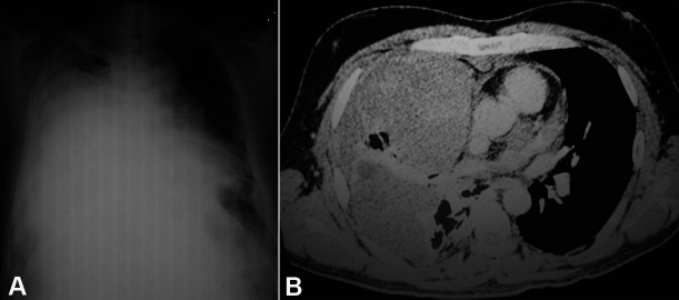
A 42-year-old previously healthy male was referred to our tertiary center complaining of high-grade fever accompanied by chills over the last 20 days. He also complained of progressively increasing breathlessness, nonproductive cough, and right-sided chest pain. There was no history of weight loss or abdominal pain. He denied smoking tobacco. On arrival at our center, he was found to be febrile and dyspneic. His pulse was 120/min, respiratory rate 28/min, and SP02 was 95% at room air. The systemic examination of the chest revealed decreased breath sounds in the right lower zone and coarse crackles in the right infrascapular region. No peripheral or deep chain lymphadenopathy was detected on physical or imaging examinations. The hematological workup showed a leukocyte count of 18,000/µl (reference range [RR]; 4000-12000/μL) with 80% polymorphs. Liver and renal function tests were within normal limits. The serum lactate dehydrogenase (LDH) was 1650 U/L (RR; 150-250U/L). The chest X-ray revealed a heterogeneous opacity in the right pulmonary lower lobe and pleural effusion, which showed to be hemorrhagic after diagnostic thoracocentesis (Figure 1A). The computed tomographic (CT)-guided Tru-cut biopsy attempted in the original-attending hospital showed necrotic tissue. The thoracic CT scan after the second hospitalization revealed a well-defined irregularly, marginated, complex, non-enhancing mass in the right lung, likely pleural based with thickened visceral pleura causing collapse and consolidation of the right lung (Figure 1B). The radiological differentials comprised malignant mesothelioma versus a synovial sarcoma.
Figure 1. A – Chest X-ray showing a homogenous opacity in right hemithorax, and contralateral mediastinal shift; B – Thoracic CT showing a pleural based large non-enhancing mass reaching the chest wall anteriorly. Note the preservation of the fat planes. Posteriorly the mass abuts and distorts the right upper lobe bronchus, stretches the ascending branch of right superior pulmonary vein, medially abuts the superior vena cava in its distal course and the ascending aorta at its proximal course and superiorly displaces the horizontal fissure causing the collapse of the middle lobe.
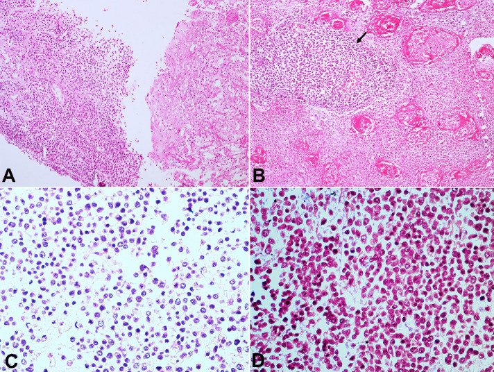
A new CT-guided biopsy was attempted and showed atypical cells consistent with an underlying malignant lesion (Figure 2A). The thoracocentesis revealed an exudative and hemorrhagic pleural effusion without malignant cells.
Figure 2. Photomicrographs of: A – Antemortem Tru-cut biopsy of the thoracic mass showing proliferating round cells (H&E stain, x200); B – Photomicrograph from post mortem lung section shows sheets of small round blue cells having peritheliomatous arrangement with extensive areas of necrosis (H&E stain, x200); C and D – the round to oval cells have stippled chromatin, indistinct nucleoli, scant cytoplasm(H&E stain, x200).
Meanwhile, the patient’s clinical condition deteriorated, he became tachypneic and developed puffiness of the face and altered sensorium and a clinical diagnosis of superior vena cava obstruction was made. He was kept on an antibiotic regimen with Piperacillin-Tazobactam, corticosteroids and controlled ventilation. Bedside 2D echocardiogram failed to evidence a pericardial tamponade, but the mass was seen compressing the right atrium and superior vena cava. On the eighth day of hospitalization, his general condition suddenly worsened, and he expired. Immediately. the autopsy was performed with the working diagnosis of a right lung mass with SVC syndrome, probably due to malignant mesothelioma or a high-grade sarcoma.
Autopsy Findings
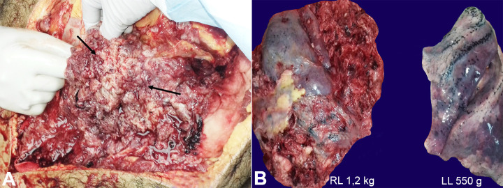
External examination revealed proptosis of eyes and midline shift of trachea to the left. On the opening of the chest cavity, the right hemithorax was replaced by a massive hemorrhagic and necrotic tumor mass, which was adherent to the chest wall. In comparison, on the left side, there were 25 ml of straw-colored pleural fluid noted with pleural sheen (Figure 3A). The right lung weighed 1200 g (RR; 360-570 g), and Left lung weighed 550 g (RR; 325-480 g). The right lung was replaced by a large tumor, which was chiefly necrotic, hemorrhagic, and pulpy. Only a portion of the posterior segment of the middle lobe was noted to be intact. The tumor mass pushed the trachea to the left. The left lung was boggy and was oozing frothy blood-stained fluid. The cut section did not show any cavities, abscesses, or tumor deposits (Figure 3B).
Figure 3. Gross view of A – the right lung mass; B – in comparison with the left lung, which appears externally normal (RL = right lung; LL = left lung).
The liver was enlarged and weighed 1800 g (RR:1500-1800 g). The external surface was smooth and glistening. No nodularity was noted. The cut surface showed a nutmeg appearance suggestive of passive venous congestion. No mass lesion was identified.
The spleen was enlarged, and weighed 230 g (RR:150-200 g). External and cut surface showed congestion. Both kidneys, pancreas, stomach, intestines, heart and brain were externally and on cut sections unremarkable.
Microscopic Findings
Histological examination of the lung depicted tumor cells in cohesive sheets, having a peritheliomatous arrangement amidst predominantly necrotic areas. The tumor cells were round to oval, had coarsely stippled chromatin with scant to mildly eosinophilic cytoplasm. Nucleoli were inconspicuous, and mitosis was brisk. Numerous apoptotic bodies were also noted (Figure 22C).
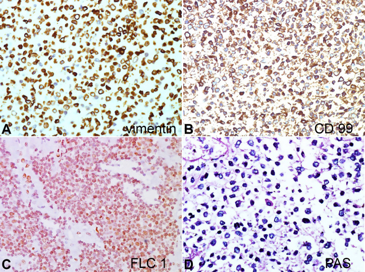
IHC reactions showed the tumor cells to be diffusely positive for Vimentin (strong cytoplasmic positivity), CD99 (strong membranous positivity), and FLI 1(strong nuclear positivity). The tumor cells were diffusely negative for calretinin, WT1, D240, synaptophysin, CD56, TTF1, Tdt, S-100, desmin, myogenin, BCL-2, LCA, and EMA. On Periodic Schiff stain (PAS), the cytoplasm of tumor cells showed strong magenta cytoplasmic positivity (Figure 4AD). Sections from the left upper and lower lobe showed normal air-filled alveoli lined by type II pneumocytes.
Figure 4. Photomicrograph of the tumor showing the immunohistochemical expression with strong positivity of tumor cells for vimentin (A), CD99 (B), FLI1 (C) and on Periodic Schiff stain (D) magenta cytoplasmic uptake. (x400 magnification).
No tumor deposits were seen. Sections from the right and left lobe of the liver showed features of passive venous congestion. No tumor deposits were noted. Sections from the spleen showed expansion of red pulp with attenuation of the white pulp. Final diagnosis was primary Ewing sarcoma of right lung causing SVC syndrome.
DISCUSSION
In 1918, Dr. Arthur Purdy Stout described an undifferentiated tumor arising from the ulnar nerve. He called it PNET because of the presence of rosettes. In 1921 James Ewing, an eminent American Pathologist, reported a tumor affecting long bones, and it was named after him as Ewing sarcoma (ES). Subsequently, these two lesions were described as different entities affecting various sites. However, by 1975, the distinction between ES and PNET began to blur, and it was realized that these two tumors represented two ends of the same histological spectrum and were grouped under the category of Ewing’s family of tumors (EFTs). 6 It was further substantiated by the fact that they share the same genetic translocation t(11;22)(q24;q12).
The EFT’s are characterized genetically by specific chromosomal translocations resulting in a fusion of the EWSR1 (22q12) gene with one of the members of the ETS family of transcription factors: the FLI1 gene (11q24) in 85% of cases and the ERG gene (21q22) in 5-10% of cases. Less frequently, EWSR1 is fused with FEV (2q36), ETV1 (7p21), or ETV4 (alias E1AF; 17q21). 7 , 8
The ES/PNET arising primarily in the lungs is extremely rare, and a high index of suspicion is required to clinch the early diagnosis and initiate timely treatment. Also, the rarity of primary ES/PNET of lung necessitates a detailed clinical and radiological examination to rule out metastasis from an extrapulmonary site. 9 Primary Lung ES/PNET was first described by Hammar et al. 10 in 1989 and a review by Deokar et al. 11 in 2015, documented only few case reports. To date, all published cases showed unilateral lung involvement, similar to our case. Histologically the ES/PNET cells are small, round with stippled chromatin, indistinct nucleoli, and scant cytoplasm. The morphological differential diagnosis, when the lung is the primary site, includes other small round cell tumors, such as rhabdomyosarcoma, lymphoma, monophasic synovial sarcoma, epithelioid malignant mesothelioma, and small cell lung carcinoma. The demographic profile, morphological features, and IHC pattern of these various lesions are enumerated in Table 1.
Table 1. Comparison of Small round cell tumors (SRCTs) arising in Lung.
| DEMOGRAPHICS/AGE | MORPHOLOGY | IHC | |
|---|---|---|---|
| Ewing Sarcoma | Children/Adolescence Male preponderance |
Densely packed small cells with extensive areas of necrosis; interspersed viable cells | Vimentin+ CD99++ FLI1 ++ |
| Rhabdomyosarcoma | Young children Peak in adolescence |
Neoplastic cells with varying degrees of resemblance to embryonic skeletal myoblasts | Desmin+ Myogenin+ MyoD+ |
| non-Hodgkin lymphoma | Average age >60yrs | Atypical lymphoid cells with centrocyte, monocytoid, plasmacytoid morphology | LCA+ |
| Synovial sarcoma | Middle aged | Epithelioid monophasic SS comprises of polygonal tumor cells in sheets Necrosis+ |
PANCK+ EMA+ BCL2+ |
| Malignant mesothelioma |
40-60yrs Occupational H/O of asbestos exposure |
Aggregates of round to polygonal cells with abundant cytoplasm & fuzzy cell borders | Calretinin+ WT1+ D240+ CK5/6+ |
| Small cell carcinoma lung | >60yrs Smoking+ |
Small blue cells, prominent nuclear molding Brisk mitosis |
CD56+ Syp + CgA+ |
CgA = Chromogranin; MyoD = Myoblast determination protein; Syp = Synaptophysin; IHC=Immunohistochemistry.
ES cells are positive for vimentin, CD99/MIC 2, and FLI1, as was seen in our case. Though CD99 is highly sensitive for ES, its specificity is low, and can also be seen in rhabdomyosarcoma, synovial sarcoma, and lymphoblastic lymphoma. Antibody against FLI1 is considered to be specific for ES/PNET. 12
Another differential to be considered, that was lower in our list was malignant melanoma, a known mimicker for various malignant entities. Hence, it is imperative to employ the full IHC panel and negate other tumors before arriving at the definitive opinion. Demonstration of t (11;22) (q24;q12) by fluorescent in situ hybridization and/or reverse transcription-polymerase chain reaction(RT-PCR) is a definitive investigation option. 13 This investigation was not available in our laboratory.
ES/PNET is an aggressive neoplasm, which is potentially lethal if not intervened timely. The deceased was referred to our hospital at a very advanced stage of the disease and had a rapid downfall in our hospital, intriguingly with a concise time of symptomatic disease. The therapeutic approach requires a combination of surgical resection, followed by chemotherapy and radiotherapy to achieve disease-free survival. Presently newer modalities of targeted immunotherapy against EWS-FLI1 chimeric fusion transcript are being explored.
Footnotes
How to cite: Gupta D, Chatterjee T, Tewari R, Trehan A, Ahuja A. Primary Ewing sarcoma/primitive neuroectodermal tumor of the lung. Autops Case Rep [Internet]. 2020;10(3):e2020199. https://doi.org/10.4322/acr.2020.199
The study was carried out in the Department of Laboratory sciences and Molecular medicine at Army Hospital( Research & Referral), New Delhi, India.
Ethics statement: The authors retain informed consent signed by the deceased’s next of kin authorizing the data publication.
Financial support: None
REFERENCES
- 1.Hancorn K, Sharma A, Shackcloth M. Primary extraskeletal Ewing’s sarcoma of the lung. Interact Cardiovasc Thorac Surg. 2010;10(5):803–804. doi: 10.1510/icvts.2009.216952. [DOI] [PubMed] [Google Scholar]
- 2.Alsit N, Fernandez C, Michel JL, Sakhri L, Derouet A, Pirvu A. Primary extraosseous Ewing sarcoma of the lung in children. E Cancer. 2013;7:312. doi: 10.3332/ecancer.2013.312. [DOI] [PMC free article] [PubMed] [Google Scholar] [Retracted]
- 3.Suárez Antelo J, Rodríguez García C, Montero Martínez C, Verea Hernando H. Pulmonary Ewing sarcoma/primitive neuroectodermal tumor: a case report and a review of the Literature. Arch Bronconeumol. 2010;46(1):44–46. doi: 10.1016/S1579-2129(10)70009-4. [DOI] [PubMed] [Google Scholar]
- 4.Takahashi D, Nagayama J, Nagatoshi Y, et al. Primary Ewing’s sarcoma family tumors of the lung a case report and review of the literature. Jpn J Clin Oncol. 2007;37(11):874–877. doi: 10.1093/jjco/hym108. [DOI] [PubMed] [Google Scholar]
- 5.Gachechiladze M, Skarda J, Ibrahim M, et al. Primitive neuroectodermal tumor (PNET) of the lung in an adult woman. World J Surg Oncol. 2014;12(1):374. doi: 10.1186/1477-7819-12-374. [DOI] [PMC free article] [PubMed] [Google Scholar]
- 6.Lee YY, Kim DH, Lee JH, et al. Primary pulmonary Ewing’s sarcoma/Primitive neuroectodermal tumour in a 67-year-old man. J Korean Med Sci. 2007;22(Suppl):S159–63. doi: 10.3346/jkms.2007.22.S.S159. [DOI] [PMC free article] [PubMed] [Google Scholar]
- 7.Turc-Carel C, Aurias A, Mugneret F, et al. Chromosomes in Ewing’s sarcoma: an evaluation of 85 cases of remarkable consistency of t(11; 22)(q24; q12) Cancer Genet Cytogenet. 1988;32(2):229–238. doi: 10.1016/0165-4608(88)90285-3. [DOI] [PubMed] [Google Scholar]
- 8.Gamberi G, Cocchi S, Benini S, et al. Molecular diagnosis in Ewing family tumors: the Rizzoli experience: 222 consecutive cases in four years. J Mol Diagn. 2011;13(3):313–324. doi: 10.1016/j.jmoldx.2011.01.004. [DOI] [PMC free article] [PubMed] [Google Scholar]
- 9.Andrei M, Cramer SF, Kramer ZB, Zeidan A, Faltas B. Adult primary pulmonary primitive neuroectodermal tumour: molecular features and translational opportunities. Cancer Biol Ther. 2013;14(2):75–80. doi: 10.4161/cbt.22635. [DOI] [PMC free article] [PubMed] [Google Scholar]
- 10.Hammar S, Bockus D, Remington F, Cooper L. The unusual spectrum of neuroendocrine lung neoplasms. Ultrastruct Pathol. 1989;13(5-6):515–560. doi: 10.3109/01913128909074534. [DOI] [PubMed] [Google Scholar]
- 11.Deokar K, Kunjir N, Ghorpade S. Primary Ewings sarcoma of the lung. J Clin Diagn Res. 2015;9(1):XD01–03. doi: 10.7860/JCDR/2015/10946.5436. [DOI] [PMC free article] [PubMed] [Google Scholar]
- 12.Folpe AL, Hill CE, Parham DM, O’Shea PA, Weiss SW. Immunohistochemical detection of FLI-1 protein expression: A study of 132 round cell tumors with emphasis on CD99-positive mimics of Ewing’s sarcoma/Primitive Neuroectodermal Tumor. Am J Surg Pathol. 2000;24(12):1657–1662. doi: 10.1097/00000478-200012000-00010. [DOI] [PubMed] [Google Scholar]
- 13.Bernstein M, Kovar H, Paulussen M, et al. Ewing’s sarcoma family of tumors: current management. Oncologist. 2006;11(5):503–519. doi: 10.1634/theoncologist.11-5-503. [DOI] [PubMed] [Google Scholar]


