Abstract
An 81-year-old man underwent F-18 fluorodeoxyglucose (FDG) positron emission tomography/computed tomography (PET/CT) to evaluate a mediastinal mass, which was discovered during the investigation for hemoptysis. The periphery of the mass abutting the aortic arch demonstrated heterogeneously increased FDG uptake, whereas most of the central portion of the mass was photopenic. The mass turned out to be an atheromatous organizing hematoma associated with contained aortic rupture.
Keywords: Thoracic aortic aneurysm, F-18 fluorodeoxyglucose, Contained rupture, Acute aortic syndrome
Introduction
Chronic contained rupture of an aortic aneurysm, also known as a sealed or contained leak, was first described by Szilagyi et al. [1]. Contained rupture is a rare subset of an aortic aneurysm, and is found in 1.5–3% of abdominal aortic aneurysms [2]. Patients with chronic contained rupture present with atypical symptoms for aneurysmal rupture, and are hemodynamically stable. The clinical presentation occasionally results in misdiagnosis, including metastatic carcinoma or infection [3]. Contained rupture of an aortic aneurysm is at risk for frank rupture, and should be identified early in the presentation.
Here we present a case of contained aortic aneurysm rupture with mural thrombi in which F-18 fluorodeoxyglucose (FDG) positron emission tomography/computed tomography (PET/CT) was used to differentiate it from malignancy.
Case Report
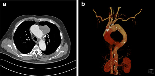
An 81-year-old man was referred for evaluation of a mediastinal mass. He visited the emergency department presenting with hemoptysis, and the mass was revealed subsequently on chest computed tomography (CT). He complained of no pain but suffered respiratory symptoms including cough and sputum. He had a history of colon cancer 7 years previously, and had been on hypertension medication for 10 years. The mass was located in the left upper thorax, with a long diameter of approximately 5.8 cm, exhibiting poor enhancement on chest CT (Fig. 1a). There was no evidence of either aortic dissection or significant steno-occlusive lesion on reconstructed CT angiography (CTA) (Fig. 1b). The clinician suspected malignancy in the mediastinum, such as a thymic carcinoma, or possible primary lung cancer and metastasis.
Fig. 1.
a Computed tomography (CT) revealed a mediastinal mass in the left upper thorax. The mass diameter was approximately 5.8 cm, and it displayed poor enhancement. b There was no evidence of aortic dissection or significant steno-occlusive lesion on reconstructed CT angiography (CTA)
The patient underwent FDG PET/CT to evaluate the mediastinal mass. PET/CT images were acquired on a Discovery MI (GE Healthcare, Milwaukee, WI, USA) at 60 min after injection of 2.96 MBq/kg of FDG. After CT (120 kVp; 10–80 mA; section thickness, 3.75 mm), an emission PET was obtained from the thigh to the skull base (1.5 min per bed). PET images were reconstructed using the Bayesian penalized likelihood reconstruction algorithm with attenuation-corrected images.
FDG PET/CT revealed heterogeneously increased FDG uptake along the periphery of the mass, with a maximum standardized uptake value (SUVmax) of 6.9 (Fig. 2). The majority of central portion of the mass was photopenic. Hematoma, rather than malignancy, was considered following the FDG PET/CT.
Fig. 2.
F-18 fluorodeoxyglucose (FDG) positron emission tomography/computed tomography (PET/CT) showed heterogeneously increased FDG uptake along the periphery of the mass; most of the central portion of the mass was photopenic
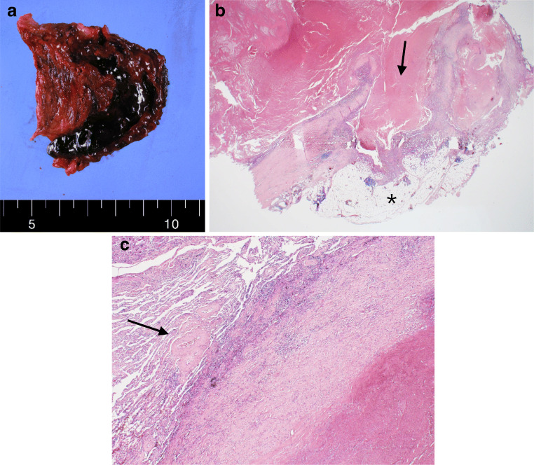
Bronchoscopy demonstrated no remarkable findings. Ascending aorta dilatation (4.16 cm) without complication was reported on a transthoracic echocardiography. The patient underwent ascending aorta and total arch replacement, as well as en bloc resection of the mass-like lesion, under low suspicion of malignancy. The surgical diagnosis was thoracic aortic aneurysm of the ascending aorta and aortic arch, contained aortic rupture with mural thrombi (Fig. 3). The mass lesion was connected to the aortic arch, and strongly attached to the adjacent lung tissue in the surgical field. The mass lesion contained necrotic, old, dark blood-colored material, corresponding to the photopenic area visualized on the FDG PET/CT. The pathology revealed no evidence of malignancy, and the mass was diagnosed as an atheromatous organizing hematoma with calcification.
Fig. 3.
The pathology revealed no evidence of malignancy, and was diagnosed as an atheromatous organizing hematoma with calcification. a Gross appearance of the mediastinal mass in the left upper thorax. b Disruption of the aortic wall (arrow) and surrounding lung tissue (asterisk) are noted. c Inflammatory infiltration to the aortic wall, corresponding to increased FDG uptake, and large vasa vasorum (arrow) are noted
Discussion
An aneurysm refers to an arterial dilatation of more than 50% of its normal diameter [4]. Approximately 95% of patients with thoracic aortic aneurysms are asymptomatic [5]. However, fatal complications including aortic dissection or rupture may occur. Acute aortic syndromes comprise a spectrum of disorders with disruption in aortic integrity, so rapid diagnosis is important [6]. CT is the imaging modality of choice for diagnosis of acute aortic syndromes (pooled sensitivity 100%, pooled specificity 98%) and also used for the differential diagnosis among acute aortic dissection, pulmonary embolism, and acute coronary syndromes in an emergency setting [6, 7].
It had been difficult to distinguish between neoplastic and vascular lesions of the thorax based on plain radiographs and clinical symptoms [8]. da Gama et al. reported a patient who was misdiagnosed as having an esophageal tumor causing his dysphagia and weight loss [9]. In that case, the patient was diagnosed with a chronic contained rupture of a descending thoracic aneurysm by CT, and was surgically treated.
The role of FDG PET/CT in aortic disorders has emerged recently. Increased FDG uptake in the abdominal aortic aneurysm wall was associated with inflammation and instability of the wall [10]. Increased FDG uptake in the aortic wall was also related to progression of acute aortic syndromes and a poor prognosis [11, 12]. However, prolonged scan time of FDG PET/CT has limited the clinical use of it in emergent settings, such as for acute aortic syndromes [13].
Primary malignant aortic tumors are very rare, and can affect any segment of the thoracoabdominal aorta [14]. Patients with primary neoplasms of the aorta can possibly present with a contained rupture of the aorta [15]. Nenoto et al. reported a case involving poorly differentiated adenocarcinoma of the lung infiltrating the aortic wall, resulting in mimicking the rupture of a thoracic aortic aneurysm [16]. FDG PET/CT could be helpful to differentiate malignant tumor from hematoma, since the former is expected to show high metabolic activity, whereas the latter exhibits a mainly metabolic defect.
There were previously reported cases that acute aortic syndrome involves the left chest. Inam et al. have reported a case of massive hemoptysis resulting from a ruptured descending thoracic aorta aneurysm to the left lung [17]. And we also found a case which reported on a mass extending into the left upper hemithorax caused by thoracic aortic rupture [18]. However, our present case was not associated with either acute chest pain or any previous episode of chest pain, which was different from most of other reported cases.
Aortic aneurysm would be included in the differential diagnosis for a thoracic periaortic mass-like lesion. Because thoracic aortic aneurysm may lead to a severe clinical course, though its exact prevalence in FDG PET/CT for investigating thoracic mass was not yet reported. It requires particular attention when the periaortic mass-like lesion shows increased FDG uptake around the metabolic defect, suggesting inflammation and instability of the aortic wall.
Conclusion
To the best of our knowledge, this is the first case which shows a FDG PET/CT image of a contained rupture of a thoracic aortic aneurysm. FDG PET/CT demonstrated increased FDG uptake around the metabolic defect, which implies aortic wall instability and hematoma. FDG PET/CT may play a role in the diagnosis of a contained rupture of an aortic aneurysm, especially in patients with atypical presentation.
Compliance with Ethical Standards
Conflict of Interest
Hyung Seok Chang, Soo Jeong Kim, and Young Hwan Kim declare that they have no conflict of interest.
Ethical Approval
All procedures performed in studies involving human participants were in accordance with the ethical standards of the institutional and/or national research committee and with the 1964 Helsinki declaration and its later amendments or comparable ethical standards.
For this type of study, formal consent is not required.
Informed Consent
The institutional review board of our institute approved this retrospective study, and the requirement to obtain informed consent was waived.
Footnotes
Publisher’s Note
Springer Nature remains neutral with regard to jurisdictional claims in published maps and institutional affiliations.
Contributor Information
Hyung Seok Chang, Email: hs712.chang@samsung.com.
Soo Jeong Kim, Email: sj12345.kim@samsung.com.
Young Hwan Kim, Email: yh27.kim@samsung.com.
References
- 1.Szilagyi DE, Smith RF, Macksood AJ, Whitcomb JG. Expanding and ruptured abdominal aortic aneurysms. Problems of diagnosis and treatment. Arch Surg. 1961;83:395–408. doi: 10.1001/archsurg.1961.01300150069009. [DOI] [PubMed] [Google Scholar]
- 2.Apter S, Rimon U, Konen E, Erlich Z, Guranda L, Amitai M, et al. Sealed rupture of abdominal aortic aneurysms: CT features in 6 patients and a review of the literature. Abdom Imaging. 2010;35:99–105. doi: 10.1007/s00261-008-9488-1. [DOI] [PubMed] [Google Scholar]
- 3.Bansal M, Bansal M, Thukral BB, Malik A. Contained rupture of a thoracoabdominal aortic aneurysm presenting as a back mass. J Thorac Imaging. 2006;21:219–221. doi: 10.1097/01.rti.0000203637.20888.1f. [DOI] [PubMed] [Google Scholar]
- 4.Goldfinger JZ, Halperin JL, Marin ML, Stewart AS, Eagle KA, Fuster V. Thoracic aortic aneurysm and dissection. J Am Coll Cardiol. 2014;64:1725–1739. doi: 10.1016/j.jacc.2014.08.025. [DOI] [PubMed] [Google Scholar]
- 5.Faiza Z, Sharman T. Thoracic aorta aneurysm. StatPearls. Treasure Island (FL): StatPearls Publishing Copyright © 2020, StatPearls Publishing LLC.; 2020. [PubMed]
- 6.Carroll BJ, Schermerhorn ML, Manning WJ. Imaging for acute aortic syndromes. Heart. 2020;106:182–189. doi: 10.1136/heartjnl-2019-314897. [DOI] [PubMed] [Google Scholar]
- 7.Bossone E, Ranieri B, Romano L, Russo V, Barbuto L, Cocchia R, et al. Acute aortic syndromes: diagnostic and therapeutic pathways. Heart Fail Clin. 2020;16:305–315. doi: 10.1016/j.hfc.2020.03.002. [DOI] [PubMed] [Google Scholar]
- 8.Shahian DM, Javid H, Faber LP, Kittle CF, Matthew GR. Lesions of the thoracic aorta and its arch branches simulating neoplasm. J Thorac Cardiovasc Surg. 1981;81:251–263. doi: 10.1016/S0022-5223(19)37634-2. [DOI] [PubMed] [Google Scholar]
- 9.da Gama AD, Martins C, Cabral G, Alves AG. Chronic contained rupture of a descending thoracic aortic aneurysm simulating an esophagus tumor. Conventional surgical management. Rev Port Cir Cardiotorac Vasc. 2009;16:157–161. [PubMed] [Google Scholar]
- 10.Adriaans BP, Wildberger JE, Westenberg JJM, Lamb HJ, Schalla S. Predictive imaging for thoracic aortic dissection and rupture: moving beyond diameters. Eur Radiol. 2019;29:6396–6404. doi: 10.1007/s00330-019-06320-7. [DOI] [PMC free article] [PubMed] [Google Scholar]
- 11.Kuehl H, Eggebrecht H, Boes T, Antoch G, Rosenbaum S, Ladd S, et al. Detection of inflammation in patients with acute aortic syndrome: comparison of FDG-PET/CT imaging and serological markers of inflammation. Heart. 2008;94:1472–1477. doi: 10.1136/hrt.2007.127282. [DOI] [PubMed] [Google Scholar]
- 12.Gorla R, Erbel R, Kuehl H, Kahlert P, Tsagakis K, Jakob H, et al. Prognostic value of (18)F-fluorodeoxyglucose PET-CT imaging in acute aortic syndromes: comparison with serological biomarkers of inflammation. Int J Cardiovasc Imaging. 2015;31:1677–1685. doi: 10.1007/s10554-015-0725-8. [DOI] [PubMed] [Google Scholar]
- 13.Kim J, Song HC. Role of PET/CT in the evaluation of aortic disease. Chonnam Med J. 2018;54:143–152. doi: 10.4068/cmj.2018.54.3.143. [DOI] [PMC free article] [PubMed] [Google Scholar]
- 14.Vacirca A, Faggioli G, Pini R, Freyrie A, Indelicato G, Fenelli C, et al. Predictors of survival in malignant aortic tumors. J Vasc Surg. 2020;71:1771–1780. doi: 10.1016/j.jvs.2019.09.030. [DOI] [PubMed] [Google Scholar]
- 15.Steinberg JB, Johnson ER, Benda JA, Lanza LA. Primary leiomyosarcoma of the thoracic aorta presenting as a contained rupture. Ann Thorac Surg. 1993;56:1387–1389. doi: 10.1016/0003-4975(93)90688-E. [DOI] [PubMed] [Google Scholar]
- 16.Nemoto K, Itoh M, Senba S, Shimizudani N, Adachi H, Kishi K, et al. A case of lung cancer mimicking rupture of a thoracic aortic aneurysm. Nihon Kokyuki Gakkai Zasshi. 2009;47:158–162. [PubMed] [Google Scholar]
- 17.Inam H, Zahid I, Khan SD, Haq SU, Fatimi S. Hemoptysis secondary to rupture of infected aortic aneurysm- a case report. J Cardiothorac Surg. 2019;14:144. doi: 10.1186/s13019-019-0959-y. [DOI] [PMC free article] [PubMed] [Google Scholar]
- 18.Ando Y, Minami H, Muramoto H, Narita M, Sakai S. Rupture of thoracic aorta caused by penetrating aortic ulcer. Chest. 1994;106:624–626. doi: 10.1378/chest.106.2.624. [DOI] [PubMed] [Google Scholar]


