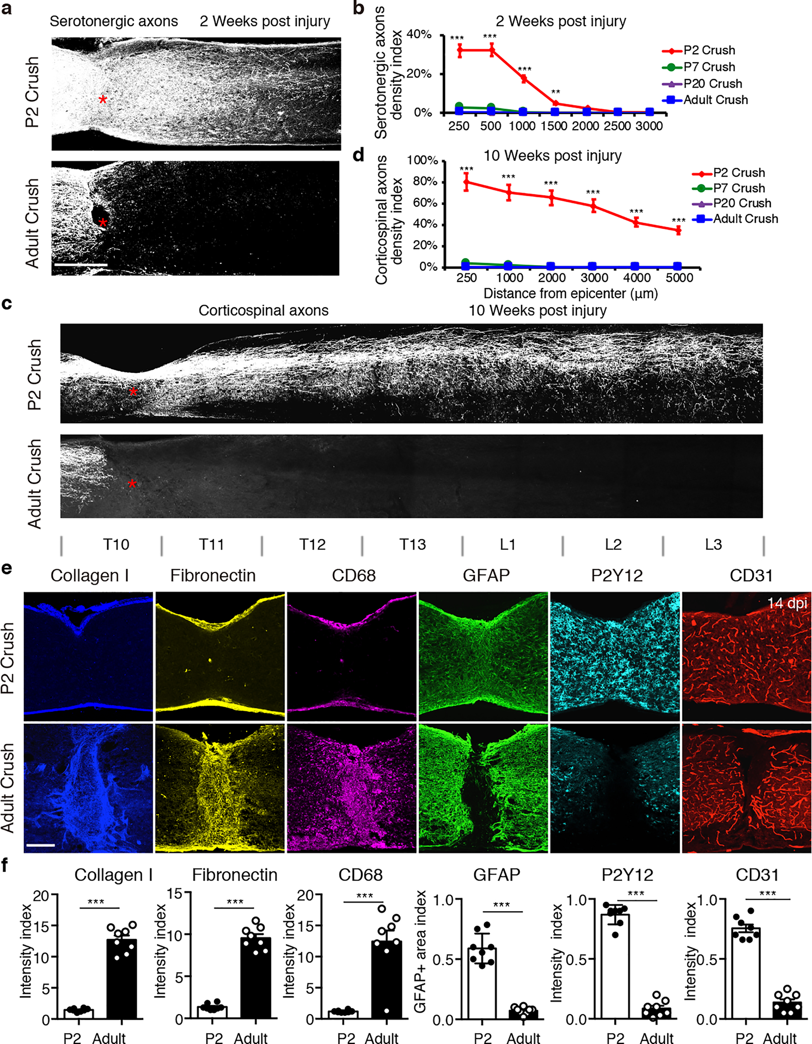
Fig. 1 |. Scar-free wound healing after neonatal spinal cord crush injury.

a, Images of spinal sections stained with anti-5HT antibody taken at 2 weeks after P2 (top) or adult (bottom) injury. Red stars indicate the lesion site. b, Quantification of serotonergic axon density (normalized to proximal of lesion) in spinal cord distal to the lesion site. n=8, 5, 5 and 8 for P2, P7, P20 and adult, respectively. **p = 0.0031; ***p < 0.0001. c, Images of spinal sections at 10 weeks post crush showing CST axons labeled by AAV-ChR2-mCherry. d, Quantification of CST axons distal to the lesion site at 10 weeks post crush (n=5). ***p < 0.0001. e, Images of spinal sections at 2 weeks after crush stained with indicated antibodies. f, Quantification of indicated immunoreactive intensity (normalized to the intact region) in the lesion site at 2 weeks after injury (n=8). ***p < 0.0001. b, d, Two-way ANOVA followed by post hoc Bonferroni correction. f, Student’s t test (two-tailed, unpaired). Data shown as mean ± s.e.m. Scale bar: 500μm (a), 1mm (b), 250μm (c, e).
