. 2020 May 14;297(3):E335–E338. doi: 10.1148/radiol.2020201955

Figure 2:

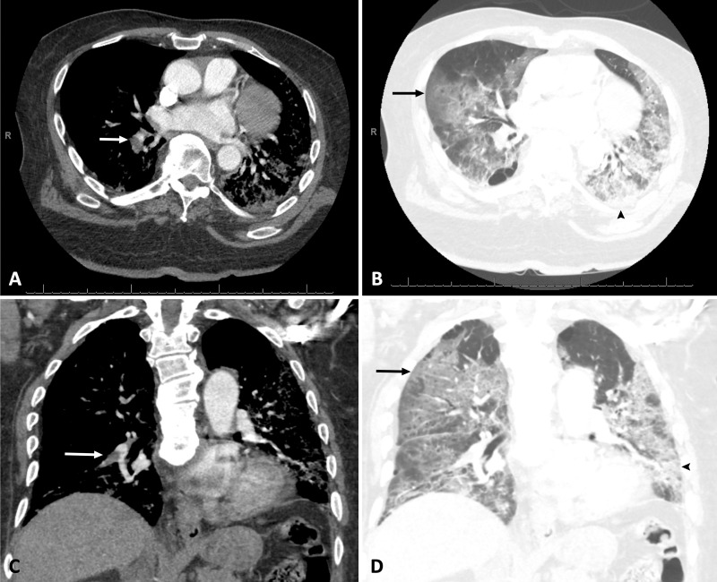
A, B, Axial and, C, D, coronal pulmonary CT angiography images in a 76-year-old African American man with body mass index of 37 kg/m2 who required admission to medical intensive care unit for acute respiratory failure secondary to coronavirus disease 2019 confirmed with reverse transcriptase polymerase chain reaction. Pulmonary CT angiography was obtained 4 days after admission and demonstrates acute pulmonary embolism in right lower lobar pulmonary artery (white arrows), bilateral ground glass opacities (black arrows), and consolidation (arrowheads).

A, B, Axial and, C, D, coronal pulmonary CT angiography images in a 76-year-old African American man with body mass index of 37 kg/m2 who required admission to medical intensive care unit for acute respiratory failure secondary to coronavirus disease 2019 confirmed with reverse transcriptase polymerase chain reaction. Pulmonary CT angiography was obtained 4 days after admission and demonstrates acute pulmonary embolism in right lower lobar pulmonary artery (white arrows), bilateral ground glass opacities (black arrows), and consolidation (arrowheads).