Abstract
Platelet-derived growth factor (PDGF) is a potent mitogen and chemoattractant that serves a role in the development of several types of solid cancer, and abnormal PDGF activity has been reported in numerous human tumors. Tumor-derived PDGF ligands are considered to act in either a paracrine or autocrine manner, serving roles in the phosphorylation of receptors on tumor and stromal cells in the tumor microenvironment. Despite the well-established association between PDGF and tumor progression, the precise mechanisms of autocrine PDGF signaling in pancreatic tumor cells remain elusive. Therefore, the present study aimed to analyze the influence of PDGF-BB in pancreatic cancer. Pancreatic adenocarcinoma BxPC-3 cells were cultured and treated with recombinant human PDGF-BB in vitro. Cell proliferation was tested using an MTT assay. Cell apoptosis was measured using flow cytometry. Tumor cell migration and invasion were examined via wound-healing and Transwell assays, respectively. The expression and subcellular localization of Yes-associated protein (YAP) was determined using western blotting and immunofluorescence. The transcriptional activity of target genes was tested using a luciferase assay and reverse transcription-quantitative PCR. The present study revealed that PDGF-BB significantly promoted cell proliferation in pancreatic adenocarcinoma BxPC-3 cells and enhanced the aggressiveness of this cell line, as demonstrated by Transwell and wound-healing assays. Anoikis resistance is an important mechanism by which metastatic cells avoid apoptosis when detaching from adjacent cells or the extracellular matrix. PDGF-BB treatment inhibited anoikis under anchorage-independent conditions. Mechanistic experiments revealed that PDGF-BB promoted the upregulation and activation of the transcriptional coactivator YAP, an effector of the Hippo signaling pathway. RhoA or protein phosphatase-1 (PP-1) inhibition partially abolished the accumulation and activation of YAP, suggesting PDGF-BB-mediated YAP dephosphorylation and transactivation via the RhoA/PP-1 cascade. Pharmacologic inhibition of the PDGF receptor directly downregulated YAP activity and the expression levels of downstream genes. Furthermore, verteporfin, a small molecular inhibitor of the Hippo/YAP signaling pathway, partially reversed the effects of PDGF-BB on cell proliferation, anoikis resistance and cell migration. In conclusion, the present study revealed that the Hippo/YAP signaling pathway may be involved in the tumor-promoting activity of PDGF-BB in pancreatic cancer.
Keywords: pancreatic cancer, PDGF, malignancy, Hippo signaling, YAP
Introduction
Platelet-derived growth factor (PDGF) is a potent mitogen and chemoattractant that stimulates cell proliferation, survival and migration in numerous types of tumors, such as bladder, breast and cervical carcinoma (1). The PDGF family consists of five disulfide-bonded dimers: PDGF-AA, -AB, -BB, -CC and -DD (2). The PDGF dimeric isoforms are synthesized as precursor molecules. PDGF-AA, -AB and -BB are cleaved and activated in secretory vesicles inside producer cells, while PDGF-CC and -DD are secreted as inactive precursor molecules that are converted into their active form by proteolytic cleavage (3). PDGF isoforms exert their cellular effects through structurally similar α- and β-tyrosine kinase receptors (PDGFRα and PDGFRβ, respectively); PDGFRα can bind all PDGF isoforms, except PDGF-DD, while PDGFRβ binds only PDGF-BB and PDGF-DD with considerable affinity (4). The binding of PDGF polypeptide chains to their receptors triggers the dimerization and autophosphorylation of PDGFRs, which in turn activate several downstream signaling pathways, such as the ERK and PI3K/AKT signaling cascades (5).
Abnormal PDGF activity is frequently detected in a number of human tumors (6–9). Tumor-derived PDGF ligands are considered to act in either a paracrine or autocrine manner, stimulating the phosphorylation of receptors on tumor and stromal cells in the tumor microenvironment (10). Previous studies suggested that tumor-derived PDGF may primarily promote tumor angiogenesis by mediating the recruitment and growth of stromal fibroblasts, perivascular cells and endothelial cells (11–14). In this way, PDGF indirectly affects tumor growth, metastatic dissemination and drug resistance. PDGF autocrine signaling may also contribute to tumorigenesis. The tumor-promoting functions of autocrine PDGF have been demonstrated in multiple non-epithelial malignancies, including squamous cell carcinoma, glioblastoma and osteosarcoma (15). Autocrine PDGF signaling is capable of modulating the malignant phenotypes of tumor cell proliferation, epithelial-to-mesenchymal transition (EMT), energy metabolism, invasion, metastasis and colonization (5). Targeting PDGF/PDGFR signaling may therefore represent a therapeutic strategy in patients with cancer (2,5).
Pancreatic cancer is one of the most lethal malignancies worldwide, currently ranking as the fourth leading cause of cancer-associated death in the USA and Europe, but is expected to be the second leading cause of death by 2020 (16). The treatment of this disease is currently problematic due to the difficulty of initial diagnosis, strong aggressive features, primary and secondary resistance to conventional chemotherapy, and high recurrence (17). Anoikis is a specialized type of apoptosis in epithelial and endothelial cells that is triggered by loss of contact with the extracellular matrix or adhesion to inappropriate locations (18). Anoikis resistance is a mechanism by which cancer cells avoid undergoing apoptosis during tumor development and metastasis (19).
Numerous studies have revealed that multiple signaling pathways are involved in the progression of pancreatic cancer, such as NF-κB, MAPK, TGFβ/Smad and Hedgehog signaling pathways (20,21). Primary and metastatic malignant endocrine pancreatic tumors express high levels of PDGFRβ compared with normal endocrine pancreatic tissues (22). PDGFRβ has been identified as a reliable prognostic marker of pancreatic adenocarcinoma, since higher levels of PDGFRβ expression are associated with a poor prognosis, as well as with lymphatic invasion and lymph node metastasis (23). Additionally, in SW1990 human pancreatic cancer xenograft models, PDGFRβ activation is observed after radioimmunotherapy or chemotherapy with imatinib (24). Transcriptional profiling and functional screening have identified PDGFRβ as both necessary and sufficient to mediate the proliferative and pro-metastatic effects of mutant p53 (25). In addition, Wnt-1/β-catenin signaling contributes to the autocrine activation of PDGF/Src signaling in pancreatic cancer (26). PDGF-BB promotes the acquisition of the EMT phenotype in pancreatic cancer AsPC-1 cells via the induction of microRNA-221 (27) and mimics the serum-induced dispersal of pancreatic epithelial cell clusters (28). Furthermore, dual-specificity phosphatase 28 and PDGF-A form an acquired autonomous autocrine signaling pathway that promotes chemoresistance and migration in pancreatic cancer (29). A neutralizing antibody directed against PDGFRβ enhances the antitumor and anti-angiogenic activity of a VEGF antagonist (30).
Despite the well-established association between PDGF and tumor progression via ERK and AKT signaling cascades, the precise mechanisms of autocrine PDGF signaling in pancreatic tumor cells remain elusive. A previous study has revealed that PDGF can affect tumorigenesis via ERK- and AKT-independent mechanisms (31). The present study aimed to study the roles of PDGF in pancreatic cancer biology, including cell proliferation, anoikis resistance and invasion, as well as the underlying mechanism through the transcriptional coactivator Yes-associated protein (YAP)/Hippo pathway.
Materials and methods
Cell culture and drugs
Pancreatic adenocarcinoma BxPC-3 cells were purchased from the Shanghai Institute of Cell Biology (Chinese Academy of Sciences) and were cultured in RPMI 1640 medium supplemented with 10% FBS (both Gibco; Thermo Fisher Scientific, Inc.), penicillin (100 U/ml), streptomycin (100 µg/ml) and 2 mM L-glutamine at 37°C in a humidified 5% CO2 incubator. Regarding drugs, PDGF-BB (R&D Systems, Inc.) with concentrations 0, 10, 25, 50 and 100 ng/ml was added and incubated at 37°C in a 5% CO2 incubator for 24 h. Verteporfin, a drug able to stop the formation of the YAP/TEAD complex in the nucleus (cat. no. HY-B0146) was purchased from MedChemExpress. Various concentrations (0.1, 0.5 and 1 µM) of Verteporfin were added into the medium and incubated at 37°C in a 5% CO2 incubator for 24 h. CP-673451, a potent selective inhibitor of PDGFR tyrosine kinase, was purchased from Selleck Chemicals (cat. no. S1536). Cells were treated with 10 nM CP-673451 at 37°C for 24 h. Rhosin and calyculin A were purchased from MedChemExpress (cat. nos. HY-12646 and HY-18983, respectively). The cells were treated with 30 µM Rhosin and 30 µM calyculin A at 37°C for 24 h. PBS was used as a control.
Transfection
Small interfering (si) RNA oligonucleotides for YAP1 and scrambled non-targeting negative control were purchased from Thermo Fisher Scientific, Inc. (Stealth RNAi™ siRNA; cat. no. AM16708). Cells (5.0×10⁵) were transfected with siRNA oligonucleotides (20 nM) using Lipofectamine® 2000 (Invitrogen; Thermo Fisher Scientific, Inc.) at 37°C for 24 h according to the manufacturer's protocol. Cell culture medium was replaced post-transfection and cells were allowed to grow for an additional 24 h before subsequent experiments.
MTT assay
Briefly, cells (5×10³ cells/well) were seeded in 96-well plates in the absence of or at 10, 25, 50 and 100 ng/ml of PDGF-BB (R&D Systems, Inc.) for 24 h at 37°C. The treatment was started 24 h after seeding. Subsequently, cells were incubated with MTT solution at 37°C for 4 h, and formazan crystals resulting from MTT reduction were dissolved by adding 100 µl DMSO in each well and gently shaking for 15 min. The absorbance of cultures was measured using a multiwell spectrophotometer at a wavelength of 560 nm. Results were calculated as the percentage of absorbance in control cultures.
Anoikis assay
Anoikis assay was performed as previously described (32). In order to induce anoikis under anchorage-independent conditions, ~1×10⁶ cells/ml were plated in an ultra-low attachment 6-well plate (cat. no. 3471; Corning, Inc.) with or without PDGF-BB addition. BxPC-3 cells were cultured under suspension or adherent conditions for 48 h and then all cells were harvested using centrifugation at 500 × g at room temperature for 5 min for apoptosis analysis.
Apoptosis assay
For cell apoptosis analysis, apoptotic cells were measured using an Annexin V-FITC Apoptosis Detection kit (Nanjing KeyGen Biotech Co., Ltd.) according to the manufacturer's protocol. Cells were harvested, washed twice with PBS and resuspended in 500 µl binding buffer. Cell suspensions were stained with 5 µl Annexin V-FITC and 5 µl PI for 30 min at room temperature in the dark. The cells were evaluated immediately via CytoFLEX LX Flow Cytometer (Beckman Coulter, Inc.). A minimum of 10,000 cells per sample was measured, and the analysis of apoptotic cells was performed using BD CellQuest Pro software v3.3 (BD Biosciences).
Wound-healing assay
Wound-healing assays were performed to examine the capacity of cell migration. Briefly, after BxPC-3 cells grew to 90–95% confluence in 6-well plates, a single scratch wound was generated using a 200-ml disposable pipette tip. Cells were washed to remove displaced and floating cells, and then incubated in fresh serum-free RPMI 1640 medium for 24 h. Wound-healing was detected at 0 and 24 h within the scratched wounds, and representative fields were photographed using an inverted light microscope with an attached digital camera (Olympus Corporation; magnification, ×200), and the distance between the borders of the wound were assessed for quantification using Image Pro-Plus 7.0 (Media Cybernetics, Inc.).
Transwell assay
Transwell chambers (EMD Millipore; 8-µm pore size) were coated with Matrigel® at 37°C for 12 h (15 µg/filter). Cells (2.0×10⁴) were plated in serum-free DMEM into the upper chambers, while the lower chambers were filled with complete medium. Cells treated with PDGF-BB or vehicle were allowed to invade across the Matrigel-coated membrane at 37°C in 5% CO2. After 24 h of incubation, the cells on the upper surface of the insert were removed, and the cells on the lower surface of the insert were fixed with 4% paraformaldehyde at room temperature for 5 min and stained with 0.1% crystal violet for 3 min at room temperature. Images of migrated cells were captured, and cell numbers in five randomly selected fields were counted under light microscopy (magnification, ×200).
Luciferase assay
The 8×GTIIC-luc plasmid was obtained from Addgene, Inc. Transient transfection was performed using Lipofectamine 2000 (Thermo Fisher Scientific, Inc.) according to the manufacturer's protocol. For the luciferase reporter assay, BxPC-3 cells were seeded in 12-well plates. The 8×GTIIC reporter plasmid containing TEA-domain (TEAD)-binding elements together with Renilla-Luc plasmids (Addgene, Inc.) were co-transfected into BxPC-3 cells. After 4 h of transfection at 37°C, cells were exposed to 0, 10, 25, 50 or 100 ng/ml PDGF-BB. After treatment for 24 h at 37°C, luciferase activities were measured using a Dual-Glo luciferase assay kit (Promega Corporation) under a Victor3 1420 plate reader (PerkinElmer, Inc.). Normalized luciferase signal was calculated by dividing the firefly luciferase signal by the Renilla luciferase signal.
Western blot analysis
Western blot analysis was performed using total cell lysates. Total cell lysates from different experiments were obtained by lysing the cells in RIPA buffer (Beyotime Institute of Biotechnology) containing protease inhibitors (100 µM phenylmethylsulfonyl fluoride, 10 µM leupeptin, 10 µM pepstatin and 2 mM EDTA). The protein content was quantitated using a BCA Protein Assay kit (Beyotime Institute of Biotechnology). Proteins (50 µg/lane) were separated via 4–20% SDS-PAGE and transferred to nitrocellulose membranes (Pall Life Sciences). Membranes were blocked with 5% non-fat milk for 1 h at room temperature and probed overnight at 4°C with primary antibodies against YAP (cat. no. 4912), phospho- (p-)YAP (Ser127; cat. no. 4911S), Macrophage Stimulating 1 (MST1; cat. no. 3682), p-MST1/2 (Thr183/180; cat. no. 3681), Large Tumor Suppressor Kinase 1 (LATS1; cat. no. 3477), p-LATS1 (Ser909; cat. no. 9157), N-cadherin (cat. no. 13116), E-cadherin (cat. no. 14472) and β-actin (cat. no. 4970), all at a dilution of 1:1,000 (Cell Signaling Technology, Inc.). Membranes were washed and incubated with horseradish peroxidase-conjugated secondary antibodies (1:5,000) (cat. nos. AS014 and AS003; ABclonal Biotech Co., Ltd.) at room temperature for 2 h, and were visualized using an enhanced chemiluminescence system (EMD Millipore).
RNA isolation and reverse transcription-quantitative (RT-q) PCR
Total RNA was isolated from cultured cells using TRIzol® reagent (Invitrogen; Thermo Fisher Scientific, Inc.). The PrimeScript RT Master Mix kit (Takara Biotechnology Co., Ltd.) was used to synthesize cDNA for mRNA detection according to the manufacturer's protocol. qPCR was performed with gene-specific primers using the SYBR Green PCR kit (Takara Biotechnology Co., Ltd.) according to the manufacturer's protocol under the following thermocycling conditions: 95°C for 5 min, followed by 40 cycles at 95°C for 10 sec for denaturation and 60°C for 30 sec for annealing/extension. The expression levels of each target gene were calculated using the 2−ΔΔCq method (33). Data were expressed as the fold-change relative to GAPDH. The primer sequences were as follows: c-MYC forward, 5′-CCCGCTTCTCTGAAAGGCTCTC-3′ and reverse, 5′-CTCTGCTGCTGCTGCTGCTGGTAG−3′; MCL-1 forward, 5′-CCAAGGCATGCTTCGGAAA-3′ and reverse, 5′-TCACAATCCTGCCCCAGTTT-3′; GAPDH forward, 5′-TCACTGGCATGGCCTTCCGTG−3′ and reverse, 5′-GCCATGAGGTCCACCACCCTG−3′; N-cadherin forward, 5′-TGAAACGGCGGGATAAAGAG-3′ and reverse, 5′-GGCTCCACAGTATCTGGTTG-3′; and E-cadherin forward, 5′-GGTTTTCTACAGCATCACCG-3′ and reverse, 5′-GCTTCCCCATTTGATGACAC-3′.
Immunofluorescence staining
After 100 ng/ml PDGF-BB treatment for 24 h at 37°C, cells cultured on slides were rinsed with PBS, fixed in 3.7% formaldehyde/PBS for 20 min at room temperature and permeabilized using 0.1% Triton X-100/PBS for 10 min at room temperature. Slides were blocked for 1 h with 1% BSA/PBS (Beyotime Institute of Biotechnology), and were incubated with antibodies against YAP (1:100; cat. no. sc-101199; Santa Cruz Biotechnology, Inc.) overnight at 4°C. Subsequently, cells were incubated with FITC-conjugated secondary antibodies (1:1,000; cat. no. F0382; Sigma-Aldrich; Merck KGaA) for 2 h at room temperature. Slides were mounted with ProLong Gold antifade reagent with DAPI (Invitrogen; Thermo Fisher Scientific, Inc.). Fluorescence images were collected using a fluorescence microscope (IX70; Olympus Corporation; magnification, ×600).
Statistical analysis
Data were expressed as the mean ± SD. Statistical analysis was performed using unpaired Student's t-test or one-way ANOVA followed by Tukey's post hoc test for comparisons among ≥3 groups using the statistical program SPSS 11.0 (SPSS, Inc.) for Windows. P<0.05 was considered to indicate a statistically significant difference.
Results
PDGF-BB promotes pancreatic cancer malignancy
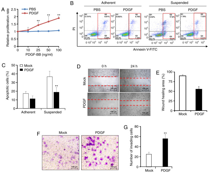
To determine the role of PDGF-BB in promoting tumor growth, cell proliferation was determined in pancreatic adenocarcinoma BxPC-3 cells using an MTT assay. After exposure to PDGF-BB (0–100 ng/ml) for 24 h, BxPC-3 cells exhibited a dose-dependent increase in proliferation (Fig. 1A). Cell proliferation was significantly increased in BxPC-3 cells treated with concentrations >25 ng/ml PDGF-BB. Anoikis is an anchorage-independent form of cell death that is initiated after the disruption of the cell matrix and cell-cell interactions; thus, anoikis resistance is an initial step in the progression of metastatic cancer (34). Subsequently, the effects of PDGF-BB on cell responsiveness to anoikis were evaluated. BxPC-3 cells were cultured under suspension or adherent conditions for 48 h, after which apoptotic cells were analyzed via flow cytometry. Compared with adherent cells, suspended BxPC-3 cells exhibited a higher rate of anoikis after culture without PDGF-BB for 48 h, with >30% apoptotic cells. Treatment with 100 ng/ml PDGF-BB significantly decreased cell apoptosis in the suspended cells, but had no significance in adherent cells, indicating enhanced anoikis resistance (Fig. 1B and C). Subsequently, the migration of BxPC-3 cells in the presence or absence of PDGF-BB was examined via wound-healing and Transwell assays. PDGF-BB significantly accelerated wound closure of BxPC-3 cells compared with that of untreated cells (Fig. 1D and E). Consistently, the Transwell assay revealed that PDGF-BB treatment increased the number of invading BxPC-3 cells (Fig. 1F and G). These results suggested that PDGF-BB may have a positive effect on cell migration. Overall, the present data revealed that PDGF-BB may promote pancreatic cancer malignancy via increasing cell proliferation, anoikis resistance and cell migration.
Figure 1.
PDGF-BB affects cell proliferation, apoptosis resistance and migration of pancreatic cancer cells. (A) MTT assay of cell proliferation after BxPC-3 cells were incubated with increasing concentrations of PDGF-BB (0–100 ng/ml) for 24 h. PBS was used as a control. (B) Flow cytometric analysis of apoptosis in BxPC-3 cells exposed to 100 ng/ml PDGF-BB or PBS and cultured in suspension for 48 h. (C) Percentage of apoptotic cells analyzed in three independent experiments. (D) Wound-healing assays of BxPC-3 cells in the presence or absence of 100 ng/ml PDGF-BB. Wounds were allowed to heal for 24 h and imaged under a microscope. (E) Wound-healing area analyzed in three independent experiments. (F) Transwell assay of cell invasion. BxPC-3 cells in the presence or absence of 100 ng/ml PDGF-BB were induced to invade through Matrigel for 24 h. (G) Number of invading cells in 6 wells was analyzed in three independent experiments. Results are presented as the mean ± SD (n≥3). *P<0.05 and **P<0.01 vs. mock/PBS. PDGF-BB, platelet-derived growth factor-BB.
PDGF-BB activates YAP signaling
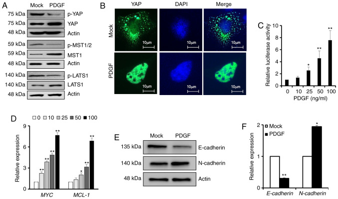
The transcriptional coactivator YAP, an effector of the Hippo signaling pathway, can act as an oncogene to promote tumor survival and metastasis if its activity is increased abnormally (35). Therefore, the present study estimated the effects of PDGF-BB on YAP activity. Compared with the mock group, the total amount of YAP, MST1 and LATS1 protein were all upregulated, while the phosphorylation of YAP at Ser127, MST1 at Thr183, and LATS1 at Ser909 as the degradation forms were markedly decreased in PDGF-BB-treated cells, indicating the aberrant activation of YAP (Fig. 2A). Immunofluorescence staining revealed the nuclear and cytoplasmic localization of YAP in BxPC-3 cells in the absence or presence of PDGF-BB, indicating that PDGF-BB caused a redistribution of YAP and enhanced its nuclear accumulation (Fig. 2B). In addition, BxPC-3 cells were transfected with a luciferase reporter plasmid (8×GTIIC-luc) containing TEAD-binding elements. PDGF-BB administration at >25 ng/ml for 24 h significantly potentiated YAP activity, as demonstrated by the luciferase assay (Fig. 2C). Accordingly, RT-qPCR revealed that PDGF-BB treatment significantly upregulated the expression of two YAP downstream genes, namely the MYC proto-oncogene and the MCL-1 apoptosis regulator (Fig. 2D). MYC has a pivotal function in growth control, while MCL-1 promotes tumor survival by enabling cells to escape apoptosis (36). Since EMT is closely associated with tumor invasion and metastasis, and is also regulated by Hippo/YAP signaling (37), the present study investigated the effect of PDGF-BB on EMT by detecting the expression levels of epithelial and mesenchymal markers. Western blot analysis suggested that the expression levels of the epithelial marker E-cadherin were decreased after PDGF-BB treatment, whereas the expression levels of the mesenchymal marker N-cadherin were increased (Fig. 2E). Similar results were obtained via RT-qPCR, indicating that PDGF-BB treatment significantly suppressed E-cadherin expression and promoted N-cadherin expression (Fig. 2F). In summary, the current results strongly suggested that PDGF-BB may stimulate YAP activity and the expression of its downstream genes.
Figure 2.
PDGF-BB affects YAP dephosphorylation, subcellular localization and downstream gene expression. (A) Western blot analysis of YAP, MST1/2 and LATS1 expression and their phosphorylation levels in BxPC-3 cells exposed to 100 ng/ml PDGF-BB or PBS. (B) Immunofluorescence staining exhibiting the subcellular localization of YAP protein after treatment of BxPC-3 cells with 100 ng/ml PDGF-BB for 24 h. (C) BxPC-3 cells were transfected with the 8×GTIIC-luc reporter and Renilla-luc plasmids and cultured under increasing concentrations of PDGF-BB (0–100 ng/ml) for 24 h. Relative firefly/Renilla luciferase activity was measured. (D) Reverse transcription-quantitative PCR analysis of the expression levels of YAP target genes (MYC and MCL-1) in BxPC-3 cells treated with PDGF-BB (0–100 ng/ml) or PBS. Relative mRNA levels were normalized to GAPDH. (E) Western blot analysis of E-cadherin and N-cadherin expression in BxPC-3 cells after PDGF-BB treatment. (F) mRNA levels of E-cadherin and N-cadherin relative to GAPDH in the mock and PDGF groups. Data are presented as the mean ± SD (n=3). *P<0.05 and **P<0.01 vs. mock or 0 ng/ml. PDGF-BB, platelet-derived growth factor-BB; YAP, Yes-associated protein; p, phosphorylated; MST1, Macrophage Stimulating 1; LATS1, Large Tumor Suppressor Kinase 1.
YAP activation contributes to PDGF-BB-enhanced pancreatic cancer malignancy
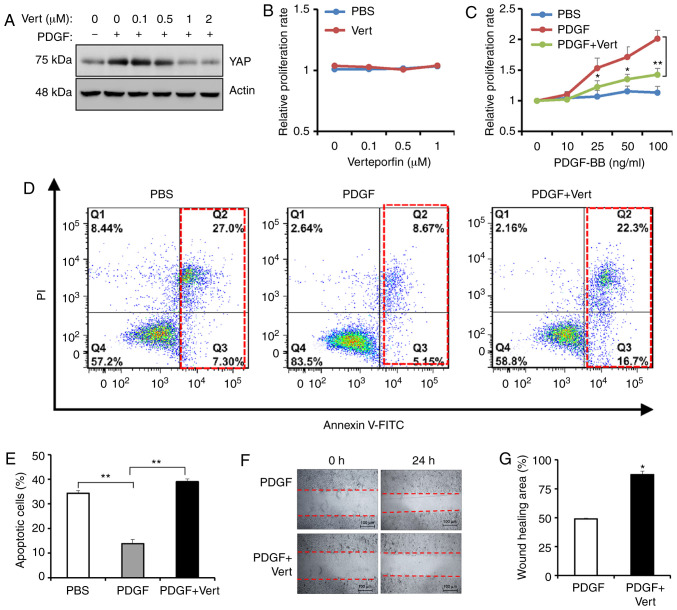
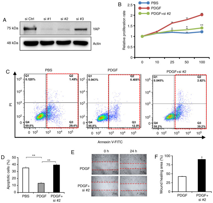
To ascertain the effects of YAP activity in PDGF-BB-induced cancer malignancy, BxPC-3 cells were treated with verteporfin, a YAP inhibitor, and PDGF-BB. Increasing concentrations of verteporfin (0.1, 0.5 and 1 µM) gradually decreased the levels of YAP protein (Fig. 3A). Treatment with verteporfin for 24 h had no effect on cell proliferation, as measured via MTT assay (Fig. 3B). Subsequently, the combined effects of 0.1 µM verteporfin (the minimum effective dose) and PDGF-BB on cell proliferation, anoikis resistance and cell migration were assessed. Notably, 0.1 µM verteporfin partially reversed the promoting effects of PDGF-BB on cell proliferation (Fig. 3C). Additionally, verteporfin increased anoikis in the presence of PDGF-BB (Fig. 3D and E). A wound-healing assay revealed that verteporfin attenuated the migration of tumor cells enhanced by PDGF-BB (Fig. 3F and G). Subsequently, YAP expression was knocked down using three siRNAs, and siRNA2 was chosen for further experiments (Fig. 4A). Similarly, YAP siRNA abrogated the cell proliferation promoting effects of PDGF-BB (Fig. 4B) and promoted anoikis in the presence of PDGF-BB (Fig. 4C and D). A wound-healing assay indicated that YAP siRNA reversed the PDGF-BB-enhanced migration of tumor cells (Fig. 4E and F). These results revealed that blockade of YAP activity with a pharmacological inhibitor partially abrogated the effects of PDGF-BB on cancer malignancy, suggesting that YAP activation may be a causal mechanism for PDGF-BB-induced tumor progression.
Figure 3.
YAP inhibition interrupts PDGF-BB-enhanced cancer malignancy. (A) Western blot analysis of YAP expression in BxPC-3 cells after treatment with 100 ng/ml PDGF-BB and increasing concentrations of verteporfin (0, 0.1, 0.5, 1 and 2 µM). MTT assay of cell proliferation after BxPC-3 cells were exposed to (B) increasing concentrations of verteporfin (0, 0.1, 0.5 and 1 µM) for 24 h and (C) 100 ng/ml PDGF-BB with or without 0.1 µM verteporfin for 24 h. (D) Analysis of cell apoptosis via flow cytometry of BxPC-3 cells exposed to 100 ng/ml PDGF-BB with or without 0.1 µM verteporfin and cultured in suspension for 48 h. (E) Percentage of apoptotic cells. (F) Wound-healing assays of BxPC-3 cells in the presence of 100 ng/ml PDGF-BB with or without 0.1 µM verteporfin for 24 h. (G) Wound-healing area in three independent experiments. Data are presented as the mean ± SD (n=3). *P<0.05 and **P<0.01. PDGF-BB, platelet-derived growth factor-BB; YAP, Yes-associated protein; Vert, verteporfin.
Figure 4.
YAP knockdown inhibits PDGF-BB-induced cancer malignancy. (A) Western blot analysis of YAP expression in BxPC-3 cells treated with YAP siRNAs for 48 h. (B) MTT assay of the proliferation of BxPC-3 cells exposed to 100 ng/ml PDGF-BB with or without YAP siRNA2 for 24 h. (C) Analysis of apoptosis via flow cytometry in BxPC-3 cells exposed to 100 ng/ml PDGF-BB with or without YAP siRNA and cultured in suspension for 48 h. (D) Percentage of apoptotic cells (E) Wound-healing assays on BxPC-3 cells in the presence of 100 ng/ml PDGF-BB with or without YAP siRNA for 24 h. (F) Wound-healing area in three independent experiments. Data are presented as the mean ± SD (n=3). *P<0.05 and **P<0.01 vs. PDGF. PDGF-BB, platelet-derived growth factor-BB; YAP, Yes-associated protein; si, small interfering; Ctrl, control.
PDGFR/RhoA/protein phosphatase-1 (PP-1) cascade participates in PDGF-BB-induced YAP activation
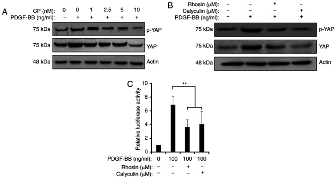
There is a complicated network regulating YAP activity. A previous study revealed that platelets mediate YAP dephosphorylation and promote its nuclear translocation via the RhoA/MYPT1/PP-1 cascade (32). Therefore, the present study explored the mechanism associated with PDGF-induced YAP activation. Firstly, CP-673451, a potent selective inhibitor of PDGFR tyrosine kinase, was used to block PDGF downstream signaling. Treatment with 10 nM CP-673451 completely abolished PDGF-BB-induced YAP stabilization and phosphorylation (Fig. 5A). Secondly, Rhosin or calyculin A were used to inhibit RhoA or PP-1 in the presence of PDGF-BB, respectively. Treatment with 30 µM Rhosin or 30 nM calyculin A partially attenuated PDGF-BB-induced YAP accumulation and dephosphorylation to different extents (Fig. 5B). Additionally, a luciferase assay demonstrated that Rhosin and calyculin A repressed the PDGF-BB-induced activity of the 8×GTIIC-luc reporter (Fig. 5C). Therefore, the current data revealed that the PDGFR/RhoA/PP-1 cascade may be involved in PDGF-BB-induced YAP activation.
Figure 5.
Blockade of the RhoA/PP-1 cascade impairs PDGF-BB-induced YAP activation. (A) Western blot analysis of YAP expression and its phosphorylation at Ser127 in BxPC-3 cells treated with 100 ng/ml PDGF-BB with or without CP-673451 for 24 h. (B) Western blot analysis of YAP accumulation and phosphorylation in BxPC-3 cells treated with 100 ng/ml PDGF-BB with 30 µM Rhosin or 30 nM calyculin A for 24 h. (C) Relative firefly/Renilla luciferase activity in BxPC-3 cells transfected with the 8×GTIIC-luc reporter and cultured under PDGF-BB treatment together with 30 µM Rhosin or 30 µM calyculin A for 24 h. Data are presented as the mean ± SD (n=3). **P<0.01. PDGF-BB, platelet-derived growth factor-BB; YAP, Yes-associated protein; p, phosphorylated.
PDGFR inhibition decreases YAP activity and cancer malignancy
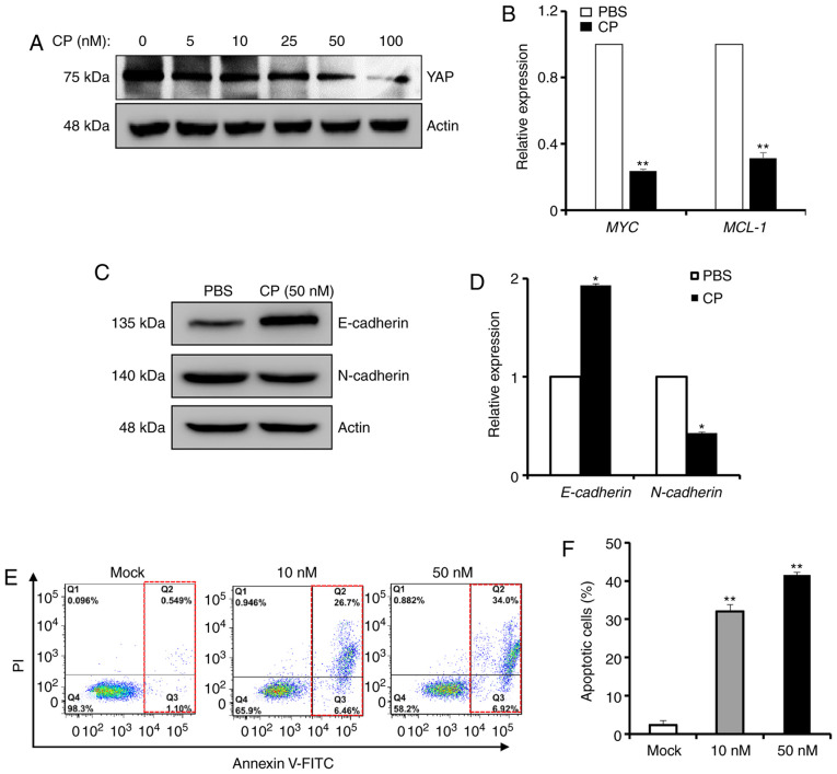
PDGF is the principal mitogen in serum and is produced by platelets and macrophages (3). In tumors, PDGF can be expressed by tumor or adjacent stroma cells, thereby acting as either a paracrine or autocrine factor (10). Therefore, the effects of CP-673451 on YAP activity and cancer malignancy were investigated. A gradual decrease in the total amount of YAP protein was observed during PDGFR inhibition with up to 100 nM CP-673451 (Fig. 6A). As a result, the expression levels of two YAP downstream genes (MYC and MCL-1) were significantly decreased after treatment with 50 µM CP-673451 for 24 h (Fig. 6B). Additionally, western blot analysis suggested that E-cadherin expression was increased after CP-673451 treatment, while N-cadherin expression was decreased (Fig. 6C). qPCR analysis indicated that E-cadherin and N-cadherin expression was regulated by CP-673451 (Fig. 6D). Furthermore, treatment with CP-673451 for 24 h alone induced BxPC-3 cells to undergo apoptosis in a dose-dependent manner, as demonstrated by flow cytometric Annexin V apoptosis analysis (Fig. 6E and F). Therefore, the present results indicated that PDGF inhibition may inhibit cancer malignancy by mediating YAP inactivation.
Figure 6.
PDGFR inhibition decreases YAP activity and cancer malignancy. (A) Western blot analysis of YAP expression in BxPC-3 cells after treatment with increasing concentrations of CP-673451 (0–100 nM) for 24 h. (B) RT-qPCR analysis of the expression levels of MYC and MCL-1 in BxPC-3 cells treated with 50 nM CP-673451 for 24 h. (C) Western blot analysis of E-cadherin and N-cadherin expression in BxPC-3 cells after CP-673451 treatment for 24 h. (D) RT-qPCR analysis of E-cadherin and N-cadherin expression in BxPC-3 cells. The results are presented as the average expression levels after normalization with GAPDH. (E) Flow cytometric analysis and (F) quantification of apoptosis in BxPC-3 cells after treatment with CP-673451 for 24 h. Data are presented as the mean ± SD (n=3). *P<0.05 and **P<0.01 vs. PBS/mock. PDGF-BB, platelet-derived growth factor-BB; YAP, Yes-associated protein; RT-qPCR, reverse transcription-quantitative PCR.
Discussion
Several studies have revealed the positive association between abnormal YAP activity and tumorigenesis (38–41). The findings of the present study suggested that PDGF-BB signaling promoted the malignancy of pancreatic cancer via YAP activation, that the RhoA/PP-1 cascade was involved in the PDGF-BB-induced dephosphorylation of YAP and that targeting PDGFR repressed YAP activity and induced tumor apoptosis.
The transcriptional coactivators YAP and its paralog TAZ are vital downstream effectors of the Hippo signaling cascade and serve versatile roles in the control of developmental transitions, organ size, cell fate and tumorigenesis (42–45). When the Hippo-signaling pathway becomes active, the MST1/2 and LATS1/2 kinases are activated by phosphorylation (46). LATS1/2 kinases phosphorylate and inhibit YAP, thereby sequestering YAP in the cytosol and limiting its transcriptional activity (46). In addition, YAP phosphorylated at Ser127 can be ubiquitinated by β-TrCP ubiquitin ligase and subsequently targeted for proteasomal degradation (47). In addition, YAP is a substrate of autophagy, and autophagy accelerates the degradation of YAP (48). Thus, the total protein forms and the phosphorylated forms of LATS1/2, MST1/2 and YAP are in a balance to maintain the activation of the Hippo signaling pathway (45). Upon silencing of Hippo signaling, YAP is dephosphorylated and active YAP is translocated to the nucleus (45). Within the nucleus, YAP functions as a transcriptional coactivator of TEAD transcription factors (49). Furthermore, YAP can interact with Smad family members and other transcription factors to regulate the expression of target genes (50). In this way, YAP is implicated in cell proliferation, apoptosis, migration, chemoresistance and angiogenesis (51,52).
YAP induces EMT and promotes the progression of cholangiocarcinoma (53). In addition, YAP participates in the development, progression and recurrence of pancreatic cancer (49). Furthermore, YAP is associated with chemoresistance and poor prognosis in pancreatic cancer (49). Verteporfin, an agent that disrupts YAP-TEAD complexes, suppresses the survival of pancreatic ductal adenocarcinoma cells (54). Verteporfin inhibits tumor angiogenesis by downregulating angiopoietin-2 and suppresses vasculogenic mimicry by decreasing the expression levels of matrix metallopeptidase 2, vascular endothelial cadherin and α-smooth muscle actin (55). YAP activation through cyclin-dependent kinase 1-mediated mitotic phosphorylation promotes pancreatic cancer cell motility, invasion and tumorigenesis (56). Considering the role of YAP in pancreatic malignancy and clinical outcome, it is reasonable to develop drugs targeting YAP for future interventions.
The results of the present study indicated that PDGF-BB induced YAP activation, contributing to cancer malignancy in pancreatic cancer cells. The YAP inhibitor verteporfin partially attenuated the effects of PDGF-BB on cell proliferation, apoptosis and migration. Additionally, PDGF-BB upregulated MYC expression, an oncogene that promotes cell division. Furthermore, PDGF-BB enhanced MCL-1 expression, which is a member of the BCL-2 family and prevents cells from undergoing apoptosis (57). Knockdown of MCL-1 by RNA interference renders B-RAF melanoma cells sensitive to anoikis, which is a unique anchorage-independent form of apoptotic cell death that occurs as a result of insufficient cell-matrix interactions (58). Anoikis resistance is a critical contributor to tumor invasion and metastasis, and malignant cells take advantage of several mechanisms to resist anoikis and thereby maintain survival (59). In addition, the present study revealed that PDGF-BB treatment altered the expression levels of E-cadherin and N-cadherin, two master regulators of EMT. Consistently, PDGF-BB increased the aggressive capability of pancreatic cancer cells, as demonstrated by Transwell and wound-healing assays. Finally, YAP inhibition effectively reversed the oncogenic effects of PDGF-BB.
Meanwhile, CP-673451, an inhibitor of PDGFR signaling, was used to block the effects of PDGF. CP-673451 downregulated YAP activation and repressed tumor malignancy. Its effects suggested that PDGF signaling may activate YAP to affect cell proliferation, survival and migration. Furthermore, the current results revealed that PDGF-BB resulted in YAP dephosphorylation and transactivation via the RhoA/PP-1 cascade, as RhoA or PP-1 inhibition abolished YAP activation. A previous study revealed that platelets can promote YAP dephosphorylation and nuclear translocation via the RhoA/MYPT1/PP-1 cascade to induce a pro-survival gene expression signature (32). Small GTPases, such as RhoA, Rac and Cdc42, can activate YAP by inhibiting its phosphorylation (60). PP-1 is a mediator of PDGF signaling in primary cultures of vascular smooth muscle cells (61). RhoA is one of the determinants of the PDGF-BB-induced migration of rat hepatic stellate cells (62). Notably, YAP is regulated by complicated mechanisms. A previous study revealed that PDGF can regulate YAP transcriptional activity via Src family kinase-dependent tyrosine phosphorylation (63). Conversely, a study on genome-wide profiling of highly aggressive Schwann cell lineage-derived sarcomas revealed that TAZ/YAP-TEAD complexes can directly activate PDGFR signaling and other oncogenic programs (64).
In conclusion, the present study suggested that there may be a convergence between the Hippo/YAP and PDGF-PDGFR signaling pathways in the malignant progression of pancreatic cancer. Therefore, the concomitant manipulation of the YAP and PDGF signaling pathways may improve the efficacy of therapy against malignant tumors.
Acknowledgements
Not applicable.
Glossary
Abbreviations
- PDGF-BB
platelet-derived growth factor-BB
- YAP
Yes-associated protein
- PP-1
protein phosphatase-1
- PDGFR
PDGF receptor
- EMT
epithelial-to-mesenchymal transition
Funding
The present study was supported by the National Natural Science Foundation of China (grant no. 81972758), the Interdisciplinary Medicine Seed Fund of Peking University (grant no. BMU2018MX018) and the Science Foundation of Peking University Cancer Hospital (grant no. 2017-23).
Availability of data and materials
The datasets used and/or analyzed during the current study are available from the corresponding author on reasonable request.
Authors' contributions
TL and TG performed the experiments, HL and HJ analyzed and interpreted the data, YW designed the study and revised the manuscript. All authors read and approved the final manuscript.
Ethics approval and consent to participate
The present study was approved by the Ethics Committee of Hainan Medical University.
Patient consent for publication
Not applicable.
Competing interests
The authors declare that they have no competing interests.
References
- 1.Bartoschek M, Pietras K. PDGF family function and prognostic value in tumor biology. Biochem Biophys Res Commun. 2018;503:984–990. doi: 10.1016/j.bbrc.2018.06.106. [DOI] [PubMed] [Google Scholar]
- 2.Heldin CH. Targeting the PDGF signaling pathway in tumor treatment. Cell Commun Signal. 2013;11:97. doi: 10.1186/1478-811X-11-97. [DOI] [PMC free article] [PubMed] [Google Scholar]
- 3.Fredriksson L, Li H, Eriksson U. The PDGF family: Four gene products form five dimeric isoforms. Cytokine Growth Factor Rev. 2004;15:197–204. doi: 10.1016/j.cytogfr.2004.03.007. [DOI] [PubMed] [Google Scholar]
- 4.Roskoski R., Jr The role of small molecule platelet-derived growth factor receptor (PDGFR) inhibitors in the treatment of neoplastic disorders. Pharmacol Res. 2018;129:65–83. doi: 10.1016/j.phrs.2018.01.021. [DOI] [PubMed] [Google Scholar]
- 5.Heldin CH, Lennartsson J, Westermark B. Involvement of platelet-derived growth factor ligands and receptors in tumorigenesis. J Intern Med. 2018;283:16–44. doi: 10.1111/joim.12690. [DOI] [PubMed] [Google Scholar]
- 6.Cao Y. Multifarious functions of PDGFs and PDGFRs in tumor growth and metastasis. Trends Mol Med. 2013;19:460–473. doi: 10.1016/j.molmed.2013.05.002. [DOI] [PubMed] [Google Scholar]
- 7.Martinho O, Longatto-Filho A, Lambros MB, Martins A, Pinheiro C, Silva A, Pardal F, Amorim J, Mackay A, Milanezi F, et al. Expression, mutation and copy number analysis of platelet-derived growth factor receptor A (PDGFRA) and its ligand PDGFA in gliomas. Br J Cancer. 2009;101:973–982. doi: 10.1038/sj.bjc.6605225. [DOI] [PMC free article] [PubMed] [Google Scholar]
- 8.Nazarenko I, Hede SM, He X, Hedrén A, Thompson J, Lindström MS, Nistér M. PDGF and PDGF receptors in glioma. Ups J Med Sci. 2012;117:99–112. doi: 10.3109/03009734.2012.665097. [DOI] [PMC free article] [PubMed] [Google Scholar]
- 9.Saito Y, Haendeler J, Hojo Y, Yamamoto K, Berk BC. Receptor heterodimerization: Essential mechanism for platelet-derived growth factor-induced epidermal growth factor receptor transactivation. Mol Cell Biol. 2001;21:6387–6394. doi: 10.1128/MCB.21.19.6387-6394.2001. [DOI] [PMC free article] [PubMed] [Google Scholar]
- 10.Andrae J, Gallini R, Betsholtz C. Role of platelet-derived growth factors in physiology and medicine. Genes Dev. 2008;22:1276–1312. doi: 10.1101/gad.1653708. [DOI] [PMC free article] [PubMed] [Google Scholar]
- 11.Paulsson J, Sjöblom T, Micke P, Pontén F, Landberg G, Heldin CH, Bergh J, Brennan DJ, Jirström K, Ostman A. Prognostic significance of stromal platelet-derived growth factor beta-receptor expression in human breast cancer. Am J Pathol. 2009;175:334–341. doi: 10.2353/ajpath.2009.081030. [DOI] [PMC free article] [PubMed] [Google Scholar]
- 12.Dhar K, Dhar G, Majumder M, Haque I, Mehta S, Van Veldhuizen PJ, Banerjee SK, Banerjee S. Tumor cell-derived PDGF-B potentiates mouse mesenchymal stem cells-pericytes transition and recruitment through an interaction with NRP-1. Mol Cancer. 2010;9:209. doi: 10.1186/1476-4598-9-209. [DOI] [PMC free article] [PubMed] [Google Scholar]
- 13.Krenzlin H, Behera P, Lorenz V, Passaro C, Zdioruk M, Nowicki MO, Grauwet K, Zhang H, Skubal M, Ito H, et al. Cytomegalovirus promotes murine glioblastoma growth via pericyte recruitment and angiogenesis. J Clin Invest. 2019;129:1671–1683. doi: 10.1172/JCI123375. [DOI] [PMC free article] [PubMed] [Google Scholar]
- 14.Ostman A. PDGF receptors-mediators of autocrine tumor growth and regulators of tumor vasculature and stroma. Cytokine Growth Factor Rev. 2004;15:275–286. doi: 10.1016/j.cytogfr.2004.03.002. [DOI] [PubMed] [Google Scholar]
- 15.Heldin CH. Autocrine PDGF stimulation in malignancies. Ups J Med Sci. 2012;117:83–91. doi: 10.3109/03009734.2012.658119. [DOI] [PMC free article] [PubMed] [Google Scholar]
- 16.Malvezzi M, Bertuccio P, Rosso T, Rota M, Levi F, La Vecchia C, Negri E. European cancer mortality predictions for the year 2015: Does lung cancer have the highest death rate in EU women? Ann Oncol. 2015;26:779–786. doi: 10.1093/annonc/mdv001. [DOI] [PubMed] [Google Scholar]
- 17.Ansari D, Tingstedt B, Andersson B, Holmquist F, Sturesson C, Williamsson C, Sasor A, Borg D, Bauden M, Andersson R. Pancreatic cancer: Yesterday, today and tomorrow. Future Oncol. 2016;12:1929–1946. doi: 10.2217/fon-2016-0010. [DOI] [PubMed] [Google Scholar]
- 18.Frisch SM, Screaton RA. Anoikis mechanisms. Curr Opin Cell Biol. 2001;13:555–562. doi: 10.1016/S0955-0674(00)00251-9. [DOI] [PubMed] [Google Scholar]
- 19.Gupta P, Gupta N, Fofaria NM, Ranjan A, Srivastava SK. HER2-mediated GLI2 stabilization promotes anoikis resistance and metastasis of breast cancer cells. Cancer Lett. 2019;442:68–81. doi: 10.1016/j.canlet.2018.10.021. [DOI] [PMC free article] [PubMed] [Google Scholar]
- 20.Khader S, Thyagarajan A, Sahu RP. Exploring signaling pathways and pancreatic cancer treatment approaches using genetic models. Mini Rev Med Chem. 2019;19:1112–1125. doi: 10.2174/1389557519666190327163644. [DOI] [PubMed] [Google Scholar]
- 21.Bai Y, Bai Y, Dong J, Li Q, Jin Y, Chen B, Zhou M. Hedgehog signaling in pancreatic fibrosis and cancer. Medicine (Baltimore) 2016;95:e2996. doi: 10.1097/MD.0000000000002996. [DOI] [PMC free article] [PubMed] [Google Scholar]
- 22.Fjällskog ML, Hessman O, Eriksson B, Janson ET. Upregulated expression of PDGF receptor beta in endocrine pancreatic tumors and metastases compared to normal endocrine pancreas. Acta Oncol. 2007;46:741–746. doi: 10.1080/02841860601048388. [DOI] [PubMed] [Google Scholar]
- 23.Yuzawa S, Kano MR, Einama T, Nishihara H. PDGFRβ expression in tumor stroma of pancreatic adenocarcinoma as a reliable prognostic marker. Med Oncol. 2012;29:2824–2830. doi: 10.1007/s12032-012-0193-0. [DOI] [PubMed] [Google Scholar]
- 24.Abe M, Kortylewicz ZP, Enke CA, Mack E, Baranowska-Kortylewicz J. Activation of PDGFr-β signaling pathway after imatinib and radioimmunotherapy treatment in experimental pancreatic cancer. Cancers (Basel) 2011;3:2501–2515. doi: 10.3390/cancers3022501. [DOI] [PMC free article] [PubMed] [Google Scholar]
- 25.Weissmueller S, Manchado E, Saborowski M, Morris JP IV, Wagenblast E, Davis CA, Moon SH, Pfister NT, Tschaharganeh DF, Kitzing T, et al. Mutant p53 drives pancreatic cancer metastasis through cell-autonomous PDGF receptor β signaling. Cell. 2014;157:382–394. doi: 10.1016/j.cell.2014.01.066. [DOI] [PMC free article] [PubMed] [Google Scholar]
- 26.Kuo TL, Cheng KH, Shan YS, Chen LT, Hung WC. β-catenin-activated autocrine PDGF/Src signaling is a therapeutic target in pancreatic cancer. Theranostics. 2019;9:324–336. doi: 10.7150/thno.28201. [DOI] [PMC free article] [PubMed] [Google Scholar]
- 27.Su A, He S, Tian B, Hu W, Zhang Z. MicroRNA-221 mediates the effects of PDGF-BB on migration, proliferation, and the epithelial-mesenchymal transition in pancreatic cancer cells. PLoS One. 2013;8:e71309. doi: 10.1371/journal.pone.0071309. [DOI] [PMC free article] [PubMed] [Google Scholar]
- 28.Hiram-Bab S, Katz LS, Shapira H, Sandbank J, Gershengorn MC, Oron Y. Platelet-derived growth factor BB mimics serum-induced dispersal of pancreatic epithelial cell clusters. J Cell Physiol. 2014;229:743–751. doi: 10.1002/jcp.24493. [DOI] [PMC free article] [PubMed] [Google Scholar]
- 29.Lee J, Lee J, Yun JH, Choi C, Cho S, Kim SJ, Kim JH. Autocrine DUSP28 signaling mediates pancreatic cancer malignancy via regulation of PDGF-A. Sci Rep. 2017;7:12760. doi: 10.1038/s41598-017-13023-w. [DOI] [PMC free article] [PubMed] [Google Scholar]
- 30.Shen J, Vil MD, Zhang H, Tonra JR, Rong LL, Damoci C, Prewett M, Deevi DS, Kearney J, Surguladze D, et al. An antibody directed against PDGF receptor beta enhances the antitumor and the anti-angiogenic activities of an anti-VEGF receptor 2 antibody. Biochem Biophys Res Commun. 2007;357:1142–1147. doi: 10.1016/j.bbrc.2007.04.075. [DOI] [PubMed] [Google Scholar]
- 31.Moench R, Grimmig T, Kannen V, Tripathi S, Faber M, Moll EM, Chandraker A, Lissner R, Germer CT, Waaga-Gasser AM, Gasser M. Exclusive inhibition of PI3K/Akt/mTOR signaling is not sufficient to prevent PDGF-mediated effects on glycolysis and proliferation in colorectal cancer. Oncotarget. 2016;7:68749–68767. doi: 10.18632/oncotarget.11899. [DOI] [PMC free article] [PubMed] [Google Scholar]
- 32.Haemmerle M, Taylor ML, Gutschner T, Pradeep S, Cho MS, Sheng J, Lyons YM, Nagaraja AS, Dood RL, Wen Y, et al. Platelets reduce anoikis and promote metastasis by activating YAP1 signaling. Nat Commun. 2017;8:310. doi: 10.1038/s41467-017-00411-z. [DOI] [PMC free article] [PubMed] [Google Scholar]
- 33.Livak KJ, Schmittgen TD. Analysis of relative gene expression data using real-time quantitative PCR and the 2(-Delta Delta C(T)) method. Methods. 2001;25:402–408. doi: 10.1006/meth.2001.1262. [DOI] [PubMed] [Google Scholar]
- 34.Jenning S, Pham T, Ireland SK, Ruoslahti E, Biliran H. Bit1 in anoikis resistance and tumor metastasis. Cancer Lett. 2013;333:147–151. doi: 10.1016/j.canlet.2013.01.043. [DOI] [PMC free article] [PubMed] [Google Scholar]
- 35.Ou C, Sun Z, Li S, Li G, Li X, Ma J. Dual roles of yes-associated protein (YAP) in colorectal cancer. Oncotarget. 2017;8:75727–75741. doi: 10.18632/oncotarget.20155. [DOI] [PMC free article] [PubMed] [Google Scholar]
- 36.Hussein MR, Haemel AK, Wood GS. Apoptosis and melanoma: Molecular mechanisms. J Pathol. 2003;199:275–288. doi: 10.1002/path.1300. [DOI] [PubMed] [Google Scholar]
- 37.Lu J, Yang Y, Guo G, Liu Y, Zhang Z, Dong S, Nan Y, Zhao Z, Zhong Y, Huang Q. IKBKE regulates cell proliferation and epithelial-mesenchymal transition of human malignant glioma via the Hippo pathway. Oncotarget. 2017;8:49502–49514. doi: 10.18632/oncotarget.17738. [DOI] [PMC free article] [PubMed] [Google Scholar]
- 38.Zygulska AL, Krzemieniecki K, Pierzchalski P. Hippo pathway-brief overview of its relevance in cancer. J Physiol Pharmacol. 2017;68:311–335. [PubMed] [Google Scholar]
- 39.Chen Q, Zhang N, Gray RS, Li H, Ewald AJ, Zahnow CA, Pan D. A temporal requirement for Hippo signaling in mammary gland differentiation, growth, and tumorigenesis. Genes Dev. 2014;28:432–437. doi: 10.1101/gad.233676.113. [DOI] [PMC free article] [PubMed] [Google Scholar]
- 40.Piccolo S, Dupont S, Cordenonsi M. The biology of YAP/TAZ: Hippo signaling and beyond. Physiol Rev. 2014;94:1287–1312. doi: 10.1152/physrev.00005.2014. [DOI] [PubMed] [Google Scholar]
- 41.Zanconato F, Cordenonsi M, Piccolo S. YAP/TAZ at the roots of cancer. Cancer Cell. 2016;29:783–803. doi: 10.1016/j.ccell.2016.05.005. [DOI] [PMC free article] [PubMed] [Google Scholar]
- 42.Hansen CG, Moroishi T, Guan KL. YAP and TAZ: A nexus for Hippo signaling and beyond. Trends Cell Biol. 2015;25:499–513. doi: 10.1016/j.tcb.2015.05.002. [DOI] [PMC free article] [PubMed] [Google Scholar]
- 43.Azzolin L, Panciera T, Soligo S, Enzo E, Bicciato S, Dupont S, Bresolin S, Frasson C, Basso G, Guzzardo V, et al. YAP/TAZ incorporation in the β-catenin destruction complex orchestrates the Wnt response. Cell. 2014;158:157–170. doi: 10.1016/j.cell.2014.06.013. [DOI] [PubMed] [Google Scholar]
- 44.Fitamant J, Kottakis F, Benhamouche S, Tian HS, Chuvin N, Parachoniak CA, Nagle JM, Perera RM, Lapouge M, Deshpande V, et al. YAP inhibition restores hepatocyte differentiation in advanced HCC, leading to tumor regression. Cell Rep. 2015;10:1692–1707. doi: 10.1016/j.celrep.2015.02.027. [DOI] [PMC free article] [PubMed] [Google Scholar]
- 45.Yuan WC, Pepe-Mooney B, Galli GG, Dill MT, Huang HT, Hao M, Wang Y, Liang H, Calogero RA, Camargo FD. NUAK2 is a critical YAP target in liver cancer. Nat Commun. 2018;9:4834. doi: 10.1038/s41467-018-07394-5. [DOI] [PMC free article] [PubMed] [Google Scholar]
- 46.Park JA, Kwon YG. Hippo-YAP/TAZ signaling in angiogenesis. BMB Rep. 2018;51:157–162. doi: 10.5483/BMBRep.2018.51.3.016. [DOI] [PMC free article] [PubMed] [Google Scholar]
- 47.Zhao B, Li L, Tumaneng K, Wang CY, Guan KL. A coordinated phosphorylation by Lats and CK1 regulates YAP stability through SCF (beta-TRCP) Genes Dev. 2010;24:72–85. doi: 10.1101/gad.1843810. [DOI] [PMC free article] [PubMed] [Google Scholar]
- 48.Lee YA, Noon LA, Akat KM, Ybanez MD, Lee TF, Berres ML, Fujiwara N, Goossens N, Chou HI, Parvin-Nejad FP, et al. Autophagy is a gatekeeper of hepatic differentiation and carcinogenesis by controlling the degradation of Yap. Nat Commun. 2018;9:4962. doi: 10.1038/s41467-018-07338-z. [DOI] [PMC free article] [PubMed] [Google Scholar]
- 49.Ansari D, Ohlsson H, Althini C, Bauden M, Zhou Q, Hu D, Andersson R. The Hippo signaling pathway in pancreatic cancer. Anticancer Res. 2019;39:3317–3321. doi: 10.21873/anticanres.13474. [DOI] [PubMed] [Google Scholar]
- 50.Ben Mimoun S, Mauviel A. Molecular mechanisms underlying TGF-β/Hippo signaling crosstalks - Role of baso-apical epithelial cell polarity. Int J Biochem Cell Biol. 2018;98:75–81. doi: 10.1016/j.biocel.2018.03.006. [DOI] [PubMed] [Google Scholar]
- 51.Dobrokhotov O, Samsonov M, Sokabe M, Hirata H. Mechanoregulation and pathology of YAP/TAZ via Hippo and non-Hippo mechanisms. Clin Transl Med. 2018;7:23. doi: 10.1186/s40169-018-0202-9. [DOI] [PMC free article] [PubMed] [Google Scholar]
- 52.Totaro A, Panciera T, Piccolo S. YAP/TAZ upstream signals and downstream responses. Nat Cell Biol. 2018;20:888–899. doi: 10.1038/s41556-018-0142-z. [DOI] [PMC free article] [PubMed] [Google Scholar]
- 53.Pei T, Li Y, Wang J, Wang H, Liang Y, Shi H, Sun B, Yin D, Sun J, Song R, et al. YAP is a critical oncogene in human cholangiocarcinoma. Oncotarget. 2015;6:17206–17220. doi: 10.18632/oncotarget.4043. [DOI] [PMC free article] [PubMed] [Google Scholar]
- 54.Gibault F, Corvaisier M, Bailly F, Huet G, Melnyk P, Cotelle P. Non-photoinduced biological properties of verteporfin. Curr Med Chem. 2016;23:1171–1184. doi: 10.2174/0929867323666160316125048. [DOI] [PubMed] [Google Scholar]
- 55.Wei H, Wang F, Wang Y, Li T, Xiu P, Zhong J, Sun X, Li J. Verteporfin suppresses cell survival, angiogenesis and vasculogenic mimicry of pancreatic ductal adenocarcinoma via disrupting the YAP-TEAD complex. Cancer Sci. 2017;108:478–487. doi: 10.1111/cas.13138. [DOI] [PMC free article] [PubMed] [Google Scholar]
- 56.Yang S, Zhang L, Purohit V, Shukla SK, Chen X, Yu F, Fu K, Chen Y, Solheim J, Singh PK, et al. Active YAP promotes pancreatic cancer cell motility, invasion and tumorigenesis in a mitotic phosphorylation-dependent manner through LPAR3. Oncotarget. 2015;6:36019–36031. doi: 10.18632/oncotarget.5935. [DOI] [PMC free article] [PubMed] [Google Scholar]
- 57.Yamaguchi R, Lartigue L, Perkins G. Targeting Mcl-1 and other Bcl-2 family member proteins in cancer therapy. Pharmacol Ther. 2019;195:13–20. doi: 10.1016/j.pharmthera.2018.10.009. [DOI] [PubMed] [Google Scholar]
- 58.Boisvert-Adamo K, Longmate W, Abel EV, Aplin AE. Mcl-1 is required for melanoma cell resistance to anoikis. Mol Cancer Res. 2009;7:549–556. doi: 10.1158/1541-7786.MCR-08-0358. [DOI] [PMC free article] [PubMed] [Google Scholar]
- 59.Su H, Si XY, Tang WR, Luo Y. The regulation of anoikis in tumor invasion and metastasis. Yi Chuan. 2013;35:10–16. doi: 10.3724/SP.J.1005.2013.00010. (In Chinese) [DOI] [PubMed] [Google Scholar]
- 60.Jang JW, Kim MK, Bae SC. Reciprocal regulation of YAP/TAZ by the Hippo pathway and the Small GTPase pathway. Small GTPases. 2018;11:280–288. doi: 10.1080/21541248.2018.1435986. [DOI] [PMC free article] [PubMed] [Google Scholar]
- 61.Zhang J, Lauf PK, Adragna NC. PDGF activates K-Cl cotransport through phosphoinositide 3-kinase and protein phosphatase-1 in primary cultures of vascular smooth muscle cells. Life Sci. 2005;77:953–965. doi: 10.1016/j.lfs.2004.08.046. [DOI] [PubMed] [Google Scholar]
- 62.Li L, Li J, Wang JY, Yang CQ, Jia ML, Jiang W. Role of RhoA in platelet-derived growth factor-BB-induced migration of rat hepatic stellate cells. Chin Med J (Engl) 2010;123:2502–2509. [PubMed] [Google Scholar]
- 63.Smoot RL, Werneburg NW, Sugihara T, Hernandez MC, Yang L, Mehner C, Graham RP, Bronk SF, Truty MJ, Gores GJ. Platelet-derived growth factor regulates YAP transcriptional activity via Src family kinase dependent tyrosine phosphorylation. J Cell Biochem. 2018;119:824–836. doi: 10.1002/jcb.26246. [DOI] [PMC free article] [PubMed] [Google Scholar]
- 64.Wu LMN, Deng Y, Wang J, Zhao C, Wang J, Rao R, Xu L, Zhou W, Choi K, Rizvi TA, et al. Programming of schwann cells by Lats1/2-TAZ/YAP signaling drives malignant peripheral nerve sheath tumorigenesis. Cancer Cell. 2018;33:292–308.e7. doi: 10.1016/j.ccell.2018.01.005. [DOI] [PMC free article] [PubMed] [Google Scholar]
Associated Data
This section collects any data citations, data availability statements, or supplementary materials included in this article.
Data Availability Statement
The datasets used and/or analyzed during the current study are available from the corresponding author on reasonable request.


