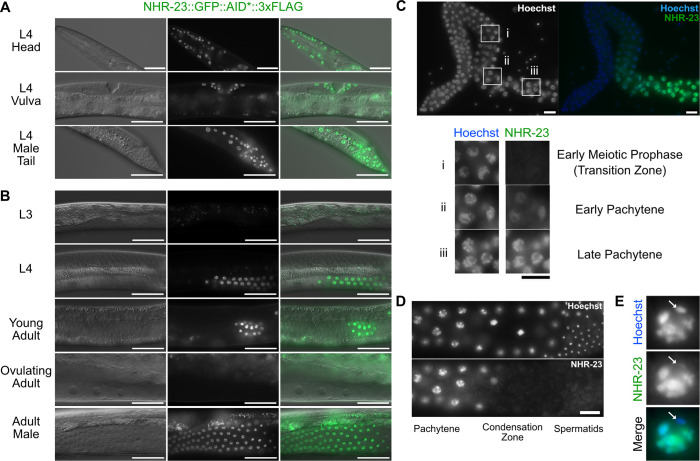
Fig. 2.
NHR-23::GFP::AID*::3xFLAG is expressed in somatic cells throughout development and in spermatogenic germlines. A strain carrying GFP::AID*::3xFLAG knocked-in to the endogenous nhr-23 gene to produce a C-terminal translational fusion to all known nhr-23 isoforms was used to monitor endogenous NHR-23 expression. (A) Representative DIC and GFP images of NHR-23::GFP::AID*::3xFLAG L4 larvae, specifically hypodermal cells of a hermaphrodite head, vulval precursor cells and seam/hypodermal cells of the male tail. (B) Representative DIC and GFP images of NHR-23::GFP::AID*::3xFLAG in pachytene cells of the germline in L3, L4, young adult, ovulating adult and adult male worms. (C) Fluorescent images of NHR-23::GFP::AID*::3xFLAG in a dissected adult male germline. (i-iii) Cells in early meiotic prophase (transition zone) (i), and in early (ii) and late (iii) pachytene. (D) Representative fluorescent images of NHR-23::GFP::AID*::3xFLAG in a late pachytene male germline. (E) Representative image of a late pachytene nucleus expressing NHR-23::GFP::AID*::3xFLAG. The location of the X chromosome is indicated with a white arrow. Scale bars: 40 µm in A,B; 10 µm in C-E. A minimum of 12 P0 animals were analyzed in A-C. Nuclei are visualized using Hoechst stain in C-E.

