Fig. 6.
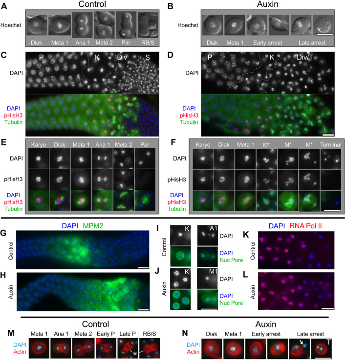
Metaphase I-like arrest in NHR-23-depleted spermatocytes. (A,B) Live spermatocytes ordered according to stage. Differential interference contrast (DIC) images of cells were overlaid by epifluorescence images of their Hoechst stained nuclei. (C-J) Isolated and fixed male gonads, and individual spermatocytes labeled with DAPI (blue) and indicated antibodies. (C,D) Gonad images show the proximal gonad from late pachytene (P) to either haploid spermatids (S) or terminal arrest (T) co-labeled with antibodies against α-tubulin (green) and phosphorylated histone H3 (ser10) (red). Controls (nhr-23::AID* without auxin or him-5 with auxin) in C; nhr-23::AID* with auxin in D. (E,F) Higher magnification images of individual spermatocytes. (G,H) Isolated control (G) and NHR-23-depleted (H) proximal male gonads (pachytene and later meiotic stages) co-labeled with antibodies against MPM2 (green), which binds diverse mitotic and meiotic phosphorylated proteins. (I,J) Staged spermatocytes co-labeled with anti-nuclear pore protein (green) in control (I) and NHR-23-depleted (J) males. (K,L) Control (K) and NHR-23-depleted (L) gonads (pachytene through karyosome) co-labeled with anti-phospho-RNA polymerase II CTD repeat (red) show switching off of global transcription in karyosome spermatocytes. (M,N) Aldehyde-fixed and staged spermatocytes with DNA pseudo-colored in cyan (DAPI) and actin microfilaments labeled with rhodamine. Arrow in N indicates the chromatin of an adjacent lysed cell. P, pachytene; K, karyosome; Div, meiotic divisions; Diak, diakinesis; Meta 1/M1, metaphase I; M*, aberrant metaphase I; Ana1/A1, anaphase I; Meta 2, metaphase II; Par, post-meiotic partitioning; RB, residual body; S, haploid spermatids; T, terminal-stage spermatocyte. Scale bars: 10 µm.

