Abstract
An increase in cork oak diseases caused by Biscogniauxia mediterranea and Diplodia corticola has been reported in the last decade. Due to the high socio-economic and ecologic importance of this plant species in the Mediterranean Basin, the search for preventive or treatment measures to control these diseases is an urgent need. Fungal endophytes were recovered from cork oak trees with different disease severity levels, using culture-dependent methods. The results showed a higher number of potential pathogens than beneficial fungi such as cork oak endophytes, even in healthy plants. The antagonist potential of a selection of eight cork oak fungal endophytes was tested against B. mediterranea and D. corticola by dual-plate assays. The tested endophytes were more efficient in inhibiting D. corticola than B. mediterranea growth, but Simplicillium aogashimaense, Fimetariella rabenhorstii, Chaetomium sp. and Alternaria alternata revealed a high potential to inhibit the growth of both. Simplicillium aogashimaense caused macroscopic and microscopic mycelial/hyphal deformations and presented promising results in controlling both phytopathogens’ growth in vitro. The evaluation of the antagonistic potential of non-volatile and volatile compounds also revealed that A. alternata compounds could be further explored for inhibiting both pathogens. These findings provide valuable knowledge that can be further explored in in vivo assays to find a suitable biocontrol agent for these cork oak diseases.
Keywords: fungi, endophytes, Biscogniauxia mediterranea, Diplodia corticola, biocontrol, cork oak
1. Introduction
Cork oak (Quercus suber L.) is an evergreen tree species that covers between 1.7 and 2.7 million ha in the western Mediterranean, distributed among Portugal, Spain, France, Italy, Morocco, Tunisia and Algeria [1]. Cork oak forests display a high socio-economic and ecological importance, being mainly explored for cork production [2]. The synthesis of cork, a homogeneous tissue of phellem cells, depends on the activity of the cork cambium, which covers the trunk and branches of cork oak. Due to the interesting and unique set of physical, biological and chemical properties, such as sealing and insulating features, cork is mainly used for the production of bottle stoppers [3]. Every year, 80% of worldwide produced cork comes from the Iberian Peninsula, in which Portugal is responsible for almost half of the total global production [2,4].
In recent years, cork oak forests have been declining in the Mediterranean region, which will be further enhanced by the combined effect of global warming and drought [5]. Indeed, the climate change predictions reveal the Mediterranean region as one of the most affected regions, and Mediterranean forests as one of the most vulnerable ecosystems to the combined effect of temperature increase and precipitation decrease [6,7]. Not only the geographic distribution of plant pathogens is predicted to be reshuffled [8], but also the severity of plant diseases and the rate at which endophytes switch from mutualistic to pathogenic behavior are expected to increase [9,10]. Indeed, during the last decades, an increase in cork oak diseases has been reported [11], including charcoal disease (caused by Biscogniauxia mediterranea (De Not.) Kuntze; Xylariales) and bot canker (caused by Diplodia corticola A.J.L. Phillips, A. Alves and J. Luque; Botryosphaeriales). Both pathogenic fungi have an endophytic lifestyle and opportunistic behavior, which account for the increase in diseases incidence in cork oak trees under environmental stress [12]. These diseases cause high economic losses due to the development of symptoms in cork oak trunk that affect both cork production and quality, and eventually lead to tree death [13].
Few preventive measures for cork oak charcoal disease and bot canker are currently known. Good phytosanitary practices in cork oak forests are essential to prevent the spreading of diseases through spore release and colonization from tree wounds derived from pruning or cork extraction [11]. Thiophanate-methyl and carbedazim fungicides have been found effective as a preventive measure against D. corticola [14,15]. However, the application of fungicides has been increasingly restricted due to the potential negative impact on human health and the environment. Therefore, more environmentally friendly strategies, such as the use of biological control agents, are now being explored for controlling many forest tree diseases [16,17]. Indeed, promising results have been obtained for restricting cork oak diseases. For example, the use of Fusarium tricinctum resulted in a reduced mortality of Q. cerris and Q. pubescens seedlings inoculated with D. corticola [18]. Further, Trichoderma spp. presented a high in vitro antagonistic potential against B. mediterranea and D. corticola [19]. In particular, a cork oak endophytic strain of Trichoderma citrinoviride produces peptaibols capable of inhibiting cork oak pathogens’ in vitro growth [20]. Spray suspensions of T. atroviride and T. viride combined with sanitation and scarification practices were recently suggested for decreasing charcoal disease incidence in Q. castaneifolia trees [21]. In the present study, we hypothesize that cork oak fungal endophytes are able to control cork oak pathogens’ (B. mediterranea and D. corticola) growth and we intend to highlight the most promising cork oak endophytes to be used as a biocontrol strategy. Specifically, we aimed to identify endophytic fungal species of cork oak that can be used to control B. mediterranea and D. corticola, by studying their antifungal activities against both pathogens and by characterizing the interactions between endophytes and pathogens.
2. Materials and Methods
2.1. Cork Oak Forests Sampling and Endophyte Recovery
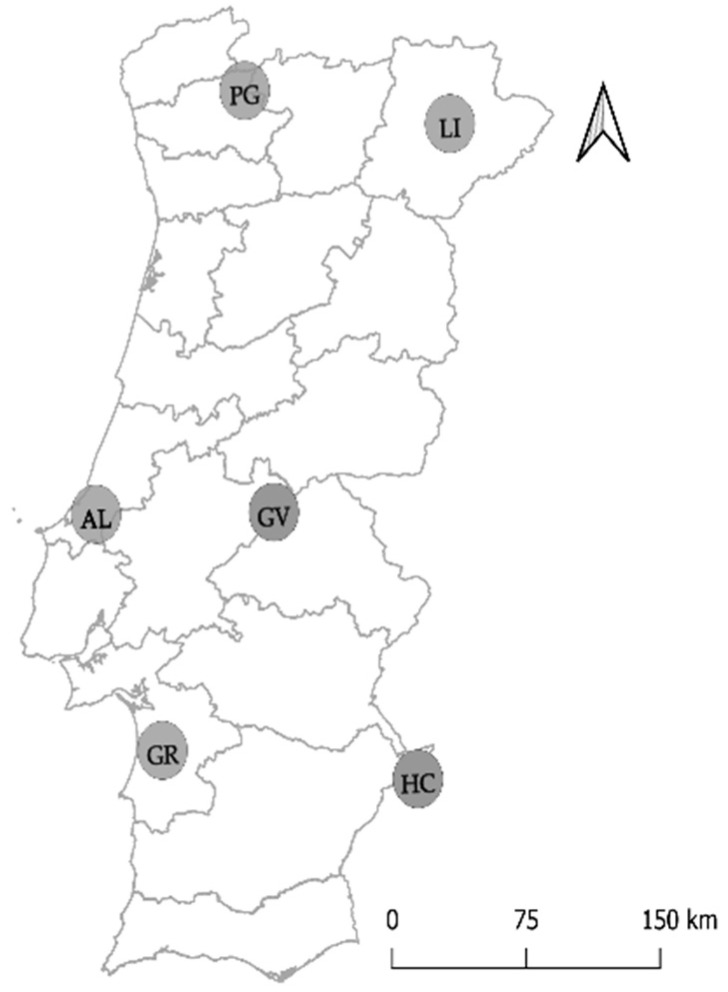
Cork oak samples were collected from eight different forests in Portugal (Table S1). Two forests were sampled from the National Park of Peneda-Gerês (PG-ER and PG-RC) and Herdade da Contenda (HC-CT and HC-MA). A single forest was sampled from Limãos (LI), Alcobaça (AL), Gavião (GV) and Grândola (GR). Between April and October of 2017, five to six trees were sampled from each cork oak forest, considering trees at different disease severity levels (Table 1; Figure 1). Disease severity levels were grouped into three categories and determined by considering defoliation (5 levels: 0–10%—no damage; 11–25%—light damage; 26–50%—moderate damage; 51–90%—severe damage; > 91%—extreme damage), as well as canopy and trunk damages (3 levels: 0—no damage; 1—moderate damage; 2—severe damage), for different factors (dried, wilting and decolorated leaves, presence of cankers, decolorated trunk, presence of exudates and visible sporulation). Trees were considered as healthy, presenting mild symptoms or declining. Healthy trees presented no or light defoliation, displaying low canopy and trunk damage (maximum of two factors with 1 damage level, but not affecting overall vigor of the tree). When tree vigor was moderately affected by a combination of factors (moderate defoliation and/or some canopy and trunk damage), trees were considered as displaying mild symptoms. Declining trees presented a clear decline in their vigor (accentuated or very accentuated defoliation that could be coupled with more than three factors classified with 1 and/or 2 damage levels).
Table 1.
Collection of cork oak branches in sampled forests. The number of trees sampled from each disease severity level is presented. For more details of sampled forests, see Table S1.
| Location | Cork Oak Stand | Collection Date | Disease Severity Level | ||
|---|---|---|---|---|---|
| Healthy | Mild Symptoms | Declining | |||
| Peneda-Gerês | PG-ER | May, 2017 | 2 | 3 | 0 |
| PG-RC | July, 2017 | 4 | 1 | 0 | |
| Limãos | LI | April, 2017 | 0 | 5 | 1 |
| Gavião | GV | July, 2017 | 0 | 4 | 2 |
| Alcobaça | AL | May, 2017 | 2 | 2 | 2 |
| Grândola | GR | May, 2017 | 0 | 3 | 3 |
| Herdade Contenda | HC-CT | October, 2017 | 2 | 2 | 2 |
| HC-MA | October, 2017 | 2 | 2 | 2 | |
Figure 1.
Distribution of cork oak stands sampled in Portugal. Letters represent sampled location (PG: Peneda-Gerês; LI: Limãos; GV: Gavião; AL: Alcobaça; GR: Grândola; HC: Herdade Contenda).
Five to seven branches were collected from each cork oak tree. Twigs were collected from each branch and thoroughly washed in tap water. To obtain only endophytes, a surface sterilization was performed based on the method described by Martins et al. [22]. Twigs were sequentially immersed in ethanol 70% (v/v) for 2 min, bleach (3–5% chlorine) for 6 min and ethanol 70% (v/v) for 1 min, followed by three washes in sterile deionized water (1 min each) and drying. Sterile twigs were cut into segments (4–5 cm) and transferred to Potato Dextrose Agar (PDA) medium (5 segments/plate). Three replicates were used for each branch. Sterilization controls were performed by spreading the last washing water (10 μL) onto PDA medium. Incubation was performed in the dark, at room temperature (21–23 °C). The outgrowing fungi were recognized as endophytic fungi and were successively subcultured in fresh PDA medium until pure cultures were obtained.
2.2. Endophytic Fungi Identification and Selection of Potential Antagonistic Fungi
Endophytic fungi were grouped into morphotypes, according to their cultural features (color, shape, elevation and margins) [23]. From each morphotype, at least three isolates were used for DNA extraction, using the Quick-DNA Fungal/Bacterial Miniprep Kit (Zymo Research, Irvine, CA, USA). The fungal rDNA-ITS region was amplified using universal primer pairs ITS1F (5’-CTTGGTCATTTAGAGGAAGTAA-3’) and ITS2 (5’-GCTGCGTTCTTCATCGATGC-3’), or ITS1F and ITS4 (5’-TCCTCCGCTTATTGATATGC-3’) [24]. PCR mixtures (25 µL) contained 1× Complete NH4 Reaction Buffer (BIORON GmbH, Germany), 200 µM of each dNTP (NZYTech, Portugal), 1 µM of each primer, 1 μL of DNA template and 1.25 U of DFS-Taq DNA Polymerase (BIORON GmbH, Germany). Amplifications were performed using the following protocol: initial denaturation 5 min at 94 °C; 35 cycles of 30 s at 94 °C, 30 s at 52 °C (or 54 °C with ITS1F-ITS4) and 60 s at 72 °C; final elongation at 72 °C for 10 min. PCR products were run on a 1% (w/v) agarose gel, stained with Green Safe Premium (NZYTech, Portugal). PCR products were purified using isopropanol 75% (v/v) and sequenced by Macrogen, Inc services (Madrid, Spain). DNA sequences were trimmed by sequencing quality and alignments were performed in Geneious 2010.4.8.5 (https://www.geneious.com). All sequences were identified using the UNITE [25] and NCBI GenBank databases and taxonomic classification was assigned for those with a similarity higher than 97%. Alignments with NCBI deposited sequences were used to confirm taxonomic classification and to attempt taxonomic classification of those sequences with similarity between 80 and 97%. Fungal identifications were supported by cultural and morphological features of fungal cultures. Fungal sequences were deposited in GenBank (www.ncbi.nlm.nih.gov/genbank/) under accession numbers MT819608–MT819946.
For understanding the ecological roles of fungal communities, the identified fungi were categorized into functional groups (pathogenic and/or beneficial), based on the bibliographic research [26,27]. Identified operational taxonomic units (OTUs) were assigned as beneficial when described as promoting plant growth and/or protecting the host against biotic or abiotic stress, while the pathogenic group contained fungi causing disease to its host and latent pathogens. Fungi belonging to other functional groups (mutualism, commensalism, etc.) were assigned to other groups and those with unknown function to unknown. Fungi belonging to more than one group (for example, pathogenic–beneficial) were added to both groups. For proceeding to the antagonism assays, endophytes were selected based on their potential beneficial role and availability. All groups were considered with the exception of those fungi exclusively described as pathogenic and all cork oak pathogens. Eight fungal isolates were selected to be tested against the pathogens in antagonistic assays due to their cultural readiness, namely Simplicillium aogashimaense isolate Gr67, Coniothyrium carteri isolate Gv5, Diaporthe passiflorae isolate Erm6, Fimetariella rabenhorstii isolate Br33, Fusarium oxysporum isolate Cab77, Chaetomium sp. isolate Erm52, Alternaria alternata isolate Cab37 and Penicillium olsonii isolate Gv63. B. mediterranea and D. corticola isolates were inoculated on cork oak stems to confirm symptoms development, completing Koch’s postulates.
2.3. Antagonistic Assay In Vitro by Dual-Plate and Categorization of Fungal Interactions
The eight selected endophytic fungi were tested in vitro against phytopathogens Biscogniauxia mediterranea isolate Gr13 and Diplodia corticola isolate Gr23, both isolated from cork oak trees showing mild symptoms. Antagonistic assays were performed by the dual-culture method in PDA medium. Fungal plugs (5 mm) of an actively growing endophyte and pathogen were placed 3 cm apart from each other (using 9 cm diameter Petri dishes) and incubated in the dark, at 28 °C (±2 °C). Those endophytes displaying a slow growth rate (<0.1 cm2/h; Table S2) were inoculated 72 h before the pathogen. In parallel, control plates were similarly prepared but using a single pathogen (or endophyte) plug. All plates were photographed 72 h after being inoculated with the endophyte and/or pathogen, and growth area was measured using ImageJ 1.50i software [28]. Growth areas were used to determine the percentage of mycelial growth inhibition, according to the formula: (Ac−Ai)/Ac × 100, where Ac is the area of fungal growth in the control plate and Ai is the area of fungal growth with the interacting fungus. Three independent experiments were performed (with at least 3 replicates each) for all endophyte/pathogen combinations. Statistical analysis was performed using ANOVA in GraphPad Prism 7.00 software (La Jolla, CA, USA) to determine the impact of endophytes on pathogen growth.
Fungal interactions between endophytes and pathogens were categorized based on Tuininga [29]. Considering that (-) corresponds to mycelial growth decrease, (+) to growth increase and (0) to similar mycelial growth, interactions among endophyte/pathogen could be defined as co-antagonism (-/-), antagonism (-/0), agonism (-/+), co-habitation (0/0), commensalism (0/+) and mutualism (+/+). For describing the fungal interactions, dual-culture plates were observed daily for 15 days. Mycelial interactions were also recorded based on Badalyan et al. [30], who defined the following classes: A—deadlock with mycelial contact; B—deadlock at distance; C—replacement, overgrowth without deadlock; CA1 and CA2—partial and complete replacement after initial deadlock with mycelial contact; and CB1 and CB2—partial and complete replacement after initial deadlock at a distance. Finally, hyphal interactions were observed using mycelia from the interacting region of dual-culture plates, after 15 days of interaction, and compared with mycelia taken from controls. Photographs were taken using a Leica MC170 HD digital camera attached to a Leica S9 D stereomicroscope (Leica Microsystems, Germany) or using an automated Leica DM5000B microscope (Leica Microsystems, Germany).
2.4. Antifungal Non-Volatile Compounds Assay
The antagonistic effect of the metabolites produced by the eight selected fungal endophytes was tested using a method adapted from Campanile et al. [18]. Three mycelial fungal plugs (5 mm) of actively growing endophytes were inoculated in Erlenmeyer flasks (250 mL) containing 50 mL of sterile Potato Dextrose Broth (BD Difco™, Switzerland). Culture flasks were placed in an orbital incubator at 28 °C (±2 °C), with 150 rpm agitation, for 12 days. Liquid cultures were transferred to falcon tubes and centrifuged for 15 min at 12,000 rpm. The supernatant was collected and filtered through a 0.22 µM membrane filter to remove hyphal residues and conidia. Fungal filtrates were added to sterile warm PDA medium [20% (v/v)] and poured into 9 cm Petri dishes. A mycelial plug of an actively growing pathogen (B. mediterranea or D. corticola) was placed in the center of the plate. Control plates were similarly prepared but containing PDA without a fungal filtrate. Mycelial growth areas were determined as previously referred and mycelial growth inhibition was determined according to the formula: (Ac−Af)/Ac × 100, where Ac is the area of fungal growth in the control plate and Af is the area of fungal growth in the presence of the filtrate. Three independent experiments were performed (with at least 3 replicates each) for all endophyte/pathogen combinations. Statistical analysis was performed using ANOVA in GraphPad Prism 7.00 software (La Jolla, CA, USA) to understand the impact of endophytic compounds on pathogen growth.
2.5. Antifungal Volatile Compounds Assay
The effect of volatile compounds produced by the eight selected fungal endophytes in inhibiting pathogen growth was evaluated by an inverted plate method [31]. For each endophyte/pathogen combination, two PDA plates were inoculated with a single mycelial plug (5 mm) of an actively growing endophyte (or pathogen). Then, the PDA plate with the pathogen was inverted on the top of the endophyte plate and both were sealed with parafilm. Incubation occurred at 28 °C (±2 °C), for 72 h. Control plates were performed without inoculation of the endophyte in the bottom plate. As previously described, those endophytes displaying a slow growth rate (<0.1 cm2/h; Table S2) were inoculated 72 h before the pathogen. The percentage of inhibition was calculated as mentioned before. Three independent experiments were performed (with at least 3 replicates each) for all endophyte/pathogen combinations. Statistical analysis was performed using ANOVA in GraphPad Prism 7.00 software (La Jolla, CA, USA) to understand the impact of endophytic volatiles on pathogen growth.
3. Results and Discussion
3.1. Endophytic Fungal Community of Cork Oak
Endophytic fungi of cork oak twigs were recovered from eight different forests in Portugal. From a total of 1117 fungal isolates, 440 were molecularly identified and grouped into 128 OTUs comprising 18 orders, 38 families, 45 genera and 39 species (Table 2). Only OTUs classified up to genus and species were considered in this work (70 OTUs). From these, and to the best of our knowledge, 54 fungal OTUs have never been reported as cork oak endophytes before (e.g., Fimetariella rabenhorstii, Discosia sp.), including 18 that have never been described as plant endophytes (e.g., Caliciopsis beckhausii, Diaporthe passiflorae, Proliferodiscus sp.) (Table 2). Among previously undescribed cork oak endophytes, 28 were only recovered from a single sampled cork oak forest, but others (26) were recovered from different locations, which strengthens their role as cork oak endophytes. For example, Discosia sp., Cryphonectria naterciae and Neocucurbitaria sp. were recovered from different Portuguese forests, or from forests displaying the highest (e.g., F. rabenhorstii) or the lowest (e.g., Plectania rhytidia) precipitation levels.
Table 2.
Endophytic fungal OTUs recovered from twigs of cork oak trees located in different forests. Taxonomic classification (with closest match identity in brackets) and their respective functional group are revealed. Reports about their endophytic behavior are referred to. When existing, references related to cork oak are presented (bold). Isolates used in this work are depicted in red. Functional groups are represented as: P—pathogenic; B—beneficial; O—other; U—unknown.
| Taxonomic Classification | Closest Match GenBank | Cork Oak Forests | Functional Group | Identified Endophyte | ||||||||
|---|---|---|---|---|---|---|---|---|---|---|---|---|
| PG-ER | PG-RC | LI | AL | GV | GR | HC-CT | HC-MA | |||||
| Ascomycota | ||||||||||||
| Amphisphaeriales | Discosia sp. | KU325138.1 (100%) | x | x | P [46]/B [47] | [48] | ||||||
| Nonappendiculata quercina | MH554025.1 (98.78%) | x | U | - | ||||||||
| Botryosphaeriales | Diplodia corticola | MT015621.1 (100%) | x | P [37] | [40] | |||||||
| Diplodia quercivora | JX894205.1 (97.72%) | x | P [38] | [11] | ||||||||
| Dothiorella iberica | MT261024.1 (100%) | x | P [49] | [12] | ||||||||
| Neofusicoccum parvum | MT645697.1 (99.3%) | x | P [35] | [50] | ||||||||
| Capnodiales | Cladosporium herbarum | LT854669.1 (99.22%) | x | P [51] | [52] | |||||||
| Cladosporium perangustum | MK111614.1 (99.10%) | x | P [53] | [54] | ||||||||
| Cladosporium sp. | MN879328.1 (100%) | x | x | P [55]/B [56] | [40] | |||||||
| Cladosporium sphaerospermum | MT645920.1 (99.51%) | x | B [57] | [57] | ||||||||
| Coryneliales | Caliciopsis beckhausii | NR_132090.1 (99.57%) | x | x | U | - | ||||||
| Caliciopsis sp. | NR_132090.1 (91.91%) | x | x | P [58] | - | |||||||
| Diaporthales | Coryneum sp. | MH674330.1 (95.54%) | x | x | x | x | P [35] | [40] | ||||
| Cryphonectria naterciae | MT645942.1 (100%) | x | x | x | x | P [36] | - | |||||
| Cytospora cedri | MN871816.1 (100%) | x | x | x | P [59] | - | ||||||
| Cytospora cinereostroma | KY051964.1 (100%) | x | x | U | - | |||||||
| Cytospora ribis | KP641138.1 (100%) | x | U | [60] | ||||||||
| Cytospora sp. | MK656248.1 (100%) | x | x | P [61] | [40] | |||||||
| Dendrostoma sp. | MN447228.1 (99.66%) | x | P [62] | - | ||||||||
| Diaporthe passiflorae | NR_120155.1 (99.82%) | x | O [63]/P [64] | - | ||||||||
| Diaporthe sp. | MT561408.1 (99.48%) | x | x | x | P [65] | [65] | ||||||
| Discula quercina | MH758705.1 (99.18%) | x | P [39] | [40] | ||||||||
| Discula sp. | KY367498.2 (94.23%) | x | P [66] | [67] | ||||||||
| Gnomoniopsis paraclavulata | MH863162.1 (100%) | x | x | U | - | |||||||
| Dothideales | Aureobasidium pullulans | MT645930.1 (99.57%) | x | O [68]/B [69] | [40] | |||||||
| Eurotiales | Penicillium glabrum | MT582777.1 (100%) | x | x | x | P [70] | [71] | |||||
| Penicillium olsonii | MT582783.1 (100%) | x | B [72] | [73] | ||||||||
| Penicillium sp. | LN901128.1 (99.54%) | x | x | x | x | x | O [74]/P [70]/B [72] | [40] | ||||
| Helotiales | Ciboria sp. | KF545322.1 (94.59%) | x | P [75] | - | |||||||
| Pezicula cinnamomea | MK907714.1 (100%) | x | P [76] | [77] | ||||||||
| Pezicula neosporulosa | KR859231.1 (100%) | x | P [78] | [79] | ||||||||
| Pezicula sporulosa | MH862573.1 (98.66%) | x | O[80] | [81] | ||||||||
| Pezicula sp. | MG098317.1 (100%) | x | x | O [82]/P [76] | [67] | |||||||
| Proliferodiscus sp. | MN901941.1 (95.50%) | x | U | - | ||||||||
| Hypocreales | Clonostachys sp. | MK789204.1 (91.84%) | x | B [83] | [84] | |||||||
| Fusarium compactum | KJ562364.1 (98.53%) | x | P [85] | [86] | ||||||||
| Fusarium oxysporum | MT530243.1 (100%) | x | x | x | x | P [87]/B [88] | [89] | |||||
| Fusarium sp. | MT645120.1 (100%) | x | x | x | x | x | x | P [87]/B [18] | [18] | |||
| Sarocladium kiliense | MK789203.1 (100%) | x | x | x | x | x | P [90]/B [91] | [92] | ||||
| Sarocladium sp. | MT645143.1 (99.36%) | x | x | x | x | P [93]/B [91] | [94] | |||||
| Simplicillium aogashimaense | MK685280.1 (99.82%) | x | x | U | - | |||||||
| Simplicillium sp. | MH859771.1 (99.12%) | x | x | x | B [95] | [95] | ||||||
| Tolypocladium sp. | KX034386.1 (100%) | x | O [96] | [97] | ||||||||
| Pezizales | Plectania rhytidia | MH003435.1 (98.99%) | x | x | U | [27] | ||||||
| Pseudoplectania ericae | MT498082.1 (99.65%) | x | U | - | ||||||||
| Pyronema sp. | MT556695.1 (100%) | x | x | O [98] | [99] | |||||||
| Phaeomoniellales | Neophaeomoniella sp. | MK646052.1 (96.14%) | x | P [100] | [101] | |||||||
| Pleosporales | Alternaria alternata | MT635274.1 (100%) | x | x | P [102]/B [103] | [40] | ||||||
| Alternaria sp. | MT557456.1 (100%) | x | x | x | x | P [104]/B [103] | [40] | |||||
| Angustimassarina sp. | MN963689.1 (100%) | x | U | - | ||||||||
| Camarosporium brabeji | LN714529.1 (97.76%) | x | x | U | [105] | |||||||
| Coniothyrium carteri | KX359604.1 (99.82%) | x | x | x | B [106] | [106] | ||||||
| Coniothyrium ferrarisianum | MH860854.1 (100%) | x | U | [107] | ||||||||
| Coniothyrium palmicola | JX681086.1 (99.53%) | x | U | - | ||||||||
| Epicoccum nigrum | MT548679.1 (100%) | x | x | x | B [27] | [40] | ||||||
| Kalmusia sp. | MK796143.1 (100%) | x | x | P [108] | [109] | |||||||
| Neocucurbitaria cava | MK796144.1 (100%) | x | x | x | U | [110] | ||||||
| Neocucurbitaria sp. | MH858303.1 (93.78%) | x | x | x | x | x | U | [110] | ||||
| Phoma sp. | KX815489.1 (100%) | x | x | P [111] | [40] | |||||||
| Preussia sp. | MN696547.1 (100%) | x | B [112] | [113] | ||||||||
| Pyrenophora biseptata | MH864748.1 (100%) | x | P [114] | - | ||||||||
| Sordariales | Chaetomium globosum | MT588864.1 (100%) | x | B [115] | [116] | |||||||
| Chaetomium sp. | MN153902.1 | x | x | x | B [117] | [118] | ||||||
| Copromyces sp. | (100%) | x | U | - | ||||||||
| Fimetariella rabenhorstii | MN555335.1 (100%) | x | x | x | O [119]/P [120] | [121] | ||||||
| Xylariales | Biscogniauxia mediterranea | MT862330.1 (100%) | x | x | x | x | x | x | x | x | P [12] | [40] |
| Daldinia sp. | MN341734.1 (97.83%) | x | B [122] | [123] | ||||||||
| Xylaria sp. | JQ761730.1 (99.78%) | x | x | P [124]/B [125] | [126] | |||||||
| Basidiomycota | ||||||||||||
| Atheliales | Athelia arachnoidea | MH860510.1 (100%) | x | P [127] | - | |||||||
Some OTUs were more widespread throughout Portuguese cork oak forests than others. B. mediterranea was the only fungus identified in all cork oak stands and Fusarium sp. was present in all forests, with the exception of LI and GR. Further, Penicillium sp., Sarocladium kiliense and Neocucurbitaria sp. were recovered from almost all cork oak stands. In contrast, there were fungi (such as Diplodia corticola, Nonappendiculata quercina, Ciboria sp. and Pezicula cinnamomea) that were only recovered from one sampling location. Since many fungi are difficult to be cultured, culture-dependent methods are known to underestimate fungal communities [32]. The used approach only targeted those endophyte fungi that can be easily cultured on artificial media and have a rapid growth rate. Furthermore, as many endophytes do not sporulate in culture, their morphotype discrimination through cultural features is challenging and might have been underestimated. For these reasons, we are aware that the performed endophyte survey through cultural methods (and based on morphotypes discrimination) misrepresented the endophyte diversity in cork oak forests, as already reported elsewhere for other plant hosts [32]. Accordingly, our study reported a strong dominance of fungi belonging to Ascomycota (only one OTU belonged to the Basidiomycota phylum), like previously reported in other studies using culture-dependent methods, e.g., [33,34]. Culture-independent methods could have provided a different picture of fungal communities. For example, in grapevine, besides Ascomycota (described using cultural methods), Basidiomycota and Zygomycota fungi were additionally detected recurring to a metabarcoding approach [33]. Despite the recognized limitations, the used culture-dependent approach provided the availability of endophyte isolates to proceed in searching for biocontrol strategies.
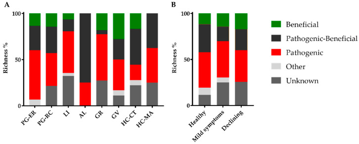
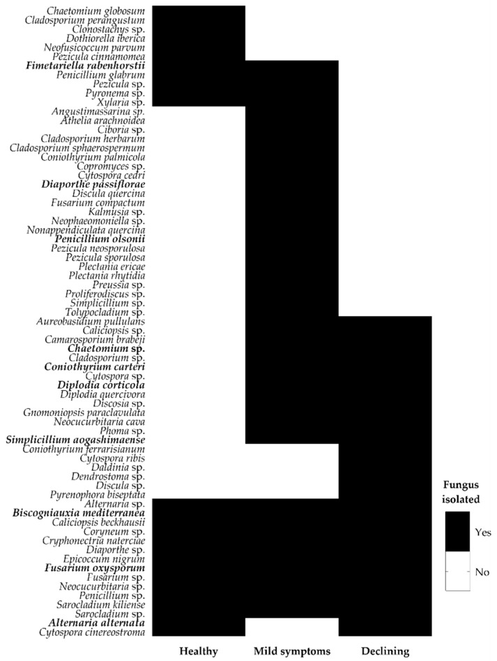
The ecological role of each identified fungal species was determined based on the literature. As the functional role of certain endophytes could change according to their plant host genotype, during different stages of the plant life cycle or in extreme conditions [26], many endophytes were included in mixed groups (such as the pathogenic–beneficial group). Furthermore, within certain genera, there are species known to be pathogenic and others beneficial, being impossible to consider a single functional role. Despite these constrains, the number of identified OTUs displaying a phytopathogenic role was higher, when compared to other functional groups (Table 2). A total of 39 OTUs were considered as displaying a phytopathogenic role (including exclusive phytopatogenic (26), phytopathogenic–beneficial (10) and phytopathogenic–other (3) functional groups), while only 21 displayed a beneficial role (including beneficial (10), phytopathogenic–beneficial (10) and beneficial–other (1) functional groups). The richness of fungi displaying a potential phytopathogenic role was also higher than any other functional group in all cork oak stands and whatever the cork oak tree disease severity level (Figure 2). Among them, few cork oak pathogens were found: B. mediterranea [12], Coryneum sp. [35], Cryphonectria naterciae [36], D. corticola [37], D. quercivora [38], Discula quercina [39] and Neofusicoccum parvum [35]. Interestingly, B. mediterranea, Coryneum sp. and C. naterciae were isolated from trees in all disease severity levels, while D. corticola, D. quercivora and Discula quercina were only isolated from trees with declining symptoms (Figure 3). In agreement, cork oak pathogens (B.mediterranea and Coryneum sp.) were isolated from healthy cork oak trees in Italy [12,40]. The presence of D. corticola, D. quercivora and Discula quercina in declining cork oak trees agrees with their role as emerging pathogens to Quercus spp. in different regions of the world [11,41], which is emphasized by their risk to cork oak health as previously reported [39,42]. Concerning the fungi displaying a beneficial role, AL forest presented the highest number of OTUs with a potential beneficial role (all described as pathogenic–beneficial), but GV forest was the richest with exclusive beneficial fungi (Figure 2A). GV forest corresponded to one of the forests displaying the highest number of declining trees (results not presented), which agrees with the finding that trees with declining symptoms displayed a higher richness of exclusive beneficial fungi (a non-significant 1.5-fold increase in relation to healthy trees; Figure 2B). The role of stress-affected plants in recruiting beneficial microorganisms is still under debate and a “cry-for-help” hypothesis was recently proposed, in which plants are able to recruit plant-protective microbes when they are under attack by pathogens [43,44]. For example, tomato plants under stress produce root exudates to signal the beneficial Trichoderma harzianum T22 strain to direct growth toward the plant host [45]. However, further studies on endophyte distribution among healthy and diseased cork oak trees are still needed for providing clear evidence that support a “cry-for-help” strategy for the sustainability of threatened cork oak forests.
Figure 2.
Cork oak endophytic fungal functional groups presented in cork oak forests (A) and disease severity level (B). Operational taxonomic units (OTUs) belonging to more than one functional group were added to both, except for the pathogenic–beneficial group that is represented as such.
Figure 3.
Endophytic fungal OTUs recovered from cork oak trees at different disease severity levels. Black/white color indicates fungal isolation/no isolation from trees with different disease severity levels. Isolates used in this work are depicted in bold.
3.2. Interactions of Endophytes against B. mediterranea and D. corticola
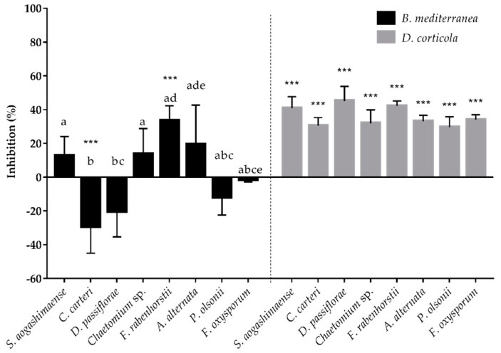
Eight endophyte isolates were selected for testing their ability to inhibit B. mediterranea and D. corticola growth, based on their potential beneficial ecological role, availability and culture readiness. D. corticola mycelial growth was persistently inhibited by the presence of these cork oak endophytes, while B. mediterranea was differentially inhibited by endophytes (Figure 4). Dual-plate experiments revealed that B. mediterranea growth was inhibited by F. rabenhorstii (33.4%, p ≤ 0.001), A. alternata (19.3%), Chaetomium sp. (13.6%) and S. aogashimaense (12.6%), whereas the other fungal endophytes promoted or did not affect pathogen growth (Figure 4). Regarding endophytes inhibiting B. mediterranea and considering the effect of pathogens on endophyte growth (Figure S1), S. aogashimaense was the only endophyte not inhibited by this pathogen, displaying a typical antagonist interaction (0/-) (Table 3). Although F. rabenhorstii strongly inhibited B. mediterranea, there was a negative impact of the pathogen on this endophyte growth (co-antagonism; -/-). Both endophytes displayed a similar interaction with the D. corticola pathogen, resulting in antagonism/co-antagonism with pathogen inhibition (40.8%, p ≤ 0.001 by S. aogashimaense; 42.2%, p ≤ 0.001 by F. rabenhorstii). In contrast, A. alternata and Chaetomium sp. increased their growth while inhibiting B. mediterranea, thus displaying agonist interactions (+/-) with this pathogen. Both endophytes displayed a distinct interaction with D. corticola. A. alternata was not affected (antagonism; 0/-) and Chaetomium sp. was inhibited by this pathogen (co-antagonism; /). Except for F. oxysporum (antagonism; 0/-), all the other tested endophytes also revealed a co-antagonism interaction with D. corticola.
Figure 4.
Inhibition of B. mediterranea and D. corticola growth caused by the tested endophytic fungi in dual-culture assay. Negative values indicate that the area of the plate covered by the interacting pathogen was higher than that of the control. Different letters represent statistical significance (p ≤ 0.005) between endophytes in each antagonistic assay and *** (p ≤ 0.001) represents statistical significance between the control and co-culture of a pathogen.
Table 3.
Classification of fungal interactions occurring between tested endophytes and pathogens on dual-culture assay. The interaction type was classified based on endophyte/pathogen growth by “+“ for higher growth, “-“ for less growth and “0“ for equal growth in relation to control. Underlined mycelial interactions denote endophyte replacement by the pathogen.
| Endophyte | B. Mediterranea | D. Corticola | ||
|---|---|---|---|---|
| Type of Interaction | Mycelial Interaction | Type of Interaction | Mycelial Interaction | |
| S. aogashimaense | antagonism (0/-) | B | antagonism (0/-) | B |
| C. carteri | agonism (-/+) | CA2 | co-antagonism (-/-) | CA2 |
| D. passiflorae | agonism (-/+) | CB1 | co-antagonism (-/-) | CB1 |
| F. rabenhorstii | co-antagonism (-/-) | A | co-antagonism (-/-) | CA1 |
| F. oxysporum | commensalism (+/0) | CA1 | antagonism (0/-) | CA1 |
| Chaetomium sp. | agonism (+/-) | CA1 | co-antagonism (-/-) | CA1 |
| A. alternata | agonism (+/-) | CA1 | antagonism (0/-) | CA1 |
| P. olsonii | agonism (-/+) | CB1 | co-antagonism (-/-) | CA1 |
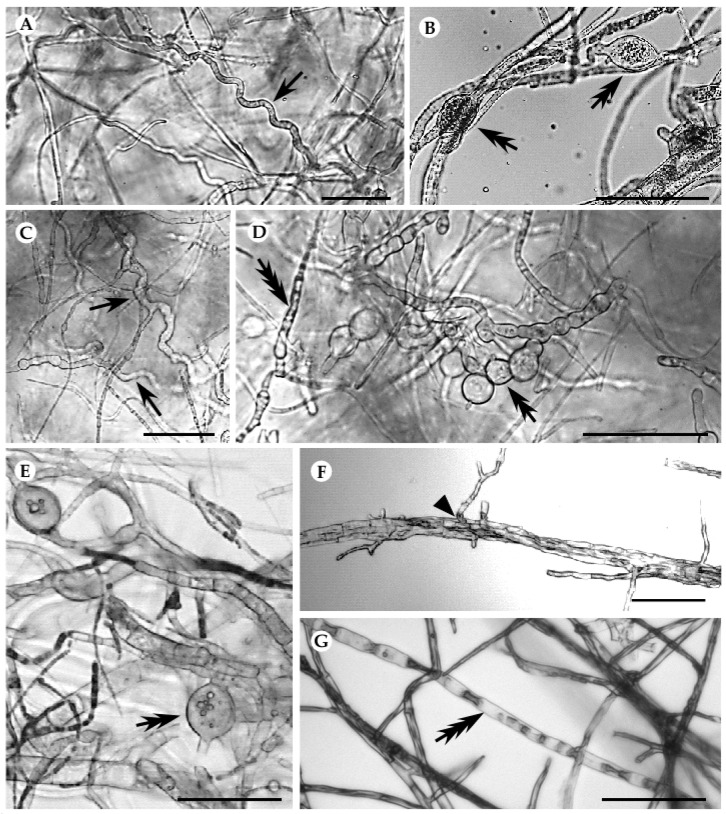
Fungal interactions were further evaluated by following the macro- and microscopic modifications of mycelia in the interaction region. When interacting with both pathogens, S. aogashimaense revealed a deadlock at distance (B) interaction type (Figure S2A,B) and caused visible modifications on hyphae of both pathogens (Figure 5A–D). In the presence of this endophyte, B. mediterranea showed typical hyphal deformations caused by the interacting partner, such as the presence of coiled hyphae and production of vesicle-like structures (Figure 5A,B). Further, D. corticola suffered mycelial modifications caused by interaction with S. aogashimaense, which included hyphal coiling and vacuolization, as well as the production of vesicle-like structures (Figure 5C,D). Such alterations have been frequently reported in different incompatibility systems [128,129] and have been related to programmed cell death (PCD) events occurring during interaction [130]. The A. alternata–B. mediterranea interaction also revealed hyphal deformations, such as hyphal vacuolization, production of vesicle-like structures and hyphal penetration that resemble a mycoparasitism interaction (Figure 5E–G). Accordingly, instead of a deadlock at distance interaction type, in this interaction, there was a partial replacement of mycelia after an initial deadlock with mycelial contact (CA1; Table 3 and Figure S2C). Interestingly, although the inhibitory activity of an A. alternata isolate from Q. cerris against D. corticola has been reported, both in dual-culture and in planta [18], we have not detected hyphal distortions in the A. alternata–D. corticola interaction. All the other studied interactions did not reveal hyphal distortions, even though similar interaction types have been detected after 15 days of interaction (partial replacement after initial deadlock with mycelial contact; CA1), as well as a deadlock with mycelial contact (A) (Table 3).
Figure 5.
Hyphal modifications produced in the interaction between S. aogashimaense and B. mediterranea (A,B), S. aogashimaense and D. corticola (C,D) and A. alternata and B. mediterranea (E–G). Single arrows designate coiled hyphae, double arrows vesicle-like structures and triple arrows hyphal vacuolization. Black line represents 50 µM scale.
3.3. Fungal Inhibitors Production by Cork Oak Endophytes
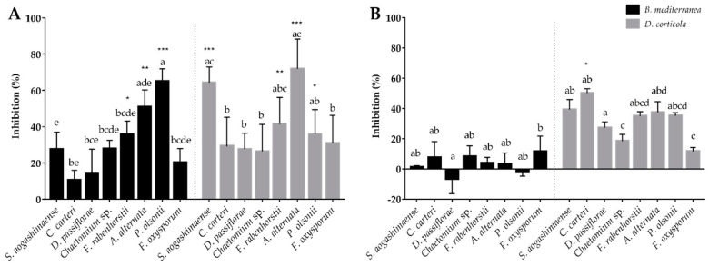
For understanding the production of inhibitors by the tested endophytes, their non-volatile and soluble compounds (produced in liquid culture) and volatile emissions were tested against B. mediterranea and D. corticola pathogens. Endophytic soluble compounds seem to have more impact on inhibiting mycelial growth than volatile compounds, mainly against B. mediterranea (Figure 6). However, this primary conclusion should be taken with some precaution as these inhibitory compounds were tested by using different strategies for their collection and assay. The endophytes that revealed the greatest inhibitory activity against B. mediterranea in the dual-culture method (F. rabenhorstii, A. alternata, Chaetomium sp. and S. aogashimaense) produced a soluble extract with high anti-fungal activity against both pathogens. These endophytes inhibited from 28% to 51% of B. mediterranea growth (F. rabenhorstii p ≤ 0.05 and A. alternata p ≤ 0.01) and from 26% to 72% of D. corticola growth (S. aogashimaense and A. alternata p ≤ 0.001; F. rabenhorstii p ≤ 0.01). The volatile emissions from the same endophytes also revealed inhibitory activities against B. mediterranea and D. corticola (ranging from 1% to 8% for B. mediterranea, and 18% to 39% for D. corticola). Multiple studies have already revealed the antifungal activity of genera from tested endophytes, in particular of Chaetomium spp., e.g., [131], Coniothyrium spp., e.g., [132], Diaporthe spp., e.g., [133], Penicillium spp., e.g., [134], and Simplicillium spp., e.g., [135], as well as their ability to produce a battery of antifungal compounds. However, less information is available on the inhibitory activity of Fimetariella spp. or the antifungal activities of A. alternaria and F. oxysporum species, which have been mainly recognized as important plant pathogens. This work provides new information related to the production of antimicrobial compounds, mainly from F. rabenhorstii and A. alternata, against cork oak pathogens.
Figure 6.
Inhibition of B. mediterranea and D. corticola growth caused by non-volatile (A) and volatile compounds (B) produced by the tested endophytic fungi. Negative values indicate that the area of the plate covered by the interacting pathogen was higher than that of the control. Different letters represent statistical significance (p < 0.005) between endophytes in each antagonistic assay and * (p ≤ 0.05), ** (p ≤ 0.01) and *** (p ≤ 0.001) represent statistical significance between the control and co-culture of a pathogen.
Interestingly, endophytes with no inhibitory activity against B. mediterranea in dual-plate methods also revealed the production of inhibitors for this phytopathogen. For example, the soluble compounds of P. olsonii revealed the highest inhibition (65%, p ≤ 0.001) of all against B. mediterranea, and volatiles from C. carteri and F. oxysporum also exhibited high inhibitory activity against the same pathogen (8% and 12%, respectively). These results suggest that the inhibitory effect of a specific fungal isolate against a phytopathogen may be due to the production of multiple compounds that could act in a synergistic or antagonistic way [136]. Accordingly, the production of inhibitory compounds such as volatiles, antibiotics and other secondary metabolites is gaining biotechnological interest for the control of phytopathogens. For example, the potential of S. coffeanum volatile compounds against Aspergillus species was reported by Gomes et al. [95] and S. lamellicola was used to produce a fungicide against Botrytis cinerea [137]. The production of inhibitory compounds is gaining interest when produced by potential pathogens. For example, the potential of Alternaria sp. was already reported for the control of fungal and bacterial growth [103], but some species are widely known as phytopathogens [104], representing a disadvantage for field application. The same is described for the F. oxysporum phytopathogen [88,138]. Therefore, the recognition of non-volatile or volatile inhibitory compounds could represent a biotechnological advantage for using those isolates as biocontrol agents.
4. Conclusions
The use of naturally adapted endophytes (for a specific plant host/environment) in a biocontrol strategy has gained increasing interest for restricting plant diseases. In this cork oak endophyte survey, we detected a high number of fungal OTUs from trees displaying different disease severity levels. The number of OTUs belonging to the pathogenic functional group (including described pathogens to other plant species) was high, even when considering healthy trees. Isolates of potential pathogens (namely, Alternaria alternata and Fusarium oxysporum) have revealed a strong in vitro inhibitory effect against cork oak pathogens (B. mediterranea and D. corticola). In particular, an A. alternata isolate revealed a high inhibitory activity against both pathogens, promoting hyphal deformations on B. mediterranea. Although this was not the case when interacting with D. corticola, this pathogen was similarly inhibited by A. alternata and F. oxysporum, displaying a mycelial interaction type based on the partial replacement of mycelia after an initial deadlock with mycelial contact. Non-volatiles and volatiles obtained from these isolates (particularly from A. alternata) revealed inhibitory activity and their potential to be used in a biocontrol strategy for restraining cork oak diseases should be further explored.
The colonization of plants by beneficial endophytes has been a useful biocontrol strategy. This work suggests Simplicillium aogashimaense as an antagonistic fungus towards B. mediterranea and D. corticola with potential to be used as a biocontrol agent against cork oak diseases. Indeed, S. aogashimaense presented promising results inhibiting both pathogens’ growth, which was reinforced by the promotion of pathogens’ hyphae deformations during interaction (deadlock at distance). A high ecological and economical value has been given to Simplicillium species due to their biocontrol role and production of bioactive compounds. For example, soybean plants when inoculated with S. lanosoniveum before infection with soybean rust pathogen (Phakopsora pachyrhizi) revealed reduced disease severity [139]. Although we believe S. aogashimaense could similarly be explored to control cork oak diseases, better understanding of its potential role as a biocontrol agent is still required. We thus conclude that cork oak endophytes could be further explored as biocontrol agents against cork oak diseases.
Acknowledgments
The authors thank Maria Carolina Varela for the help in identification of disease severity levels of trees and Pinto Moreira (HC forests), Eng. Carlos Barroso Dias (PNPG), Eng. Luís Dias (Herdade das Barradas da Serra—GR) and José Lino Neto (GV forest) for providing access to cork oak forests.
Supplementary Materials
The following are available online at https://www.mdpi.com/2309-608X/6/4/287/s1, Table S1: Characterization of sampled cork oak stands; Table S2: Growth rates of cork oak endophytic fungi used for the antagonistic assays; Figure S1: Inhibition of fungal endophytes growth caused by the B. mediterranea and D. corticola in dual culture assay; Figure S2: Interaction in dual culture of S. aogashimaense-B. mediterranea (A), S. aogashimaense-D. corticola (B) and A. alternata- B. mediterranea (C) 72 h and 15 days after inoculation.
Author Contributions
Conceived and designed the experiments: T.L.-N., P.B., R.M.T. and D.C. Sampling: T.L.-N. and D.C. Conducted the main experiments: D.C. Analyzed the data: T.L.-N. and D.C. Wrote the original draft: D.C. Reviewed, edited and prepared the MS for submission: T.L.-N., P.B. and R.M.T. All authors have read and agreed to the published version of the manuscript.
Funding
This work was supported by FEDER funds through COMPETE (Programa Operacional Factores de Competitividade) and by national funds by FCT (Fundação para a Ciência e a Tecnologia) in the framework of the project POCI-01-0145-FEDER-028635, as well as for financial support to BioISI (UIDB/04046/2020) and CIMO (UID/AGR/00690/2020) by national funds by FCT/MCTES/PIDDAC. D. Costa thanks FCT for PhD grant SFRH/BD/120516/2016.
Conflicts of Interest
The authors declare no conflict of interest.
Footnotes
Publisher’s Note: MDPI stays neutral with regard to jurisdictional claims in published maps and institutional affiliations.
References
- 1.Gauquelin T., Michon G., Joffre R., Duponnois R., Génin D., Fady B., Dagher-Kharrat M.B., Derridj A., Slimani S., Badri W., et al. Mediterranean forests, land use and climate change: A social-ecological perspective. Reg. Environ. Chang. 2018;18:623–636. doi: 10.1007/s10113-016-0994-3. [DOI] [Google Scholar]
- 2.FAO and Plan Bleu . State of Mediterranean Forests 2018. Food and Agriculture Organization of the United Nations, Food and Agriculture Organization of the United Nations, Rome and Plan Bleu; Marseille, France: 2018. [Google Scholar]
- 3.Costa R., Lourenço A., Oliveira V., Pereira H. Chemical characterization of cork, phloem and wood from different Quercus suber provenances and trees. Heliyon. 2019;5:02910. doi: 10.1016/j.heliyon.2019.e02910. [DOI] [PMC free article] [PubMed] [Google Scholar]
- 4.APCOR . APCOR’s Cork Yearbook 2018/2019. Portuguese Cork Association; Santa Maria de Lamas, Portugal: 2019. [Google Scholar]
- 5.Touhami I., Chirino E., Aouinti H., El Khorchani A., Elaieb M.T., Khaldi A., Nasr Z. Decline and dieback of cork oak (Quercus suber L.) forests in the Mediterranean basin: A case study of Kroumirie, Northwest Tunisia. J. For. Res. 2020;31:1461–1477. doi: 10.1007/s11676-019-00974-1. [DOI] [Google Scholar]
- 6.Giorgi F. Climate change hot-spots. Geophys. Res. Lett. 2006;33 doi: 10.1029/2006GL025734. [DOI] [Google Scholar]
- 7.Rego F.C., Rocha M.S. Climatic patterns in the Mediterranean region. Ecol. Mediterr. 2014;40:49–59. doi: 10.3406/ecmed.2014.1269. [DOI] [Google Scholar]
- 8.Shaw M.W., Osborne T.M. Geographic distribution of plant pathogens in response to climate change. Plant Pathol. 2011;60:31–43. doi: 10.1111/j.1365-3059.2010.02407.x. [DOI] [Google Scholar]
- 9.La Porta N., Capretti P., Thomsen I.M., Kasanen R., Hietala A.M., Von Weissenberg K. Forest pathogens with higher damage potential due to climate change in Europe. Can. J. Plant Pathol. 2008;30:177–195. doi: 10.1080/07060661.2008.10540534. [DOI] [Google Scholar]
- 10.Elad Y., Pertot I. Climate Change Impacts on Plant Pathogens and Plant Diseases. J. Crop. Improv. 2014;28:99–139. doi: 10.1080/15427528.2014.865412. [DOI] [Google Scholar]
- 11.Moricca S., Linaldeddu B.T., Ginetti B., Scanu B., Franceschini A., Ragazzi A. Endemic and Emerging Pathogens Threatening Cork Oak Trees: Management Options for Conserving a Unique Forest Ecosystem. Plant Dis. 2016;100:2184–2193. doi: 10.1094/PDIS-03-16-0408-FE. [DOI] [PubMed] [Google Scholar]
- 12.Linaldeddu B.T., Sirca C., Spano D., Franceschini A. Variation of endophytic cork oak-associated fungal communities in relation to plant health and water stress. For. Pathol. 2011;41:193–201. doi: 10.1111/j.1439-0329.2010.00652.x. [DOI] [Google Scholar]
- 13.Linaldeddu B.T., Sirca C., Spano D., Franceschini A. Physiological responses of cork oak and holm oak to infection by fungal pathogens involved in oak decline. For. Pathol. 2009;39:232–238. doi: 10.1111/j.1439-0329.2008.00579.x. [DOI] [Google Scholar]
- 14.Luque J., Pera J., Parladé J. Evaluation of fungicides for the control of Botryosphaeria corticola on cork oak in Catalonia (NE Spain) For. Pathol. 2008;38:147–155. doi: 10.1111/j.1439-0329.2007.00526.x. [DOI] [Google Scholar]
- 15.Serrano M.S., Romero M.A., Jiménez J.J., De Vita P., Ávila A., Trapero A., Sánchez M.E. Preventive control of Botryosphaeria canker affecting Quercus suber in southern Spain. Forestry. 2015;88:500–507. doi: 10.1093/forestry/cpv016. [DOI] [Google Scholar]
- 16.Terhonen E., Kovalchuk A., Zarsav A., Asiegbu F.O. Endophytes of Forest Trees. Springer; Cham, Switzerland: 2018. Biocontrol potential of forest tree endophytes; pp. 283–318. [DOI] [Google Scholar]
- 17.Martín-García J., Zas R., Solla A., Woodward S., Hantula J., Vainio E.J., Mullett M., Morales-Rodríguez C., Vannini A., Martínez-Álvarez P., et al. Environmentally friendly methods for controlling pine pitch canker. Plant Pathol. 2019;68:843–860. doi: 10.1111/ppa.13009. [DOI] [Google Scholar]
- 18.Campanile G., Ruscelli A., Luisi N. Antagonistic activity of endophytic fungi towards Diplodia corticola assessed by in vitro and in planta tests. Eur. J. Plant Pathol. 2007;117:237–246. doi: 10.1007/s10658-006-9089-1. [DOI] [Google Scholar]
- 19.Linaldeddu B.T., Maddau L., Franceschini A. Preliminary in vitro investigation on the interactions among endophytic fungi isolated from Quercus spp. IOBC WPRS Bull. 2005;28:101. [Google Scholar]
- 20.Maddau L., Cabras A., Franceschini A., Linaldeddu B.T., Crobu S., Roggio T., Pagnozzi D. Occurrence and characterization of peptaibols from Trichoderma citrinoviride, an endophytic fungus of cork oak, using electrospray ionization quadrupole time-of-flight mass spectrometry. Microbiology. 2009;155:3371–3381. doi: 10.1099/mic.0.030916-0. [DOI] [PubMed] [Google Scholar]
- 21.Karami J., Kavosi M.R., Babanezhad M., Kiapasha K. Integrated management of the charcoal disease by silviculture, chemical and biological methods in forest parks. J. Sustain. For. 2018;37:429–444. doi: 10.1080/10549811.2017.1416642. [DOI] [Google Scholar]
- 22.Martins F., Pereira J.A., Bota P., Bento A., Baptista P. Fungal endophyte communities in above- and belowground olive tree organs and the effect of season and geographic location on their structures. Fungal Ecol. 2016;20:193–201. doi: 10.1016/j.funeco.2016.01.005. [DOI] [Google Scholar]
- 23.Costa D., Tavares R., Baptista P., Lino-Neto T. Diversity of fungal endophytic community in Quercus suber L. under different climate scenarios. Rev. Ciências Agrárias. 2018;41:41–50. doi: 10.19084/RCA.17063. [DOI] [Google Scholar]
- 24.White T.J., Bruns T., Lee SJ W.T., Taylor J.L. Amplification and direct sequencing of fungal ribosomal rna genes for phylogenetics. PCR Protoc. Guid. Methods Appl. 1990;18:315–322. doi: 10.1016/B978-0-12-372180-8.50042-1. [DOI] [Google Scholar]
- 25.Nilsson R.H., Larsson K.-H., Taylor A.F.S., Bengtsson-Palme J., Jeppesen T.S., Schigel D., Kennedy P., Picard K., Glöckner F.O., Tedersoo L., et al. The UNITE database for molecular identification of fungi: Handling dark taxa and parallel taxonomic classifications. Nucleic Acids Res. 2019;47:D259–D264. doi: 10.1093/nar/gky1022. [DOI] [PMC free article] [PubMed] [Google Scholar]
- 26.Hardoim P.R., Van Overbeek L.S., Berg G., Pirttilä A.M., Compant S., Campisano A., Döring M., Sessitsch A. The Hidden World within Plants: Ecological and Evolutionary Considerations for Defining Functioning of Microbial Endophytes. Microbiol. Mol. Biol. Rev. 2015;79:293–320. doi: 10.1128/MMBR.00050-14. [DOI] [PMC free article] [PubMed] [Google Scholar]
- 27.Gomes T., Pereira J.A., Lino-Neto T., Bennett A.E., Baptista P. Bacterial disease induced changes in fungal communities of olive tree twigs depend on host genotype. Sci. Rep. 2019;9:1–10. doi: 10.1038/s41598-019-42391-8. [DOI] [PMC free article] [PubMed] [Google Scholar]
- 28.Schneider C.A., Rasband W.S., Eliceiri K.W. NIH Image to ImageJ: 25 years of image analysis. Nat. Methods. 2012;9:671–675. doi: 10.1038/nmeth.2089. [DOI] [PMC free article] [PubMed] [Google Scholar]
- 29.Tuininga A. Interspecific interaction terminology: From mycology to general ecology. In: Dighton J., White J.P.O., editors. The Fungal Community: Its Organization and Role in the Ecosystem. CRC Press; Boca Raton, FL, USA: 2005. pp. 274–280. [Google Scholar]
- 30.Badalyan S.M., Innocenti G., Garibyan N.G. Antagonistic Activity of Xylotrophic Mushrooms against Pathogenic Fungi of Cereals in Dual Culture. Phytopathol. Mediterr. 2002;41:220–225. doi: 10.14601/Phytopathol_Mediterr-1668. [DOI] [Google Scholar]
- 31.Dennis C., Webster J. Antagonistic properties of species-groups of Trichoderma. Trans. Br. Mycol. Soc. 1971;57:41–48. doi: 10.1016/S0007-1536(71)80078-5. [DOI] [Google Scholar]
- 32.Wu B., Hussain M., Zhang W., Stadler M., Liu X., Xiang M. Current insights into fungal species diversity and perspective on naming the environmental DNA sequences of fungi. Mycology. 2019;10:127–140. doi: 10.1080/21501203.2019.1614106. [DOI] [PMC free article] [PubMed] [Google Scholar]
- 33.Dissanayake A.J., Purahong W., Wubet T., Hyde K.D., Zhang W., Xu H., Zhang G., Fu C., Liu M., Xing Q., et al. Direct comparison of culture-dependent and culture-independent molecular approaches reveal the diversity of fungal endophytic communities in stems of grapevine (Vitis vinifera) Fungal Divers. 2018;90:85–107. doi: 10.1007/s13225-018-0399-3. [DOI] [Google Scholar]
- 34.Gomes T., Pereira J.A., Benhadi J., Lino-Neto T., Baptista P. Endophytic and Epiphytic Phyllosphere Fungal Communities Are Shaped by Different Environmental Factors in a Mediterranean Ecosystem. Microb. Ecol. 2018;76:668–679. doi: 10.1007/s00248-018-1161-9. [DOI] [PubMed] [Google Scholar]
- 35.Bragança H., Machado H., Inácio L., Henriques J., Diogo E., Moreira C. Abstracts of the Congresso Florestal Nacional. Vila Real/Bragança; Bragança, Portugal: 2013. Detecção de agentes potencialmente patogénicos em sobreiro e azinheira. [Google Scholar]
- 36.Smahi H., Belhoucine-Guezouli L., Bouhraoua R.T., Franceschini A., Linaldeddu B.T. First Report of Branch Canker and Dieback Caused by Cryphonectria naterciae on Quercus suber in Algeria. Plant Dis. 2018;102:251. doi: 10.1094/PDIS-07-17-1130-PDN. [DOI] [Google Scholar]
- 37.Luque J., Parladé J., Pera J. Pathogenicity of fungi isolated from Quercus suber in Catalonia (NE Spain) For. Pathol. 2000;30:247–263. doi: 10.1046/j.1439-0329.2000.00208.x. [DOI] [Google Scholar]
- 38.Bragança H., Neno J., Henriques J., Diogo E., Alves A. First Report of Diplodia quercivora Causing Dieback on Quercus suber and in Europe. Plant Dis. 2016;100:2166. doi: 10.1094/PDIS-03-16-0380-PDN. [DOI] [Google Scholar]
- 39.Ragazzi A., Turco E., Marianelli L., Dellavalle I., Moricca S. Disease gradient of the anthracnose agent Apiognomonia quercina in a natural oak stand. Phytopathol. Mediterr. 2007;46:295–303. doi: 10.14601/Phytopathol_Mediterr-2240. [DOI] [Google Scholar]
- 40.Franceschini A., Linaldeddu B.T., Marras F. Occurrence and distribution of fungal endophytes in declining cork oak forests in Sardinia (Italy) IOBC WPRS Bull. 2005;28:67–74. [Google Scholar]
- 41.Ferreira S.L., Stauder C.M., Martin D., Kasson M.T. Morphological and Phylogenetic Resolution of Diplodia corticola and D. quercivora, Emerging Canker Pathogens of Oak (Quercus spp.), in the United States. Plant Dis. 2020 doi: 10.1094/PDIS-05-20-0977-RE. [DOI] [PubMed] [Google Scholar]
- 42.Linaldeddu B.T., Franceschini A., Alves A., Phillips A.J.L. Diplodia quercivora sp. nov.: A new species of Diplodia found on declining Quercus canariensis trees in Tunisia. Mycologia. 2013;105:1266–1274. doi: 10.3852/12-370. [DOI] [PubMed] [Google Scholar]
- 43.Bakker P.A., Pieterse C.M., De Jonge R., Berendsen R.L. The Soil-Borne Legacy. Cell. 2018;172:1178–1180. doi: 10.1016/j.cell.2018.02.024. [DOI] [PubMed] [Google Scholar]
- 44.Liu H., Macdonald C.A., Cook J., Anderson I.C., Singh B.K. An Ecological Loop: Host Microbiomes across Multitrophic Interactions. Trends Ecol. Evol. 2019;34:1118–1130. doi: 10.1016/j.tree.2019.07.011. [DOI] [PubMed] [Google Scholar]
- 45.Lombardi N., Vitale S., Turrà D., Reverberi M., Fanelli C., Vinale F., Marra R., Ruocco M., Pascale A., D’Errico G., et al. Root Exudates of Stressed Plants Stimulate and Attract Trichoderma Soil Fungi. Mol. Plant-Microbe Interact. 2018;31:982–994. doi: 10.1094/MPMI-12-17-0310-R. [DOI] [PubMed] [Google Scholar]
- 46.Crous P., Wingfield M.J., Guarro J., Cheewangkoon R., Van Der Bank M., Swart W.J., Stchigel A.M., Cano-Lira J.F., Roux J., Madrid H., et al. Fungal Planet description sheets: 154–213. Persoonia Mol. Phylogeny Evol. Fungi. 2013;31:188–296. doi: 10.3767/003158513X675925. [DOI] [PMC free article] [PubMed] [Google Scholar]
- 47.Rahi P., Vyas P., Sharma S., Gulati A., Gulati A. Plant growth promoting potential of the fungus Discosia sp. FIHB 571 from tea rhizosphere tested on chickpea, maize and pea. Indian J. Microbiol. 2009;49:128–133. doi: 10.1007/s12088-009-0026-9. [DOI] [PMC free article] [PubMed] [Google Scholar]
- 48.Szink I., Davis E.L., Ricks K.D., Koide R.T. New evidence for broad trophic status of leaf endophytic fungi of Quercus gambelii. Fungal Ecol. 2016;22:2–9. doi: 10.1016/j.funeco.2016.04.003. [DOI] [Google Scholar]
- 49.Smahi H., Belhoucine-Guezouli L., Berraf-Tebbal A., Chouih S., Arkam M., Franceschini A., Linaldeddu B.T., Phillips A.J.L. Molecular characterization and pathogenicity of Diplodia corticola and other Botryosphaeriaceae species associated with canker and dieback of Quercus suber in Algeria. Mycosphere. 2017;8:1261–1272. doi: 10.5943/mycosphere/8/2/10. [DOI] [Google Scholar]
- 50.Li H., Li Z., Ruan G., Yu Y., Liu X. Asymmetric reduction of acetophenone into R-(+)-1-phenylethanol by endophytic fungus Neofusicoccum parvum BYEF07 isolated from Illicium verum. Biochem. Biophys. Res. Commun. 2016;473:874–878. doi: 10.1016/j.bbrc.2016.03.142. [DOI] [PubMed] [Google Scholar]
- 51.Barbosa M.A.G., Rehn K.G., Menezes M., Mariano R.D.L.R. Antagonism of Trichoderma species on Cladosporium herbarum and their enzimatic characterization. Braz. J. Microbiol. 2001;32:98–104. doi: 10.1590/S1517-83822001000200005. [DOI] [Google Scholar]
- 52.Larran S., Perelló A., Simón M.R., Moreno V. The endophytic fungi from wheat (Triticum aestivum L.) World J. Microbiol. Biotechnol. 2007;23:565–572. doi: 10.1007/s11274-006-9266-6. [DOI] [Google Scholar]
- 53.Oliveira R.R., Aguiar R.L., Tessmann D.J., Nunes W.M.C., Santos A.F., Vida J.B. First Report of Leaf Spot Caused by Cladosporium perangustum on Syagrus oleracea in Brazil. Plant Dis. 2014;98:280. doi: 10.1094/PDIS-06-13-0595-PDN. [DOI] [PubMed] [Google Scholar]
- 54.Ashkezari S.J., Fotouhifar K.-B. Diversity of endophytic fungi of common yew (Taxus baccata L.) in Iran. Mycol. Prog. 2017;16:247–256. doi: 10.1007/s11557-017-1274-4. [DOI] [Google Scholar]
- 55.Jones D.A., Thomas C.M., Hammond-Kosack K.E., Balint-Kurti P.J., Jones J.D.G. Isolation of the tomato Cf-9 gene for resistance to Cladosporium fulvum by transposon tagging. Science. 1994;266:789–793. doi: 10.1126/science.7973631. [DOI] [PubMed] [Google Scholar]
- 56.Wang X., Gul W., Taráwneh A.H., Gao J., Wedge D.E., Rosa L.H., Cutler H.G., Cutler S.J. Antifungal Activity against Plant Pathogens of Metabolites from the Endophytic Fungus Cladosporium cladosporioides. J. Agric. Food Chem. 2013;61:4551–4555. doi: 10.1021/jf400212y. [DOI] [PMC free article] [PubMed] [Google Scholar]
- 57.Hamayun M., Khan S.A., Ahmad N., Tang D.-S., Kang S.-M., Na C.-I., Sohn E.-Y., Hwang Y.-H., Shin D.-H., Lee B.-H., et al. Cladosporium sphaerospermum as a new plant growth-promoting endophyte from the roots of Glycine max (L.) Merr. World J. Microbiol. Biotechnol. 2009;25:627–632. doi: 10.1007/s11274-009-9982-9. [DOI] [Google Scholar]
- 58.Pascoe I., (Maher) P.M., Smith I., Dinh S.-Q., Edwards J. Caliciopsis pleomorpha sp. nov. (Ascomycota: Coryneliales) causing a severe canker disease of Eucalyptus cladocalyx and other eucalypt species in Australia. Fungal Syst. Evol. 2018;2:45. doi: 10.3114/fuse.2018.02.04. [DOI] [PMC free article] [PubMed] [Google Scholar]
- 59.Panteleev S.V., Baranov O.Y., Rubel I.E., Yarmolovich V.A., Dishuk N.G., Seredich M.O. Diseases of Container-Grown Conifers in the Nurseries of Mogilev Area According to Molecular Phytopathological Survey. Proceedings of BSTU. 2016;1:95–97. [Google Scholar]
- 60.Alidadi A., Kowsari M., Javan-Nikkhah M., Jouzani G.R.S., Rastaghi M.E. New pathogenic and endophytic fungal species associated with Persian oak in Iran. Eur. J. Plant Pathol. 2019;155:1017–1032. doi: 10.1007/s10658-019-01830-y. [DOI] [Google Scholar]
- 61.Lawrence D.P., Travadon R., Pouzoulet J., Rolshausen P.E., Wilcox W.F., Baumgartner K. Characterization of Cytospora isolates from wood cankers of declining grapevine in North America, with the descriptions of two newCytosporaspecies. Plant Pathol. 2016;66:713–725. doi: 10.1111/ppa.12621. [DOI] [Google Scholar]
- 62.Jiang N., Fan X.-L., Crous P.W., Tian C. Species of Dendrostoma (Erythrogloeaceae, Diaporthales) associated with chestnut and oak canker diseases in China. MycoKeys. 2019;48:67–96. doi: 10.3897/mycokeys.48.31715. [DOI] [PMC free article] [PubMed] [Google Scholar]
- 63.Li H., Yu S., Tang W., Miao M., Liu Y. First Report of Diaporthe passiflorae and Diaporthe nobilis Causing a Postharvest Kiwifruit Rot in Sichuan Province, China. Plant Dis. 2019;103:771. doi: 10.1094/PDIS-07-18-1220-PDN. [DOI] [Google Scholar]
- 64.Elfar K., Torres R., Díaz G.A., Latorre B.A. Characterization of Diaporthe australafricana and Diaporthe spp. Associated with Stem Canker of Blueberry in Chile. Plant Dis. 2013;97:1042–1050. doi: 10.1094/PDIS-11-12-1030-RE. [DOI] [PubMed] [Google Scholar]
- 65.Gomes R.R., Glienke C., Videira S.I.R., Lombard L., Groenewald J.Z., Crous P. Diaporthe: A genus of endophytic, saprobic and plant pathogenic fungi. Pers. Mol. Phylogeny Evol. Fungi. 2013;31:1–41. doi: 10.3767/003158513X666844. [DOI] [PMC free article] [PubMed] [Google Scholar]
- 66.Venkatasubbaiah P., Chilton W.S. Toxins Produced by the Dogwood Anthracnose Fungus Discula sp. J. Nat. Prod. 1991;54:1293–1297. doi: 10.1021/np50077a009. [DOI] [Google Scholar]
- 67.Ganley R.J., Brunsfeld S.J., Newcombe G. A community of unknown, endophytic fungi in western white pine. Proc. Natl. Acad. Sci. USA. 2004;101:10107–10112. doi: 10.1073/pnas.0401513101. [DOI] [PMC free article] [PubMed] [Google Scholar]
- 68.Castoria R., De Curtis F., Lima G., Caputo L., Pacifico S., De Cicco V. Aureobasidium pullulans (LS-30) an antagonist of postharvest pathogens of fruits: Study on its modes of action. Postharvest Biol. Technol. 2001;22:7–17. doi: 10.1016/S0925-5214(00)00186-1. [DOI] [Google Scholar]
- 69.Wachowska U., Głowacka K. Antagonistic interactions between Aureobasidium pullulans and Fusarium culmorum, a fungal pathogen of winter wheat. BioControl. 2014;59:635–645. doi: 10.1007/s10526-014-9596-5. [DOI] [Google Scholar]
- 70.Bardas G.A., Tzelepis G.D., Lotos L., Karaoglanidis G.S. First Report of Penicillium glabrum Causing Fruit Rot of Pomegranate (Punica granatum) in Greece. Plant Dis. 2009;93:1347. doi: 10.1094/PDIS-93-12-1347A. [DOI] [PubMed] [Google Scholar]
- 71.Hammerschmidt L., Wray V., Lin W., Kamilova E., Proksch P., Aly A.H. New styrylpyrones from the fungal endophyte Penicillium glabrum isolated from Punica granatum. Phytochem. Lett. 2012;5:600–603. doi: 10.1016/j.phytol.2012.06.003. [DOI] [Google Scholar]
- 72.Demirci E., Dane E., Eken C. In vitro antagonistic activity of fungi isolated from sclerotia on potato tubers against Rhizoctonia solani. Turk. J. Biol. 2011;35:457–462. doi: 10.3906/biy-1004-98. [DOI] [Google Scholar]
- 73.Vega F.E., Posada F., Peterson S.W., Gianfagna T.J., Chaves F. Penicillium species endophytic in coffee plants and ochratoxin A production. Mycologia. 2006;98:31–42. doi: 10.1080/15572536.2006.11832710. [DOI] [PubMed] [Google Scholar]
- 74.Stierle A.A., Stierle D.B. Studies in Natural Products Chemistry. Volume 24. Elsevier; Amsterdam, The Netherlands: 2000. Bioactive Compounds from four Endophytic Penicillium sp. of a Northwest Pacific Yew Tree; pp. 933–977. [Google Scholar]
- 75.Whetzel H.H., Wolf F.A. The Cup Fungus, Ciboria carunculoides, Pathogenic on Mulberry Fruits. Mycologia. 1945;37:476. doi: 10.1080/00275514.1945.12024007. [DOI] [Google Scholar]
- 76.Kehr R.D. Pezicula canker of Quercus rubra L., caused by Pezicula cinnamomea (DC.) Sacc. I. Symptoms and pathogenesis. Eur. J. For. Pathol. 1991;21:218–233. doi: 10.1111/j.1439-0329.1991.tb00973.x. [DOI] [Google Scholar]
- 77.Bissegger M., Sieber T.N. Assemblages of Endophytic Fungi in Coppice Shoots of Castanea sativa. Mycologia. 1994;86:648. doi: 10.1080/00275514.1994.12026464. [DOI] [Google Scholar]
- 78.Chen C., Verkley G.J.M., Sun G., Groenewald J.Z., Crous P.W. Redefining common endophytes and plant pathogens in Neofabraea, Pezicula, and related genera. Fungal Biol. 2016;120:1291–1322. doi: 10.1016/j.funbio.2015.09.013. [DOI] [PubMed] [Google Scholar]
- 79.Yuan Z., Verkley G.J.M. Pezicula neosporulosa sp. nov. (Helotiales, Ascomycota), an endophytic fungus associated with Abies spp. in China and Europe. Mycoscience. 2015;56:205–213. doi: 10.1016/j.myc.2014.06.004. [DOI] [Google Scholar]
- 80.McMullin D.R., Green B.D., Prince N.C., Tanney J.B., Miller J.D. Natural Products of Picea Endophytes from the Acadian Forest. J. Nat. Prod. 2017;80:1475–1483. doi: 10.1021/acs.jnatprod.6b01157. [DOI] [PubMed] [Google Scholar]
- 81.Liu K.H., Ding X., Deng B.W., Chen W. Isolation and characterization of endophytic taxol-producing fungi from Taxus chinensis. J. Ind. Microbiol. Biotechnol. 2009;36:1171–1177. doi: 10.1007/s10295-009-0598-8. [DOI] [PubMed] [Google Scholar]
- 82.Schulz B., Sucker J., Aust H.J., Krohn K., Ludewig K., Jones P.G., Döring D. Biologically active secondary metabolites of endophytic Pezicula species. Mycol. Res. 1995;99:1007–1015. doi: 10.1016/S0953-7562(09)80766-1. [DOI] [Google Scholar]
- 83.Xue A.G. Biological Control of Pathogens Causing Root Rot Complex in Field Pea Using Clonostachys rosea Strain ACM941. Phytopathology. 2003;93:329–335. doi: 10.1094/PHYTO.2003.93.3.329. [DOI] [PubMed] [Google Scholar]
- 84.Cannon P.F., Simmons C.M. Diversity and host preference of leaf endophytic fungi in the Iwokrama Forest Reserve, Guyana. Mycologia. 2002;94:210–220. doi: 10.1080/15572536.2003.11833226. [DOI] [PubMed] [Google Scholar]
- 85.Madar Z., Kimchi M., Solel Z. Fusarium canker of Italian cypress. Eur. J. For. Pathol. 1996;26:107–112. doi: 10.1111/j.1439-0329.1996.tb00715.x. [DOI] [Google Scholar]
- 86.Manici L.M., Kelderer M., Franke-Whittle I.H., Rühmer T., Baab G., Nicoletti F., Caputo F., Topp A., Insam H., Naef A. Relationship between root-endophytic microbial communities and replant disease in specialized apple growing areas in Europe. Appl. Soil Ecol. 2013;72:207–214. doi: 10.1016/j.apsoil.2013.07.011. [DOI] [Google Scholar]
- 87.Di Pietro A., García-Maceira F.I., Méglecz E., Roncero M.I.G. A MAP kinase of the vascular wilt fungus Fusarium oxysporum is essential for root penetration and pathogenesis. Mol. Microbiol. 2001;39:1140–1152. doi: 10.1111/j.1365-2958.2001.02307.x. [DOI] [PubMed] [Google Scholar]
- 88.Bolwerk A., Lagopodi A.L., Lugtenberg B.J.J., Bloemberg G.V. Visualization of Interactions Between a Pathogenic and a Beneficial Fusarium Strain During Biocontrol of Tomato Foot and Root Rot. Mol. Plant-Microbe Interact. 2005;18:710–721. doi: 10.1094/MPMI-18-0710. [DOI] [PubMed] [Google Scholar]
- 89.Kour A., Shawl A.S., Rehman S., Sultan P., Qazi P.H., Suden P., Khajuria R.K., Verma V. Isolation and identification of an endophytic strain of Fusarium oxysporum producing podophyllotoxin from Juniperus recurva. World J. Microbiol. Biotechnol. 2008;24:1115–1121. doi: 10.1007/s11274-007-9582-5. [DOI] [Google Scholar]
- 90.Fernández-Silva F., Capilla J., Mayayo E., Sutton D., Guarro J. In VitroEvaluation of Antifungal Drug Combinations against Sarocladium (Acremonium) kiliense, an Opportunistic Emergent Fungus Resistant to Antifungal Therapies. Antimicrob. Agents Chemother. 2014;58:1259–1260. doi: 10.1128/AAC.02131-13. [DOI] [PMC free article] [PubMed] [Google Scholar]
- 91.Campos L.A. Caracterização de Leveduras Promotoras do Crescimento de Plantas. RIUFSC; Florianópolis, Brazil: 2017. [Google Scholar]
- 92.Yuan W.H., Jiang N., Dong C.H., Wei Z.W., Wu H.K., Chen C.F., Zhao Y.X., Zhou S.L., Zhang M.M., Zheng W.F. Lasiodiplodin analogues from the endophytic fungus Sarocladium kiliense. Chem. Pharm. Bull. 2013;61:363–365. doi: 10.1248/cpb.c12-00898. [DOI] [PubMed] [Google Scholar]
- 93.Tschen J.S.M., Chen L.L., Hsieh S.T., Wu T.S. Isolation and phytotoxic effects of helvolic acid from plant pathogenic fungus Sarocladium oryzae. Bot. Bull. Acad. Sin. 1997;38:251–256. [Google Scholar]
- 94.Potshangbam M., Indira S., Sahoo D., Strobel G.A. Functional Characterization of Endophytic Fungal Community Associated with Oryza sativa L. and Zea mays L. Front. Microbiol. 2017;8:325. doi: 10.3389/fmicb.2017.00325. [DOI] [PMC free article] [PubMed] [Google Scholar]
- 95.Gomes A.A.M., Pinho D.B., Cardeal Z.D.L., Menezes H.C., De Queiroz M.V., Pereira O.L. Simplicillium coffeanum, a new endophytic species from Brazilian coffee plants, emitting antimicrobial volatiles. Phytotaxa. 2018;333:188–198. doi: 10.11646/phytotaxa.333.2.2. [DOI] [Google Scholar]
- 96.Herrero N., Zabalgogeazcoa I. Mycoviruses infecting the endophytic and entomopathogenic fungus Tolypocladium cylindrosporum. Virus Res. 2011;160:409–413. doi: 10.1016/j.virusres.2011.06.015. [DOI] [PubMed] [Google Scholar]
- 97.Hanada R.E., Pomella A.W.V., Costa H.S., Bezerra J.L., Loguercio L.L., Pereira J.O. Endophytic fungal diversity in Theobroma cacao (cacao) and T. grandiflorum (cupuaçu) trees and their potential for growth promotion and biocontrol of black-pod disease. Fungal Biol. 2010;114:901–910. doi: 10.1016/j.funbio.2010.08.006. [DOI] [PubMed] [Google Scholar]
- 98.Deng Z., Li C., Luo D., Teng P., Guo Z., Tu X., Zou K., Gong D. A new cinnamic acid derivative from plant-derived endophytic fungus Pyronema sp. Nat. Prod. Res. 2017;31:2413–2419. doi: 10.1080/14786419.2017.1311890. [DOI] [PubMed] [Google Scholar]
- 99.Botella L., Diez J.J. Phylogenic diversity of fungal endophytes in Spanish stands of Pinus halepensis. Fungal Divers. 2011;47:9–18. doi: 10.1007/s13225-010-0061-1. [DOI] [Google Scholar]
- 100.Ferreira A.B.M., Leite L.G., Hernandes J.L., Harakava R., Padovani C.R., Bueno C.J. Colonization of vines by Petri disease fungi, susceptibility of rootstocks to Phaeomoniella chlamydospora and their disinfection. Arquivos Instituto Biológico. 2018;85 doi: 10.1590/1808-1657000882017. [DOI] [Google Scholar]
- 101.Lacerda L.T., Gusmão L.F.P., Rodrigues A. Diversity of endophytic fungi in Eucalyptus microcorys assessed by complementary isolation methods. Mycol. Prog. 2018;17:719–727. doi: 10.1007/s11557-018-1385-6. [DOI] [Google Scholar]
- 102.Peever T.L., Ibañez A., Akimitsu K., Timmer L.W. Worldwide Phylogeography of the Citrus Brown Spot Pathogen, Alternaria alternata. Phytopathology. 2002;92:794–802. doi: 10.1094/PHYTO.2002.92.7.794. [DOI] [PubMed] [Google Scholar]
- 103.Soltani J., Moghaddam M.S.H. Antiproliferative, Antifungal, and Antibacterial Activities of Endophytic Alternaria Species from Cupressaceae. Curr. Microbiol. 2014;69:349–356. doi: 10.1007/s00284-014-0594-8. [DOI] [PubMed] [Google Scholar]
- 104.Thomma B.P.H.J. Alternaria spp.: From general saprophyte to specific parasite. Mol. Plant Pathol. 2003;4:225–236. doi: 10.1046/j.1364-3703.2003.00173.x. [DOI] [PubMed] [Google Scholar]
- 105.Jinu M., Jayabaskaran C. Diversity and anticancer activity of endophytic fungi associated with the medicinal plant Saraca asoca. Curr. Res. Environ. Appl. Mycol. 2015;5:169–179. doi: 10.5943/cream/5/3/2. [DOI] [Google Scholar]
- 106.Qadri M., Rajput R., Abdin M.Z., Vishwakarma R.A., Riyaz-Ul-Hassan S. Diversity, Molecular Phylogeny, and Bioactive Potential of Fungal Endophytes Associated with the Himalayan Blue Pine (Pinus wallichiana) Microb. Ecol. 2014;67:877–887. doi: 10.1007/s00248-014-0379-4. [DOI] [PubMed] [Google Scholar]
- 107.Ibrahim A., Sørensen D., Jenkins H.A., Ejim L., Capretta A., Sumarah M.W. Epoxynemanione A, nemanifuranones A–F, and nemanilactones A–C, from Nemania serpens, an endophytic fungus isolated from Riesling grapevines. Phytochemistry. 2017;140:16–26. doi: 10.1016/j.phytochem.2017.04.009. [DOI] [PubMed] [Google Scholar]
- 108.Abed-Ashtiani F., Narmani A., Arzanlou M. Analysis of Kalmusia variispora associated with grapevine decline in Iran. Eur. J. Plant Pathol. 2019;154:787–799. doi: 10.1007/s10658-019-01702-5. [DOI] [Google Scholar]
- 109.Ghobad-Nejhad M., Asgari B., Dokhaharani S.C. Notes on some endophytic fungi isolated from Quercus brantii in Dena Region of Kohgiluyeh and Boyer-Ahmad Province. Iran. Mycol. Iran. 2017;4:1–12. doi: 10.22043/mi.2018.115893. [DOI] [Google Scholar]
- 110.Kwaśna H., Szewczyk W., Behnke-Borowczyk J. Fungal root endophytes of Quercus robur subjected to flooding. For. Pathol. 2015;46:35–46. doi: 10.1111/efp.12212. [DOI] [Google Scholar]
- 111.Strobel G., Singh S.K., Riyaz-Ul-Hassan S., Mitchell A., Geary B., Sears J. An endophytic/pathogenic Phoma sp. from creosote bush producing biologically active volatile compounds having fuel potential. FEMS Microbiol. Lett. 2011;320:87–94. doi: 10.1111/j.1574-6968.2011.02297.x. [DOI] [PubMed] [Google Scholar]
- 112.Weber H.A., Gloer J.B. The preussomerins: Novel antifungal metabolites from the coprophilous fungus Preussia isomera Cain. J. Org. Chem. 1991;56:4355–4360. doi: 10.1021/jo00014a007. [DOI] [Google Scholar]
- 113.Pulina M.A., Linaldeddu B.T., Franceschini A. Topoclimats et communautés des champignons endophytiques dans des bois de chênes-lièges dépéris et non dépéris en Sardaigne (Italie); Proceddings of XIXe COLLOQUE INTERNATIONAL DE CLIMATOLOGIE; Epernay, France. 6–9 September 2006; pp. 474–479. [Google Scholar]
- 114.Summerell B.A. Diseases of Proteaceae. In: McGovern R.E.W., editor. Handbook of Plant Disease Management. Springer; Cham, Switzerland: 2017. pp. 1–20. [Google Scholar]
- 115.Zhao S.-S., Zhang Y.-Y., Yan W., Cao L.-L., Xiao Y., Ye Y.-H. Chaetomium globosum CDW7, a potential biological control strain and its antifungal metabolites. FEMS Microbiol. Lett. 2016 doi: 10.1093/femsle/fnw287. [DOI] [PubMed] [Google Scholar]
- 116.Qin C., Tao J., Liu T., Liu Y., Xiao N., Li T., Gu Y., Yin H., Meng D. Responses of phyllosphere microbiota and plant health to application of two different biocontrol agents. AMB Express. 2019;9:1–13. doi: 10.1186/s13568-019-0765-x. [DOI] [PMC free article] [PubMed] [Google Scholar]
- 117.Tveit M., Wood R.K.S. The control of Fusarium blight in oat seedlings with antagonistic species of chaetomium. Ann. Appl. Biol. 1955;43:538–552. doi: 10.1111/j.1744-7348.1955.tb02503.x. [DOI] [Google Scholar]
- 118.Fisher P., Petrini O., Petrini L. Endophytic ascomycetes and deuteromycetes in roots of Pinus sylvestris. Nov. Hedwig. 1991;52:11–15. [Google Scholar]
- 119.Deng L., Niu S., Liu X., Che Y., Li E. Coniochaetones E–I, new 4H-chromen-4-one derivatives from the Cordyceps-colonizing fungus Fimetariella sp. Fitoterapia. 2013;89:8–14. doi: 10.1016/j.fitote.2013.04.011. [DOI] [PubMed] [Google Scholar]
- 120.Bashiri S., Abdollahzadeh J., Di Lecce R., Alioto D., Górecki M., Pescitelli G., Masi M., Evidente A. Rabenchromenone and Rabenzophenone, Phytotoxic Tetrasubstituted Chromenone and Hexasubstituted Benzophenone Constituents Produced by the Oak-Decline-Associated Fungus Fimetariella rabenhorstii. J. Nat. Prod. 2020;83:447–452. doi: 10.1021/acs.jnatprod.9b01017. [DOI] [PMC free article] [PubMed] [Google Scholar]
- 121.Tao M.H., Li D.-L., Zhang W.-M., Tan J.-W., Wei X.-Y. [Study on the chemical constituents of endophytic fungus Fimetariella rabenhorstii isolated from Aquilaria sinensis] Zhong Yao Cai. 2011;34:221–223. [PubMed] [Google Scholar]
- 122.Liarzi O., Bar E., Lewinsohn E., Ezra D. Use of the Endophytic Fungus Daldinia cf. concentrica and Its Volatiles as Bio-Control Agents. PLoS ONE. 2016;11:e0168242. doi: 10.1371/journal.pone.0168242. [DOI] [PMC free article] [PubMed] [Google Scholar]
- 123.Higginbotham S.J., Arnold A.E., Ibañez A., Spadafora C., Coley P.D., Kursar T.A. Bioactivity of Fungal Endophytes as a Function of Endophyte Taxonomy and the Taxonomy and Distribution of Their Host Plants. PLoS ONE. 2013;8:e73192. doi: 10.1371/journal.pone.0073192. [DOI] [PMC free article] [PubMed] [Google Scholar]
- 124.Ko W.H., Kunimoto R.K. Quick decline of macadamia trees: Association with Xylaria arbuscula. Plant Pathol. 1991;40:643–644. doi: 10.1111/j.1365-3059.1991.tb02432.x. [DOI] [Google Scholar]
- 125.Park J.H., Choi G.J., Lee H.B., Kim K.M., Jung H.S., Lee S.W., Jang K.S., Cho K.Y., Kim J.C. Griseofulvin from Xylaria sp. Strain F0010, an endophytic fungus of Abies holophylla and its antifungal activity against plant pathogenic fungi. J. Microbiol. Biotechnol. 2005;15:112–117. [Google Scholar]
- 126.Liu X., Dong M., Chen X., Jiang M., Lv X., Yan G. Antioxidant activity and phenolics of an endophytic Xylaria sp. from Ginkgo biloba. Food Chem. 2007;105:548–554. doi: 10.1016/j.foodchem.2007.04.008. [DOI] [Google Scholar]
- 127.Adams G.C., Kropp B.R. Athelia arachnoidea, the sexual state of Rhizoctonia carotae, a pathogen of carrot in cold storage. Mycologia. 1996;88:459–472. doi: 10.1080/00275514.1996.12026672. [DOI] [Google Scholar]
- 128.Kotasthane A.S., Agrawal T., Kushwah R., Rahatkar O.V. In-vitro antagonism of Trichoderma spp. against Sclerotium rolfsii and Rhizoctonia solani and their response towards growth of cucumber, bottle gourd and bitter gourd. Eur. J. Plant Pathol. 2014;141:523–543. doi: 10.1007/s10658-014-0560-0. [DOI] [Google Scholar]
- 129.Ujor V.C., Adukwu E.C., Okonkwo C.C. Fungal wars: The underlying molecular repertoires of combating mycelia. Fungal Biol. 2018;122:191–202. doi: 10.1016/j.funbio.2018.01.001. [DOI] [PubMed] [Google Scholar]
- 130.Escano-Calderón C., Rotem N., Harris R., Vela-Corcía D., Levy M. Pseudozyma aphidis activates reactive oxygen species production, programmed cell death and morphological alterations in the necrotrophic fungusBotrytis cinerea. Mol. Plant Pathol. 2019;20:562–574. doi: 10.1111/mpp.12775. [DOI] [PMC free article] [PubMed] [Google Scholar]
- 131.Fatima N., Muhammad S.A., Khan I., Qazi M.A., Shahzadi I., Mumtaz A., Hashmi M.A., Khan A.K., Ismail T. Chaetomium endophytes: A repository of pharmacologically active metabolites. Acta Physiol. Plant. 2016;38:1–18. doi: 10.1007/s11738-016-2138-2. [DOI] [Google Scholar]
- 132.Yang N. Secondary Metabolites Isolated from Coniothyrium Species. Nat. Prod. J. 2017;7:248–254. doi: 10.2174/2210315507666170613102007. [DOI] [Google Scholar]
- 133.Tanapichatsakul C., Monggoot S., Gentekaki E., Pripdeevech P. Antibacterial and Antioxidant Metabolites of Diaporthe spp. Isolated from Flowers of Melodorum fruticosum. Curr. Microbiol. 2018;75:476–483. doi: 10.1007/s00284-017-1405-9. [DOI] [PubMed] [Google Scholar]
- 134.Gandía M., Monge A., Garrigues S., Orozco H., Giner-Llorca M., Marcos J.F., Manzanares P. Novel insights in the production, activity and protective effect of Penicillium expansum antifungal proteins. Int. J. Biol. Macromol. 2020;164:3922–3931. doi: 10.1016/j.ijbiomac.2020.08.208. [DOI] [PubMed] [Google Scholar]
- 135.Chen R.-S., Huang C.-C., Li J.-C., Tsay J.-G. Evaluation of characteristics of Simplicillium lanosoniveum on pathogenicity to aphids and in vitro antifungal potency against plant pathogenic fungi. Int. J. Environ. Agric. Res. 2017;3:55–61. [Google Scholar]
- 136.Caesar L.K., Cech N.B. Synergy and antagonism in natural product extracts: When 1 + 1 does not equal 2. Nat. Prod. Rep. 2019;36:869–888. doi: 10.1039/C9NP00011A. [DOI] [PMC free article] [PubMed] [Google Scholar]
- 137.Shin T.S., Yu N.H., Lee J., Choi G.J., Kim J.-C., Shin C.S. Development of a Biofungicide Using a Mycoparasitic Fungus Simplicillium lamellicola BCP and Its Control Efficacy against Gray Mold Diseases of Tomato and Ginseng. Plant Pathol. J. 2017;33:337–344. doi: 10.5423/PPJ.FT.04.2017.0087. [DOI] [PMC free article] [PubMed] [Google Scholar]
- 138.Fravel D., Olivain C., Alabouvette C. Fusarium oxysporum and its biocontrol. New Phytol. 2003;157:493–502. doi: 10.1046/j.1469-8137.2003.00700.x. [DOI] [PubMed] [Google Scholar]
- 139.Gauthier N.A.W., Robertson C.L., Chanda A.K., Schneider R.W. Effects of Simplicillium lanosoniveum on Phakopsora pachyrhizi, the Soybean Rust Pathogen, and Its Use as a Biological Control Agent. Phytopathology. 2012;102:749–760. doi: 10.1094/phyto-01-11-0031. [DOI] [PubMed] [Google Scholar]
Associated Data
This section collects any data citations, data availability statements, or supplementary materials included in this article.


