PLOS ONE logoLink to PLOS ONE
. 2020 Dec 3;15(12):e0243325. doi: 10.1371/journal.pone.0243325

Analysis of the effects of a tricyclic antidepressant on secondary sleep disturbance induced by chronic pain in a preclinical model

Hisakatsu Ito 1,*, Yoshinori Takemura 1,, Yuta Aoki 1,#, Mizuki Hattori 1,#, Hideyo Horikawa 1,, Mitsuaki Yamazaki 1,#
Editor: Rosanna Di Paola2
PMCID: PMC7714178  PMID: 33270791

Abstract

Chronic pain and sleep have a bidirectional relationship that promotes a vicious circle making chronic pain more difficult to treat. Therefore, pain and sleep should be treated simultaneously. In our previous study, we suggested that hyperactivation of ascending serotonergic neurons could cause secondary sleep disturbance in chronic pain. This study aimed to demonstrate the effects of a tricyclic antidepressant (amitriptyline) and a selective 5-hydroxy-tryptamine 2A (5-HT2A) antagonist (MDL 100907) that adjust serotonergic transmission, on secondary sleep disturbance induced in a preclinical chronic pain model. We produced a chronic neuropathic pain model by partial sciatic nerve ligation in mice, analyzed their electroencephalogram (EEG) and electromyogram (EMG) using the SleepSign software, and evaluated the sleep condition of the pain model mice after administration of amitriptyline or MDL 100907. Amitriptyline improved thermal hyperalgesia and the amount of sleep, especially non-REM sleep. Time change of normalized power density of δ wave in the nerve ligation group with amitriptyline administration showed a normal pattern that was similar to sham mice. In addition, MDL 100907 normalized sleep condition similar to amitriptyline, without improvement in pain threshold. In conclusion, amitriptyline could improve sleep quantity and quality impaired by chronic pain. 5-HT2A receptor antagonism could partially contribute to this sleep improvement, but is not associated with pain relief.

Introduction

Sleep disturbance in chronic pain patients is considered an important public health problem. 50–80% of chronic pain patients report some kind of sleep problem [14]. On the other hand, sleep loss causes increased pain intensity and spontaneous pain [57]. This bidirectional relationship between sleep and pain promotes a vicious circle that makes the pathology of chronic pain more intractable and complex. Furthermore, sleep loss impairs cognition, decision making, motor function, and mood [8, 9]. In addition, poor sleep cause obesity, diabetes, and hypertension, that are considered risk factors for stroke and fatal cardiovascular disease [1012]. Therefore, the exploration of sleep treatment in chronic pain patients has clinical significance.

Commonly prescribed sedatives, such as benzodiazepines and non-benzodiazepines, induce sleep through the allosteric regulation of gamma-aminobutyric acid A (GABAA) receptors. It has been reported that this pharmacological effect could be limited in cases of secondary sleep disturbance induced by pain [13]. Indiscriminate use of these drugs can cause a number of side effects, such as sedation, daytime sleepiness, dizziness, and cognitive and motor impairments [14, 15].

Amitriptyline, a tricyclic antidepressant, is a first-line drug for neuropathic pain, and is commonly prescribed for the treatment of neuropathic pain. A previous clinical study showed that amitriptyline, which adjusts serotoninergic transmission in the brain, could be effective in sleep disturbance due to chronic neuropathic pain [16]. However, it is not known whether this sleep-improving effect is an indirect effect due to pain improvement or a direct pharmacological effect of amitriptyline.

Amitriptyline has several mechanisms of action, such as serotonin receptor antagonism, adrenalin α1 receptor antagonism, histamine-1 receptor antagonism, muscarine-1 receptor antagonism, and sodium channel blocker [1720]. There is a possibility that these mechanisms directly improve sleep, especially of 5-hydroxytryptamine 2A (5-HT2A) receptor antagonism by amitriptyline. [21, 22]. The reticular activating system in the brain stem is a crucial neuronal network that controls sleep and wakefulness. Five neurotransmitters, serotonin, dopamine, noradrenaline, histamine, and acetylcholine are considered to be associated with wakefulness. We suggested previously that hyperactivation of ascending serotonergic neurons could cause secondary sleep disturbance in chronic pain [23]. Therefore, we hypothesized that antagonism of excitatory 5-HT2A receptors by amitriptyline would improve pain-related sleep disturbance directly. In the present study, we investigated the effects of amitriptyline on secondary sleep disturbance induced in a preclinical model of chronic neuropathic pain, by analyzing the electroencephalogram (EEG) and electromyogram (EMG). Furthermore, we investigated the effects of 5-HT2A receptor antagonism, which is one of the pharmacological effects of amitriptyline, on sleep condition in the same pain model.

Materials and methods

Animals

This study was carried out in strict accordance with the Act on Welfare and Management of Animals and the recommendations in the Guidelines for Proper Conduct of Animal Experiments of the Science Council of Japan. The protocol was approved by the Committee of Ethics in Animal Experiments of the University of Toyama (Protocol Number: A2014NED-14). All surgeries were performed under Isoflurane anesthesia, and all efforts were made to minimize suffering. Male C57BL/6J mice (8 weeks; Sankyo Labo Service, Japan) were used in this study. The animals were housed in a room maintained at constant temperature and humidity (24±2°C、50±10%), with a 12 h light -dark cycle (7:00, 19:00). Food and water were available ad libitum. Every effort was made to minimize the numbers and any suffering of animals used in the experiments. Each animal was used only once.

Chronic neuropathic pain mice model

We produced a chronic neuropathic pain model by partial sciatic nerve ligation under general anesthesia with 3% isoflurane as described previously by Seltzer et al. [24]. Only half of the thickness of the common sciatic nerve on the right-side hind thigh of C57BL/6J male mice were ligated by 8–0 surgical silk. In sham operated mice, the sciatic nerve was only exposed without ligation.

Evaluation of hyperalgesia

We performed plantar test to evaluate the thermal hyperalgesia as reported in our previous study [23]. The tests were performed following vehicle administration on the 7th post-operative day, and following administration of amitriptyline or the selective 5-HT2A receptor antagonist, MDL 100907, on the 8th post-operative day. We placed mice in an acrylic cylinder (height: 15 cm, Diameter: 8 cm) following vehicle or drug administration. Mice were habituated for 1 hour before measurement. The mice’s hind paws were stimulated by infrared heat device (IITC Life Science Inc., USA) and the paw withdrawal latency was measured three times and the mean value was calculated. The paw withdrawal latency was defined as the time from thermal stimulation until the hind paw was withdrawn. The minimum interval between consecutive tests was 3 minutes. Paw movements with weight shift were excluded. The intensity of infrared radiation was adjusted so that the paw withdrawal latency was about 8–10 seconds in normal mice. We tested only the ipsilateral paw in the plantar tests to confirm the effects of sciatic nerve ligation surgery and drug administrations on pain in the injured paw.

EEG and EMG recording

Under general anesthesia with 3% isoflurane, we implanted the electrodes and devices to record EEG and EMG and fixed them on the mice’s skulls using a stereotaxic apparatus. Four EEG electrodes were placed on the dura mater through holes drilled in the skull, 1.5 mm lateral to the sagittal suture on either side of, and 1 mm anterior to coronal and lambda sutures. Two wire electrodes to record EMG were inserted under the trapezius muscle. They were fixed using a head mount device (Pinnacle Technology, USA) with acrylic lysine. Mice were administrated amitriptyline, MDL 100907, or vehicle, at 6:40 am and 18:40. EEG and EMG of the mice were recorded for 24 hours (7:00 am to next 7:00 am) through preamplifier and cable with a low-torque commutator, which allowed the mice unencumbered freedom of movement.

Analysis of sleep condition

We analyzed the EEG and EMG using the SleepSign software (Kissei Comtec, Japan) and evaluated the sleep condition of the mice. 5 seconds were defined as 1 epoch and classified into awake, REM sleep, or non-REM sleep stages. Sleep stage was decided according to low and stable integral value of EMG amplitude. Further, the sleep stage was classified into REM sleep or non-REM sleep stage according to the ratio of θ waves (5.0–10.0 Hz) or power density of δ waves (0.65–4.5 Hz). In brief, non—REM sleep (low EMG and high EEG amplitude, high δ wave activity), and REM sleep (low EMG and low EEG amplitude, high rate of θ wave over 80%) were determined for each 5 s epoch, as described in our previous study [23]. Non-REM sleep time, REM sleep time, and awake time per 24 hours for each animal were calculated by SleepSign. The values were summed for each group and divided by the number of animals to calculate the mean for each group. Finally, we calculated the mean time per hour. Furthermore, we evaluated the time change of power density of δ wave to estimate the quality of sleep. Normalized power density of δ wave was calculated as a value every 3 hours for the total power density of δ wave during non-REM sleep per day.

Drugs

A tricyclic antidepressant, amitriptyline (Sigma-Aldrich, USA) was dissolved in 0.9% saline just before injection. The dose of amitriptyline was decided as 10 mg/kg as suggested in previous reports [25, 26]. Mice received intraperitoneal injection of amitriptyline or vehicle (0.9% saline). Selective 5-HT2A receptor antagonist, MDL 100907 (Wako Pure Chemical Industries, Japan) was dissolved in ethanol and diluted with 0.9% saline to 3% ethanol concentration. The dose of MDL 100907 was decided as 3 mg/kg as suggested in a previous report [27]. Mice received intraperitoneal injection of MDL 100907 or vehicle (3% ethanol).

Statistical analysis

All data were represented as the mean with standard error of the mean (SEM) or the mean only. The statistically significant differences between the groups were assessed with one-way ANOVA, or two-way ANOVA followed by the Bonferroni multiple comparisons test. All statistical analyses were performed with Prism software, version 6.0 b (GraphPad Software, USA).

Results

Thermal hyperalgesia in neuropathic pain model

We evaluated the thermal hyperalgesia in neuropathic pain model and sham operated group at 7, 8 and 9 days after nerve ligation surgery, using the plantar test. The paw withdrawal latency was decreased in the nerve ligation group at any time points compared with the sham group (Fig 1). From this result, we confirmed that sufficient pain was persistent in this model during the experimental period.

Fig 1. Change of thermal hyperalgesia in neuropathic pain model mice.
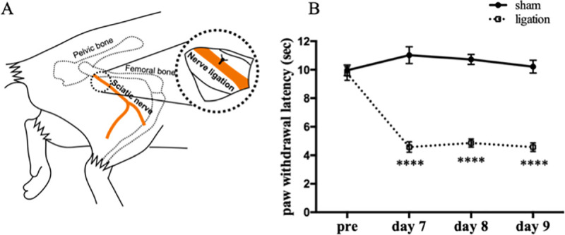
Fig 1

Schematic diagram of sciatic nerve ligation (A). Plantar tests were performed before nerve ligation surgery, 7, 8 and 9 days after surgery. Data are expressed as means ± SEM. Data are analyzed using two-way ANOVA followed by Bonferroni’s post hoc test and considered statistically significant at ****P < 0.0001 when comparing the sham (n = 8) and nerve ligation groups (n = 8) (B).

Changes in hyperalgesia by amitriptyline

We produced the neuropathic pain model mice as described above on test day 0. Plantar tests were performed before nerve ligation surgery on test day 0, after vehicle administration on test day 7, and after administration of amitriptyline on test day 8 (Fig 2A). In the results, amitriptyline significantly improved the reduction of paw withdrawal latency by sciatic nerve ligation (Fig 2B).

Fig 2. Amitriptyline improved thermal hyperalgesia in neuropathic pain model.
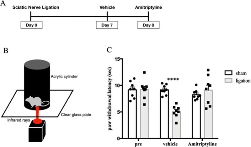
Fig 2

Plantar tests were performed before nerve ligation surgery, test day 7 with vehicle, and test day 8 with amitriptyline administration (n = 8) (A). Schematic diagram of plantar test (B). Paw withdrawal latencies were plotted for every test (C). Data are expressed as means ± SEM. Data are analyzed using two-way ANOVA followed by Bonferroni’s post hoc test and considered statistically significant at ****P < 0.0001 when comparing the sham and ligation groups.

Effects of amitriptyline on sleep condition

Mice were implanted EEG/EMG recording head-mount and electrodes at 5 days after nerve ligation or sham surgery, and habituated in a special cage for at least 24 hours before recording. We injected vehicle (0.9% saline, i.p.) at test day 7, and amitriptyline (10 mg/kg, i.p.) at test day 9, twice a day (at 7:00 and 19:00) (Fig 3A). In the results, the nerve ligation group showed significant increase in wake time and decrease in non-REM sleep time compared with sham group, when they received vehicle. Amitriptyline significantly reduced the wake time (2308 s/h vs. 1789 s/h; Ligation-vehicle vs. Ligation-amitriptyline, p < 0.001) and increased in non-REM sleep time (1175 s/h vs. 1654 s/h; Ligation-vehicle vs. Ligation-amitriptyline, p < 0.001) in the nerve ligation group. However, amitriptyline did not change the amount of sleep and wake time in the sham group (Fig 3B).

Fig 3. Amitriptyline improved quantity of sleep in neuropathic pain model.
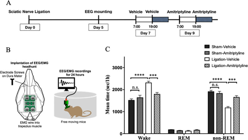
Fig 3

EEG and EMG were recorded for 24 hours with administration of vehicle (test day 7) and amitriptyline (test day 9) (A). Schematic diagram of EEG/EMG implantation (B). The mean value per hour of wake, REM sleep, and non-REM sleep time are represented for the sham (n = 7) and nerve ligation groups (n = 7) (C). Data are expressed as means ± SEM. Data are analyzed using two-way ANOVA followed by Bonferroni’s post hoc test and considered statistically significant at ***P < 0.001, and ****P < 0.0001 when comparing the groups.

We analyzed the full frequency of EEG power density in non-REM and REM sleep. There were slight differences in peak of EEG power density during non-REM sleep when comparing the sham-vehicle and ligation-vehicle (Fig 4A). We analyzed the time change of normalized power density of δ wave during non-REM sleep to evaluate the quality of sleep. Sham-vehicle mice showed high δ wave power, that indicated deep sleep at the beginning of the light phase. Then, their sleep became lighter towards the end of the light phase. On the other hand, sleep of nerve ligation mice was shallow in the beginning and deepened gradually until 16:00. However, there was no significant difference in the dark phase. With administration of amitriptyline, nerve ligation mice got deep sleep at first, in the inactive phase. Although there were no significant differences, amitriptyline tended to reduce the δ wave power in the active phase (Fig 4B).

Fig 4. Amitriptyline improved quality of sleep in neuropathic pain model.
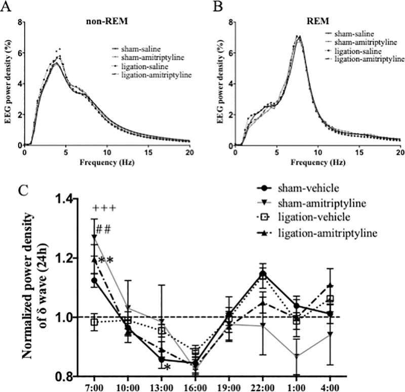
Fig 4

Distribution of EEG power density in each frequency during non-REM and REM sleep. Data are expressed as means only. *P < 0.05 at 4.2 Hz and 4.7 Hz when comparing sham-vehicle and ligation-vehicle groups (A). Time changes of the normalized power density of δ wave were plotted every 3 hours in sham (n = 5–7) and nerve ligation (n = 7) mice. Data are expressed as means ± SEM (B) and are analyzed using two-way ANOVA followed by Bonferroni’s post hoc test and considered statistically significant at **P < 0.01 when comparing sham-vehicle and ligation-vehicle groups, ##P < 0.01 when comparing ligation-vehicle and ligation amitriptyline group, and +++P < 0.001 when comparing the sham-amitriptyline and ligation-vehicle groups.

Changes in hyperalgesia by 5-HT2A receptor antagonist

Plantar tests were performed before sciatic nerve ligation surgery on test day 0, after vehicle administration on test day 7, and after administration of MDL 100907 on test day 8 (Fig 5A). In the results, MDL 100907 did not change the paw withdrawal latency in the neuropathic pain model mice (Fig 5B).

Fig 5. Selective 5HT2A receptor antagonist improved thermal hyperalgesia in neuropathic pain model.
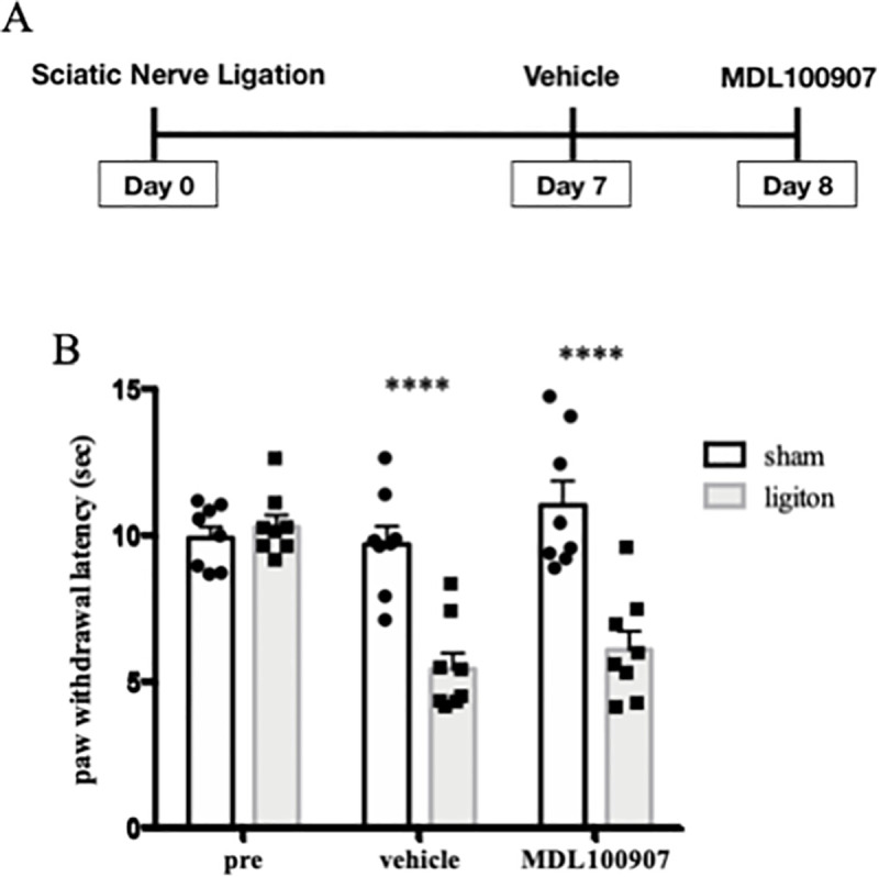
Fig 5

Plantar tests were performed before nerve ligation surgery, on test day 7 with vehicle, and on test day 8 with MDL 100907 administration (n = 8) (A). Paw withdrawal latencies were plotted for every test. Data are expressed as means ± SEM. Data are analyzed using two-way ANOVA followed by Bonferroni’s post hoc test and considered statistically significant at ****P < 0.0001 when comparing the sham and ligation groups (B).

Effects of 5-HT2A receptor antagonist on sleep condition

We injected vehicle on test day 7, and MDL 100907 (3 mg/kg, i.p.) on test day 9, twice a day (at 7:00 and 19:00) (Fig 6A). The nerve ligation group showed a significant increase in wake time and decrease in non-REM sleep time compared with the sham group, similar to the amitriptyline experiment. MDL 100907 significantly reduced the wake time (2171 s/h vs. 1526 s/h; Ligation-vehicle vs. Ligation-MDL, p < 0.001) and improved non-REM sleep time (1347 s/h vs. 1910 s/h; Ligation-vehicle vs. Ligation-MDL, p < 0.001) in the nerve ligation group (Fig 6B). In addition, MDL 100907 significantly increased REM sleep time in both sham and nerve ligation groups (Fig 6B).

Fig 6. Selective 5HT2A receptor antagonist improved quantity of sleep in neuropathic pain model.
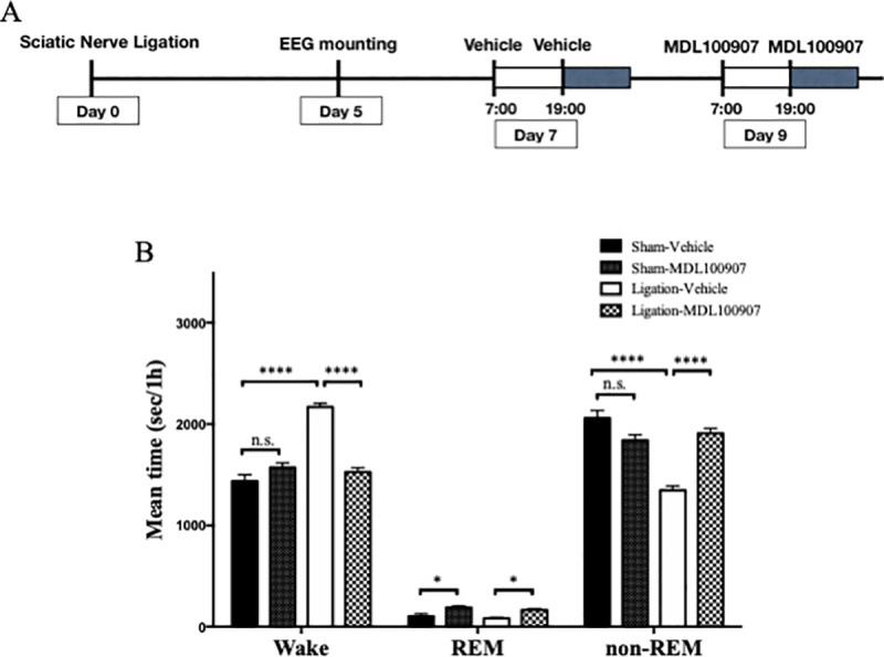
Fig 6

EEG and EMG were recorded for 24 hours with administration of vehicle (test day 7) and MDL 100907 (test day 9) (A). The mean value per hour of wake, REM sleep, and non-REM sleep time are represented for sham (n = 7) and nerve ligation groups (n = 7). Data are expressed as means ± SEM. Data are analyzed using two-way ANOVA followed by Bonferroni’s post hoc test and considered statistically significant at ***P < 0.001, and ****P < 0.0001 when comparing all groups (B).

The peak of EEG power density during non-REM sleep tended to be high in ligation-vehicle, but not significantly (Fig 7A). The result of normalized power density of δ wave during non-REM sleep showed that administration of MDL 100907 improved the sleep quality of nerve ligation mice, to obtain deep sleep at first and go to light sleep gradually, similar to amitriptyline (Fig 7B).

Fig 7. Selective 5HT2A receptor antagonist improved quality of sleep in neuropathic pain model.
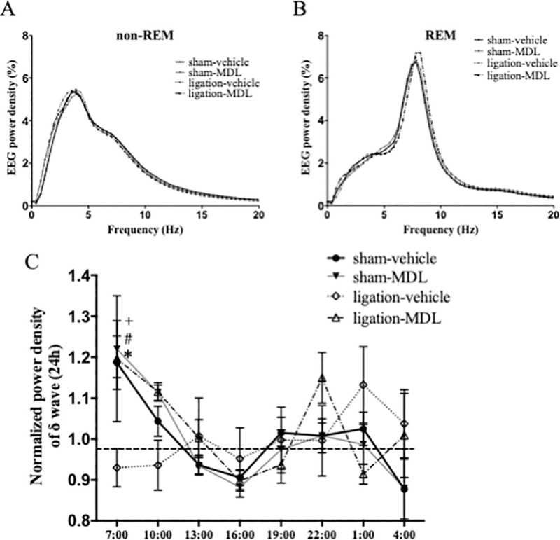
Fig 7

Distribution of EEG power density in each frequency during non-REM and REM sleep. Data are expressed as means only (A). Time changes of the normalized power density of δ wave are plotted every 3 hours in sham (n = 6) and nerve ligation (n = 5) mice. Data are expressed as means ± SEM (B). Data are analyzed using two-way ANOVA followed by Bonferroni’s post hoc test and considered statistically significant at *P < 0.05 when comparing the sham-vehicle and ligation-vehicle groups, #P < 0.05 when comparing the ligation-vehicle and ligation-MDL 100907 groups, and +P < 0.05 when comparing the sham-amitriptyline and ligation-vehicle groups.

Discussion

In the present study, the analysis of EEG and EMG showed that the neuropathic pain model mice exhibited increased wake time and decreased non-REM sleep in the chronic pain state. A tricyclic antidepressant, amitriptyline improved both increased arousal time and reduced non-REM sleep that were secondary to the pain, but did not show a significant change in the sham group. Hyperactivity of monoamine neurons is one of the mechanisms for secondary sleep disturbance [23, 28]. Since amitriptyline inhibits the re-uptake of serotonin and norepinephrine, it increases the concentrations of these neurotransmitters in the synaptic cleft. However, amitriptyline antagonizes excitatory 5-HT2A receptors coupled with Gq protein that induce spontaneous excitatory synaptic transmission. On the other hand, amitriptyline has little effect on the inhibitory 5-HT1A receptors coupled with Gi protein [21, 22]. Therefore, we believe that amitriptyline may suppress the neuronal activity as an overall effect. Amitriptyline and selective 5-HT2A receptor antagonist MDL 100907 inhibit the binding of serotonin to 5-HT2A receptors [29]. This could result in a relative increase in serotonin binding to the inhibitory 5-HT1A receptor. These additive effects may suppress the hyperactivity of serotonergic neurons and obtain sleep normalization. This may be one of the reasons why amitriptyline does not cause over-sedation in normal subjects without pain.

According to the time change of normalized power density of δ wave during non-REM sleep, in the nerve ligation group with vehicle administration, the non-REM sleep is not deep enough in the beginning of the inactive phase. On the other hand, the nerve ligation group with amitriptyline administration showed a time change of normalized power density of δ wave similar to the sham-vehicle group in the inactive phase. These findings suggest that amitriptyline could provide near-normal quality of sleep in chronic pain. Amitriptyline tended to decrease the delta power at 22:00, though not significantly, in both the sham and ligation groups. Therefore, amitriptyline may induce shallow sleep in the active phase. However, since there is less sleep in the active phase, the data dispersion is large, and further research on this aspect is needed.

We performed additional experiments using the selective 5-HT2A receptor antagonist, Administration of 3 mg/kg MDL 100907 did not show inhibitory effects on hyperalgesia in the neuropathic pain model. However, the same dose of MDL 100907 showed a sleep-improving effect in the neuropathic pain model, similar to amitriptyline. Therefore, 5-HT2A antagonism was assumed to improve sleep directly, regardless of pain inhibition.

MDL 100907 slightly increased the time of REM sleep in both the ligation and sham groups in this study. There are few reports examining the effects of 5-HT2A receptor antagonists on REM sleep, and their results are controversial. Jaime M. et al reported that administration of 5-HT2A antagonist reversed the reduction of REM sleep caused by microinjection of 5-HT2A/2C receptor agonist into the dorsal raphe nucleus in rats [30]. However, Stephen R. et al reported that 5-HT2A antagonist tended to increase REM sleep compared with vehicle administration, but not significantly [27].

There were slight differences in peak of EEG power density during non-REM sleep when comparing sham-vehicle and ligation-vehicle in the amitriptyline experiment (Fig 4A), but only a tendency in the MDL-experiment (Fig 7A). The reduction in sleep time with nerve injury may increase the absolute value of δ power density slightly, as a homeostatic compensation [31]. However, the results were not reproducible in our study.

This study has several limitations, First, as we did not find a significant abnormal behavior induced by the administration of these drugs, we cannot evaluate the neurological findings of the test in detail. Consequently, the possibility of neurological side effects such as suppression of motor coordination and activity cannot be ruled out. Second, the side effects of repeated administration were also not verified. Third, it is unknown whether these drugs had any effect on the ascending serotonergic neurons in brain stem reticular formation, because we only tested the effect of systemic administration. Fourth, only male mice were used for this study. The mechanisms of chronic pain have been shown to differ between males and females [32]; therefore, we need to examine them separately in pain studies. We consider that similar studies on female mice are also important as another study. Finally, we set the recovery time from EEG mounting surgery as 2 days, which was the same as in previous studies [13, 23]. However, we could not prove the presence or absence of any effects of stress caused by the surgery and anesthesia.

Despite these limitations, our study has important implications for sleep improvement and pain relief. Further studies using advanced genetic engineering such as optogenetics or chemogenetics are required, to verify the neuronal mechanism in detail.

Conclusions

This study showed that amitriptyline could improve sleep quantity and quality impaired by chronic pain. However, 5-HT2A receptor antagonism could not be associated with pain but was shown to be closely related to sleep improvement.

Acknowledgments

We would like to thank Editage (www.editage.com) for English language editing.

Data Availability

All relevant data are within the manuscript files.

Funding Statement

HI received grant below. Grant numbers: 26893092 Grant name:Grant-in-Aid for Research Activity start-up from Japan Society for the Promotion of Science Website: https://kaken.nii.ac.jp/ja/ The funders had no role in study design, data collection and analysis, decision to publish, or preparation of the manuscript.

References

  • 1.Alsaadi SM, McAuley JH, Hush JM, Maher CG. Erratum to: Prevalence of sleep disturbance in patients with low back pain. Eur Spine J. 2012;21:554–60. 10.1007/s00586-011-1954-8 [DOI] [PMC free article] [PubMed] [Google Scholar]
  • 2.Emery PC, Wilson KG, Kowal J. Major depressive disorder and sleep disturbance in patients with chronic pain. Pain Res Manag. 2014;19:35–41. 10.1155/2014/480859 [DOI] [PMC free article] [PubMed] [Google Scholar]
  • 3.McCracken LM, Williams JL, Tang NK. Psychological flexibility may reduce insomnia in persons with chronic pain: a preliminary retrospective study. Pain Med. 2011;12:904–12. 10.1111/j.1526-4637.2011.01115.x [DOI] [PubMed] [Google Scholar]
  • 4.Tang NK, Wright KJ, Salkovskis PM. Prevalence and correlates of clinical insomnia co-occurring with chronic back pain. J Sleep Res. 2007;16:85–95. 10.1111/j.1365-2869.2007.00571.x [DOI] [PubMed] [Google Scholar]
  • 5.Simpson NS, Scott-Sutherland J, Gautam S, Sethna N, Haack M. Chronic exposure to insufficient sleep alters processes of pain habituation and sensitization. Pain. 2018;159:33–40. 10.1097/j.pain.0000000000001053 [DOI] [PMC free article] [PubMed] [Google Scholar]
  • 6.Krause AJ, Prather AA, Wager TD, Lindquist MA, Walker MP. The pain of sleep loss: A brain characterization in humans. J Neurosci. 2019;39:2291–2300. 10.1523/JNEUROSCI.2408-18.2018 [DOI] [PMC free article] [PubMed] [Google Scholar]
  • 7.Alexandre C, Latremoliere A, Ferreira A, Miracca G, Yamamoto M, Scammell TE, et al. Decreased alertness due to sleep loss increases pain sensitivity in mice. Nat Med. 2017;23:768–774. 10.1038/nm.4329 [DOI] [PMC free article] [PubMed] [Google Scholar]
  • 8.Dinges DF, Pack F, Williams K, Gillen KA, Powell JW, Ott GE, et al. Cumulative sleepiness, mood disturbance, and psychomotor vigilance performance decrements during a week of sleep restricted to 4–5 hours per night. Sleep. 1997;20:267–277. [PubMed] [Google Scholar]
  • 9.Killgore WD, Balkin TJ, Wesensten NJ. Impaired decision making following 49 h of sleep deprivation. J Sleep Res. 2006;15:7–13. 10.1111/j.1365-2869.2006.00487.x [DOI] [PubMed] [Google Scholar]
  • 10.Mims KN, Kirsch D. Sleep and stroke. Sleep Med Clin. 2016;11:39–51. 10.1016/j.jsmc.2015.10.009 [DOI] [PubMed] [Google Scholar]
  • 11.Buxton OM, Marcelli E. Short and long sleep are positively associated with obesity, diabetes, hypertension, and cardiovascular disease among adults in the United States. Soc Sci Med. 2010;71:1027–1036. 10.1016/j.socscimed.2010.05.041 [DOI] [PubMed] [Google Scholar]
  • 12.Sofi F, Cesari F, Casini A, Macchi C, Abbate R, Gensini GF. Insomnia and risk of cardiovascular disease: a meta-analysis. Eur J Prev Cardiol. 2014;21:57–64. 10.1177/2047487312460020 [DOI] [PubMed] [Google Scholar]
  • 13.Narita M, Niikura K, Nanjo-Niikura K, Narita M, Furuya M, Yamashita A, et al. Sleep disturbances in a neuropathic pain-like condition in the mouse are associated with altered GABAergic transmission in the cingulate cortex. Pain. 2011;152:1358–1372. 10.1016/j.pain.2011.02.016 [DOI] [PubMed] [Google Scholar]
  • 14.Holbrook AM, Crowther R, Lotter A, Cheng C, King D. Meta-analysis of benzodiazepine use in the treatment of insomnia. CMAJ. 2000;162:225–233. [PMC free article] [PubMed] [Google Scholar]
  • 15.Nijs J, Mairesse O, Neu D, Leysen L, Danneels L, Cagnie B, et al. Sleep disturbances in chronic pain: neurobiology, assessment, and treatment in physical therapist practice. Phys Ther. 2018;98:325–335 10.1093/ptj/pzy020 [DOI] [PubMed] [Google Scholar]
  • 16.Boyle J, Eriksson ME, Gribble L, Gouni Ravi, Johnsen S, Coppini DV, et al. Randomized, placebo-controlled comparison of amitriptyline, duloxetine, and pregabalin in patients with chronic diabetic peripheral neuropathic pain: impact on pain, polysomnographic sleep, daytime functioning, and quality of life. Diabetes Care. 2012;35:2451–2458. 10.2337/dc12-0656 [DOI] [PMC free article] [PubMed] [Google Scholar]
  • 17.Choi A, Mitchelson F. Variation in the affinity of amitriptyline for muscarine receptor subtypes. Pharmacology. 1994;48:293–300. 10.1159/000139192 [DOI] [PubMed] [Google Scholar]
  • 18.Atkin T, Comai S, Gobbi G. Drugs for insomnia beyond benzodiazepines: pharmacology, clinical applications, and discovery. Pharmacol Rev. 2018;70:197–245. 10.1124/pr.117.014381 [DOI] [PubMed] [Google Scholar]
  • 19.Vande Griend JP, Anderson SL. Histamine-1 receptor antagonism for treatment of insomnia. J Am Pharm Assoc. 2012;52:e210–219. 10.1331/JAPhA.2012.12051 [DOI] [PubMed] [Google Scholar]
  • 20.Sawynok J, Reid AR, Esser MJ. Peripheral antinociceptive action of amitriptyline in the rat formalin test: involvement of adenosine. Pain. 1999;80:45–55. 10.1016/s0304-3959(98)00195-x [DOI] [PubMed] [Google Scholar]
  • 21.Peroutka SJ. Antimigraine drug interactions with serotonin receptor subtypes in human brain. Ann Neurol. 1988;23:500–504. 10.1002/ana.410230512 [DOI] [PubMed] [Google Scholar]
  • 22.Cusack B, Nelson A, Richelson E. Binding of antidepressants to human brain receptors: focus on newer generation compounds. Psychopharmacology. 1994;114:559–565. 10.1007/BF02244985 [DOI] [PubMed] [Google Scholar]
  • 23.Ito H, Yanase M, Yamashita A, Kitabatake C, Hamada A, Suhara Y, et al. Analysis of sleep disorders under pain using an optogenetic tool: possible involvement of the activation of dorsal raphe nucleus-serotonergic neurons. Mol Brain 2013; 6:59 10.1186/1756-6606-6-59 [DOI] [PMC free article] [PubMed] [Google Scholar]
  • 24.Seltzer Z, Dubner R, Shir Y. A novel behavioral model of neuropathic pain disorders produced in rats by partial sciatic nerve injury. Pain 1990;43:205–218. 10.1016/0304-3959(90)91074-s [DOI] [PubMed] [Google Scholar]
  • 25.Sanna MD, Ghelardini C, Galeotti N. Spinal astrocytic c-Jun N-terminal kinase (JNK) activation as counteracting mechanism to the amitriptyline analgesic efficacy in painful peripheral neuropathies. Eur J Pharmacol. 2017;798:85–93. 10.1016/j.ejphar.2017.01.025 [DOI] [PubMed] [Google Scholar]
  • 26.Mika J, Jurga AM, Starnowska J, Wasylewski M, Rojewska E, Makuch W, et al. Effects of chronic doxepin and amitriptyline administration in naïve mice and in neuropathic pain mice model. Neuroscience. 2015;294:38–50. 10.1016/j.neuroscience.2015.03.003 [DOI] [PubMed] [Google Scholar]
  • 27.Morairty SR, Hedley L, Flores J, Martin R, Kilduff TS. Selective 5HT2A and 5HT6 receptor antagonists promote sleep in rats. Sleep. 2008;31:34–44. 10.1093/sleep/31.1.34 [DOI] [PMC free article] [PubMed] [Google Scholar]
  • 28.Koh K, Hamada A, Hamada Y, Yanase M, Sakaki M, Someya K, et al. Possible involvement of activated locus coeruleus-noradrenergic neurons in pain-related sleep disorders. Neurosci Lett. 2015;589:200–206. 10.1016/j.neulet.2014.12.002 [DOI] [PubMed] [Google Scholar]
  • 29.Monti JM, Jantos H. Effects of the serotonin 5-HT2A/2C receptor agonist DOI and of the selective 5-HT2A or 5-HT2C receptor antagonists EMD 281014 and SB-243213, respectively, on sleep and waking in the rat. Eur J Pharmacol. 2006;553:163–170. 10.1016/j.ejphar.2006.09.027 [DOI] [PubMed] [Google Scholar]
  • 30.Monti JM, Jantos H. Effects of activation and blockade of 5-HT2A/2C receptors in the dorsal raphe nucleus on sleep and waking in the rat. Prog Neuropsychopharmacol Biol Psychiatry. 2006;30:1189–1195. 10.1016/j.pnpbp.2006.02.013 [DOI] [PubMed] [Google Scholar]
  • 31.Vyazovskiy VV, Delogu A. NREM and REM sleep: complementary roles in recovery after wakefulness. Neuroscientist. 2014;20:203–219. 10.1177/1073858413518152 [DOI] [PubMed] [Google Scholar]
  • 32.Dance A. Why the sexes don't feel pain the same way. Nature. 2019;567:448–450. 10.1038/d41586-019-00895-3 [DOI] [PubMed] [Google Scholar]

Decision Letter 0

Jianhong Zhou

11 Sep 2020

PONE-D-20-20832

Analysis of the effects of tricyclic antidepressant on secondary sleep disturbance induced by chronic pain

PLOS ONE

Dear Dr. Ito,

Thank you for submitting your manuscript to PLOS ONE. After careful consideration, we feel that it has merit but does not fully meet PLOS ONE’s publication criteria as it currently stands. Therefore, we invite you to submit a revised version of the manuscript that addresses the points raised during the review process.

Please submit your revised manuscript by Oct 26 2020 11:59PM. If you will need more time than this to complete your revisions, please reply to this message or contact the journal office at plosone@plos.org. When you're ready to submit your revision, log on to https://www.editorialmanager.com/pone/ and select the 'Submissions Needing Revision' folder to locate your manuscript file.

Please include the following items when submitting your revised manuscript:

  • A rebuttal letter that responds to each point raised by the academic editor and reviewer(s). You should upload this letter as a separate file labeled 'Response to Reviewers'.

  • A marked-up copy of your manuscript that highlights changes made to the original version. You should upload this as a separate file labeled 'Revised Manuscript with Track Changes'.

  • An unmarked version of your revised paper without tracked changes. You should upload this as a separate file labeled 'Manuscript'.

If you would like to make changes to your financial disclosure, please include your updated statement in your cover letter. Guidelines for resubmitting your figure files are available below the reviewer comments at the end of this letter.

If applicable, we recommend that you deposit your laboratory protocols in protocols.io to enhance the reproducibility of your results. Protocols.io assigns your protocol its own identifier (DOI) so that it can be cited independently in the future. For instructions see: http://journals.plos.org/plosone/s/submission-guidelines#loc-laboratory-protocols

We look forward to receiving your revised manuscript.

Kind regards,

Jianhong Zhou

Associate Editor

PLOS ONE

Journal Requirements:

When submitting your revision, we need you to address these additional requirements.

1. Please ensure that your manuscript meets PLOS ONE's style requirements, including those for file naming. The PLOS ONE style templates can be found at

https://journals.plos.org/plosone/s/file?id=wjVg/PLOSOne_formatting_sample_main_body.pdf and

https://journals.plos.org/plosone/s/file?id=ba62/PLOSOne_formatting_sample_title_authors_affiliations.pdf

2. Thank you for including your ethics statement:  "The present study was conducted in accordance with the Guiding Principles for the Care and Use of Laboratory Animals at University of Toyama (Toyama, Japan), as adopted by the Committee on Animal Research of University of Toyama.

Approval number: A2014NED-14

Methods of anesthesia: general anesthesia using isoflurane

Methods for euthanasia of animals: Carbon dioxide and Nerve destruction".   

Please amend your current ethics statement to confirm that your named ethics committee specifically approved this study.

For additional information about PLOS ONE submissions requirements for ethics oversight of animal work, please refer to http://journals.plos.org/plosone/s/submission-guidelines#loc-animal-research  

Once you have amended this/these statement(s) in the Methods section of the manuscript, please add the same text to the “Ethics Statement” field of the submission form (via “Edit Submission”).

[Note: HTML markup is below. Please do not edit.]

Reviewers' comments:

Reviewer's Responses to Questions

Comments to the Author

1. Is the manuscript technically sound, and do the data support the conclusions?

The manuscript must describe a technically sound piece of scientific research with data that supports the conclusions. Experiments must have been conducted rigorously, with appropriate controls, replication, and sample sizes. The conclusions must be drawn appropriately based on the data presented.

Reviewer #1: Yes

Reviewer #2: Partly

Reviewer #3: Yes

**********

2. Has the statistical analysis been performed appropriately and rigorously?

Reviewer #1: Yes

Reviewer #2: No

Reviewer #3: Yes

**********

3. Have the authors made all data underlying the findings in their manuscript fully available?

The PLOS Data policy requires authors to make all data underlying the findings described in their manuscript fully available without restriction, with rare exception (please refer to the Data Availability Statement in the manuscript PDF file). The data should be provided as part of the manuscript or its supporting information, or deposited to a public repository. For example, in addition to summary statistics, the data points behind means, medians and variance measures should be available. If there are restrictions on publicly sharing data—e.g. participant privacy or use of data from a third party—those must be specified.

Reviewer #1: Yes

Reviewer #2: No

Reviewer #3: No

**********

4. Is the manuscript presented in an intelligible fashion and written in standard English?

PLOS ONE does not copyedit accepted manuscripts, so the language in submitted articles must be clear, correct, and unambiguous. Any typographical or grammatical errors should be corrected at revision, so please note any specific errors here.

Reviewer #1: No

Reviewer #2: No

Reviewer #3: Yes

**********

5. Review Comments to the Author

Please use the space provided to explain your answers to the questions above. You may also include additional comments for the author, including concerns about dual publication, research ethics, or publication ethics. (Please upload your review as an attachment if it exceeds 20,000 characters)

Reviewer #1: The authors have provided an interesting study that contributes to the growing field of knowledge surrounding the relationship between disrupted sleep and chronic pain. Specifically, the authors sought to examine the role of serotonergic components of the ascending reticular activating system in sleep disruption induced by chronic pain. To accomplish this, a partial sciatic nerve ligation model was selected and a tricyclic antidepressant (amitriptyline) and a selective 5-HT2A antagonist (MDL 100907) were deployed. Using the plantar test and EMG/EEG recording, the authors observed that amitriptyline improved both hyperalgesia symptoms and sleep quantity, whereas MDL100907 only normalized sleep quantity but had no effects on examined hyperalgesia symptoms. From this study, we can conclude that that amitriptyline’s effects on improved sleep quality in rodents during chronic pain is likely due to at least some antagonism of 5-HT2AR, but that its ability to ameliorate hyperalgesia is mediated by another mechanism unrelated to 5-HT2AR antagonism. The authors provide a strong list of limitations to the interpretation of this study.

However, the authors need to seek out a native English speaker to improve the quality of the writing and grammar, need to explain why several groups appear to have been excluded from analysis (See Results comments), why proper controls were not used throughout (See Results comments), and why only males were used for the study (and please do not say because of the cyclic female sex steroid hormones unless they address the males’ pulsatile testosterone secretion patterns).

Comments:

Introduction:

- In the introduction, the authors should provide rationale for why they selected amitriptyline as the TCA of choice from the cited study (16). It may also be useful for the reader to know that it is commonly prescribed in the treatment of neuropathic pain.

- In the introduction, the rationale for using MDR 100907 should be explained. The third paragraph of the discussion may function best if moved to the introduction.

- Authors should provide justification for using only males in the study.

Methods:

- Did the mice receive any analgesic after surgery? If so, then the authors should describe this.

- The methods describing the evaluation of hyperalgesia are not consistent with the timelines in figures 2 & 3. Specifically, “Immediately after the administration of amitriptyline, selective 5-HT2A receptor antagonist, MDL100907, or vehicle, mice were placed in the acrylic cylinder (height: 15cm, Diameter: 8cm) and habituated for 1 hour before measurement.” These discrepancies should be corrected.

- What was the interstimulus interval for the repeated measurements of paw withdrawal latency? (E.g., 3-minutes were given between each test).

- The authors do not discuss ipsilateral or contralateral paw tests. Considering this information is not provided, we must assume that only the ipsilateral paw was tested. This should be clarified by the authors and a justification for not testing the contralateral paw should be provided.

- Though commonly used, the authors should cite the methods of classifying REM vs. NREM via Theta/Delta ratios, (e.g. Grosmark et. Al 2012 Cell).

Results:

- Figure 2 and figure 5 do not contain proper controls (i.e. a sham PSL group). The authors should provide a strong justification as to why this group was not included and a within-group historic control was used instead. Additionally, the authors should provide justification as to why a saline injection was given to the same group for a within-group historical comparison as opposed to having a full-factorial study design.

- Authors should delete word “clearly” from results section 1.

- Authors should provide rationale for why the sham-drug groups were excluded from figures 4 and 7.

- Authors should explain that delta power was increased during the inactive but not active phase. Reword this sentence “By administration of amitriptyline, nerve ligation mice got deep sleep at first and went to light sleep as same as sham mice” in Results section 3.

Discussion:

- Authors should reword the following sentence “According to the time change of normalized power density of δ wave during non-REM sleep, nerve ligation group with vehicle administration did not get enough had less deep sleep at an early sleep phase during the beginning of the inactive phase”

Reviewer #2: Antidepressants are reported to have analgesic effects, especially on chronic pain, and tricyclic antidepressants are used to treat chronic pain in combination with serotonin-noradrenaline reuptake inhibitors. Here, Ito and colleagues used the amitriptyline, a tricyclic antidepressant, and MDL 100907, a selective 5-HT2A antagonist, affect the reduction of chronic pain-induced sleep disturbance.

My major concern regarding this manuscript is about the experimental design. The authors confirmed the paw withdrawal latency of thermal hyperalgesia in neuropathic pain model mice in a weekly manner (7 days apart). However, in the subsequent experiments, they recorded the effect either on the next day (day 7, 8) or after of gap of 2 days (day7, 9) of the vehicle administration. Authors can follow any of the following three alternatives: Providing evidence that the mouse showed the same paw withdrawal latency within the days; or re-perform the experiment as a crossover manner, and/or collect the data weekly. Moreover, the authors have given less than 2 days for recovery after EEG mounting which might not be enough. The data presented to show the effect of the drug on sleep is also not convincing. Please provide a full frequency of EEG analysis in each vigilance stage of vehicle administration. Please provide the details on how they calculate the mean time used in each figure.

Minor Comments:

Please discuss the increase of REM sleep by MDL 100907 by providing possible explanations or other references. Several papers indicated the increase in NREM sleep in rats and humans, but there was no change in REM sleep.

There was no schematic diagram of sciatic nerve ligation, planter test, and especially EEG EMG implantation.

Please correct Figure 5 legend and some other spelling.

In Figure 4 there was a visible difference (not significant) at 22:00 between vehicle and amitriptyline group. How did the authors define this? However, the authors defined nerve ligation mice showed light sleep from the beginning and kept sleep pattern flat but if we observe from 13:00 to 22:00 time period, this is not correct, and please explain more precisely about this.

Reviewer #3: The manuscript "Analysis of the effect of tricyclic antidepressant on secondary sleep disturbance induced by chronic pain" by Ito et al., addresses the important connection between chronic pain and sleep. They use a mouse model (sciatic nerve ligation) to induce chronic pain and find a persistent increase in pain sensitivity in these mice after one week. The authors show that this increase in pain sensitivity can be reduced with the treatment of the tricyclic antidepressant Amitriptyline. Furthermore, they show that mice with sciatic nerve ligation spend more time awake and less time in NREM sleep over a 24h period than fellow control mice. Administration of Amitriptyline significantly decreases the amount of time spent in wake and increased NREM sleep amounts, showing that Amitriptyline can increase NREM sleep quantity. Next, Ito and co-workers show that Amitriptyline also improves sleep quality in that the drug increases EEG delta power in ligated mice during the first 3 hours of the light phase. Lastly, to try to elucidate some of the mechanisms through which Amitriptyline might affect sleep and pain, the authors use a selective 5HT2A receptor antagonist (MDL100907) and show that this drug also improves sleep quantity and quality in ligated mice, but fails to decrease pain sensitivity. The authors provide a step by step framework for their experiments that is easy to follow.

Major points:

1) While on first glance well executed, this study does not provide truly new insights into the interrelationship between sleep and pain or into the role of Amitriptyline. Previous studies (e.g. Boyle et al., 2012 Diabetes Care) already show that Amitriptyline is effective at treating pain and improving sleep in humans. The new information here is that Amitriptyline might work though the 5-HTA2 receptor to achieve its effects on sleep and pain. However, the selective 5HT2A receptor antagonist MDL100907 used here to support this claim does not improve pain sensitivity, thereby casting doubts on the involvement of the 5-HT2A involvement.

2) The authors stress the idea that Amitriptyline might increase NREM sleep and reduce pain sensitivity by blocking the 5-HTA2 receptor, but do not discuss that Amitriptyline is a tricyclic anti-depressant and is thought to primarily inhibit the re-uptake of Serotonin and Norepinephrine (thereby increasing the concentrations of these neurotransmitters in the synaptic cleft). This important point needs to be addressed and citations for the antagonistic effect of Amitriptyline on the 5-HTA2 receptor need to be added.

3) The authors mention in the methods that the implantation of the EEG/EMG leads happened 2 days before baseline sleep recordings with a 24h acclimation period to the recording chamber and cables. This time frame seems seriously short considering the severity of the surgery and the discomfort experienced thereafter. Never mind that animals were then connected to EEG/EMG cables to which they only had 24h to acclimate. Another point is the effect of anesthesia (such as Isoflurane) on subsequent sleep/wake architecture soon after the exposure (e.g. Pick et al., 2011 Anesthesiiology; Zhang et al., 2016 Sleep). Considering how susceptible sleep is to environmental influences (including pain and discomfort), it would have been better to first perform the EEG/EMG surgery, acclimate the mice to the cage and cable for 1-2 weeks total (as is otherwise customary in mouse sleep research) and then perform the sciatic nerve ligation and drug applications.

Minor:

Figure 2 – please show individual data points to allow a more informative assessment of the results

Figure 4 – shows “normalized delta power density’ for each mouse and demonstrates a flat beginning for vehicle-treated ligation mice. However, it would also be informative to compare the delta amplitude in more absolute values to see whether nerve ligation as such decreased delta during NREM sleep as compared to the sham surgery. Also, the “sham+amitriptyline” group is missing from this graph.

Please format references to show complete spelling of last names, rather than first names (this happened in several references)

To improve readability of the manuscript , the authors should work with a language coach to address spelling and sentence structure throughout the manuscript

Some important references could be added in the introduction e.g. Alexandre et al., 2017 Nat Med

**********

6. PLOS authors have the option to publish the peer review history of their article (what does this mean?). If published, this will include your full peer review and any attached files.

If you choose “no”, your identity will remain anonymous but your review may still be made public.

Do you want your identity to be public for this peer review? For information about this choice, including consent withdrawal, please see our Privacy Policy.

Reviewer #1: No

Reviewer #2: No

Reviewer #3: No

[NOTE: If reviewer comments were submitted as an attachment file, they will be attached to this email and accessible via the submission site. Please log into your account, locate the manuscript record, and check for the action link "View Attachments". If this link does not appear, there are no attachment files.]

While revising your submission, please upload your figure files to the Preflight Analysis and Conversion Engine (PACE) digital diagnostic tool, https://pacev2.apexcovantage.com/. PACE helps ensure that figures meet PLOS requirements. To use PACE, you must first register as a user. Registration is free. Then, login and navigate to the UPLOAD tab, where you will find detailed instructions on how to use the tool. If you encounter any issues or have any questions when using PACE, please email PLOS at figures@plos.org. Please note that Supporting Information files do not need this step.

PLoS One. 2020 Dec 3;15(12):e0243325. doi: 10.1371/journal.pone.0243325.r002

Author response to Decision Letter 0


25 Oct 2020

Journal Requirements:

1. Please ensure that your manuscript meets PLOS ONE's style requirements, including those for file naming. The PLOS ONE style templates can be found at

https://journals.plos.org/plosone/s/file?id=wjVg/PLOSOne_formatting_sample_main_body.pdf and https://journals.plos.org/plosone/s/file?id=ba62/PLOSOne_formatting_sample_title_authors_affiliations.pdf

A. We have checked our manuscripts to meet PLOS ONE's style.

2. Thank you for including your ethics statement: "The present study was conducted in accordance with the Guiding Principles for the Care and Use of Laboratory Animals at University of Toyama (Toyama, Japan), as adopted by the Committee on Animal Research of University of Toyama.

Approval number: A2014NED-14

Methods of anesthesia: general anesthesia using isoflurane

Methods for euthanasia of animals: Carbon dioxide and Nerve destruction".

Please amend your current ethics statement to confirm that your named ethics committee specifically approved this study.

A. We have amended our ethics statement. (p.5 Line 75)

Response to Reviewers

Reviewer #1: The authors have provided an interesting study that contributes to the growing field of knowledge surrounding the relationship between disrupted sleep and chronic pain. Specifically, the authors sought to examine the role of serotonergic components of the ascending reticular activating system in sleep disruption induced by chronic pain. To accomplish this, a partial sciatic nerve ligation model was selected and a tricyclic antidepressant (amitriptyline) and a selective 5-HT2A antagonist (MDL 100907) were deployed. Using the plantar test and EMG/EEG recording, the authors observed that amitriptyline improved both hyperalgesia symptoms and sleep quantity, whereas MDL100907 only normalized sleep quantity but had no effects on examined hyperalgesia symptoms. From this study, we can conclude that that amitriptyline’s effects on improved sleep quality in rodents during chronic pain is likely due to at least some antagonism of 5-HT2AR, but that its ability to ameliorate hyperalgesia is mediated by another mechanism unrelated to 5-HT2AR antagonism. The authors provide a strong list of limitations to the interpretation of this study.

However, the authors need to seek out a native English speaker to improve the quality of the writing and grammar, need to explain why several groups appear to have been excluded from analysis (See Results comments), why proper controls were not used throughout (See Results comments), and why only males were used for the study (and please do not say because of the cyclic female sex steroid hormones unless they address the males’ pulsatile testosterone secretion patterns).

Comments:

Introduction:

1. In the introduction, the authors should provide rationale for why they selected amitriptyline as the TCA of choice from the cited study (16). It may also be useful for the reader to know that it is commonly prescribed in the treatment of neuropathic pain.

A. Thank you for highlighting this point. According to your advice, we added the following statement: “Amitriptyline, a tricyclic antidepressant, is a first-line drug for neuropathic pain, and is commonly prescribed for the treatment of neuropathic pain.” in the introduction section of our manuscript. (p.4 Line 51)

2. In the introduction, the rationale for using MDR 100907 should be explained. The third paragraph of the discussion may function best if moved to the introduction.

A. Thank you for your advice. We have moved the third paragraph of the discussion to the introduction. (p.4 Line 57)

- Authors should provide justification for using only males in the study.

A. Thank you for the helpful suggestion. As you rightly indicated, the investigation of females is equally important. However, as the mechanisms of chronic pain have been shown to differ between males and females, we need to examine them separately. Although we investigated using only males in this study, we consider that similar studies on females should also be conducted. We mentioned this as a limitation of this study in the discussion section. (p.20 Line 36)

Methods:

- Did the mice receive any analgesic after surgery? If so, then the authors should describe this.

A. We did not use any analgesics after surgery in this study.

- The methods describing the evaluation of hyperalgesia are not consistent with the timelines in figures 2 & 3. Specifically, “Immediately after the administration of amitriptyline, selective 5-HT2A receptor antagonist, MDL100907, or vehicle, mice were placed in the acrylic cylinder (height: 15cm, Diameter: 8cm) and habituated for 1 hour before measurement.” These discrepancies should be corrected.

A. Thank you for pointing out the discrepancy. We have specified the timeline of plantar tests according to the figures. (p.7 Line 95)

- What was the inter-stimulus interval for the repeated measurements of paw withdrawal latency? (E.g., 3-minutes were given between each test).

A. We specified that we had set the test interval of 3 minutes or more. (p.7 Line 102)

- The authors do not discuss ipsilateral or contralateral paw tests. Considering this information is not provided, we must assume that only the ipsilateral paw was tested. This should be clarified by the authors and a justification for not testing the contralateral paw should be provided.

A. We specified that we tested only the ipsilateral paw in the plantar tests. (p.7 Line 104) The most important aim of the plantar test in this study was to confirm the effects of sciatic nerve ligation surgery and drug administrations on pain in the injured paw. The ipsilateral assessment is sufficient to determine whether the sciatic nerve is successful or if the drug improves the pain. We have determined that the contralateral test is less useful in this study and such less needed painful tests are unethical.

- Though commonly used, the authors should cite the methods of classifying REM vs. NREM via Theta/Delta ratios, (e.g. Grosmark et. Al 2012 Cell).

A. We added a brief description of the methods of classifying REM and non-REM in brief, along with a relevant citation [23] (p.8 Line 125).

Results:

- Figure 2 and figure 5 do not contain proper controls (i.e. a sham PSL group). The authors should provide a strong justification as to why this group was not included and a within-group historic control was used instead. Additionally, the authors should provide justification as to why a saline injection was given to the same group for a within-group historical comparison as opposed to having a full-factorial study design.

A. As you indicated, it is necessary to show the sham data. We have added the data from sham experiments in figure 2 and 5.

Because sciatic nerve ligation causes stressful chronic pain in animals, we are required to use the minimum number of animals due to ethical issues. To overcome this restriction, we administered vehicle and drug to the same group for historical comparison. Following the advice of reviewer 2, we confirmed that the mouse showed the same hyperalgesia on the test days (days 7, 8, and 9). We changed figure 1 to show these data.

- Authors should delete word “clearly” from results section 1.

A. We have deleted “clearly” from this section.

- Authors should provide rationale for why the sham-drug groups were excluded from figures4 and 7.

A. Thank you pointing out the omission. We have added the data from sham-drugs groups in figures 4 and 7.

- Authors should explain that delta power was increased during the inactive but not active phase. Reword this sentence “By administration of amitriptyline, nerve ligation mice got deep sleep at first and went to light sleep as same as sham mice” in Results section 3.

A. Thank you for your advice. We reworded the sentences, “With administration of amitriptyline, nerve ligation mice got deep sleep at first, in the inactive phase. Although there were no significant differences, amitriptyline tended to reduce the δ wave power in the active phase” in results section 3 (p.13, Line 205)

Discussion:

- Authors should reword the following sentence “According to the time change of normalized power density of Δ wave during non-REM sleep, nerve ligation group with vehicle administration did not get enough had less deep sleep at an early sleep phase during the beginning of the inactive

phase”

A. We have reworded the sentence pointed out and the sentence following it as follows,

“According to the time change of normalized power density of δ wave during non-REM sleep, in the nerve ligation group with vehicle administration, the non-REM sleep is not deep enough in the beginning of the inactive phase. On the other hand, the nerve ligation group with amitriptyline administration showed a time change of normalized power density of δ wave similar to the sham-vehicle group in the inactive phase. These findings suggest that amitriptyline could provide near-normal quality of sleep in chronic pain.” (p.18, Line 281)

Reviewer #2: Antidepressants are reported to have analgesic effects, especially on chronic pain, and tricyclic antidepressants are used to treat chronic pain in combination with serotonin-noradrenaline reuptake inhibitors. Here, Ito and colleagues used the amitriptyline, a tricyclic antidepressant, and MDL 100907, a selective 5-HT2Aantagonist, affect the reduction of chronic pain-induced sleep disturbance.

My major concern regarding this manuscript is about the experimental design.

The authors confirmed the paw withdrawal latency of thermal hyperalgesia in neuropathic pain model mice in a weekly manner (7 days apart). However, in the subsequent experiments, they recorded the effect either on the next day (day 7, 8) or after of gap of 2 days (day7, 9) of the vehicle administration.

Authors can follow any of the following three alternatives:

-Providing evidence that the mouse showed the same paw withdrawal latency within the days; or re-perform the experiment as a crossover manner, and/or collect the data weekly.

A. Thank you for your pointing out these discrepancies. We re-conducted plantar tests as per the schedule provided by you and changed figure 1 to show the same paw withdrawal latency from day 7 to day 9.

Moreover, the authors have given less than 2 days for recovery after EEG mounting which might not be enough.

A. In previous studies, which used the same EEG recording devices (Pinnacle technology, USA), recovery time given was 2 days (references 12, 15). This EEG mounting surgery is less invasive and only needs a short anesthesia time < 15 minutes. Therefore, we set the recovery time from surgery as 2 days, which was the same as in previous studies. However, we could not confirm the presence or absence of any effects of stress caused by the surgery and anesthesia.

We added the above text in the discussion section as a limitation. (p.20 Line 316)

The data presented to show the effect of the drug on sleep is also not convincing. Please provide a full frequency of EEG analysis in each vigilance stage of vehicle administration.

A. Thank you very much for your advice. We have added the results of full frequency of EEG analysis in result section 3 (p.13 Line 198) and 5 (p.16 Line 249) and included new figures 4A and 7A.

“There were slight differences in peak of EEG power density during non-REM sleep when comparing sham-vehicle and ligation-vehicle in the amitriptyline experiment (Figure 4A), but only a tendency in the MDL experiment (Figure 7A). The reduction of sleep time with nerve injury may increase the absolute value of δ power density slightly, as homeostatic compensation [31]. However, the results were not reproducible in our study.”

We have added the above text in the discussion section (p.19 Line 302).

Please provide the details on how they calculate the mean time used in each figure.

A. non-REM sleep time, REM sleep time, and awakening time per 24 hours for each animal were calculated by SleepSign. The values were summed for each group and divided by the number of animals to calculate the mean for each group. Finally, we calculated the mean time per hour.

We described this calculation of mean time in methods section 5. (p.8 Line 127)

Minor Comments:

Please discuss the increase of REM sleep by MDL 100907 by providing possible explanations or other references. Several papers indicated the increase in NREM sleep in rats and humans, but there was no change in REM sleep.

A. “5-HT2A receptor antagonist slightly increased the time of REM sleep in both the ligation and sham groups in this study. There are few reports examining the effects of 5-HT2A receptor antagonists on REM sleep, and their results are controversial. Jaime M. et al reported that the administration of 5-HT2A antagonist reversed the reduction of REM sleep caused by microinjection of 5-HT2A/2C receptor agonist into the dorsal raphe nucleus in rat [30]. However, Stephen R. et al reported that 5-HT2A antagonist tended to increase REM sleep compared with vehicle administration, but not significantly [27].” This is contrary to what you have mentioned

We added above contents in discussion. (p.19 Line 295)

There was no schematic diagram of sciatic nerve ligation, planter test, and especially EEG EMG implantation.

A. Following your advice, we added schematic diagrams of sciatic nerve ligation (Fig 1A), plantar test (Fig 2B), and EEG EMG implantation (Fig 3B).

Please correct Figure 5 legend and some other spelling.

A. We have corrected them. A professional English language editing service also have checked all.

In Figure 4 there was a visible difference (not significant) at 22:00 between vehicle and amitriptyline group. How did the authors define this?

A. As you pointed out, there was a visible difference at 22:00 in the amitriptyline group. We added the following sentences in the second paragraph of the discussion.

“Amitriptyline tended to decrease the delta power at 22:00, though not significantly, in both sham and ligation groups. Therefore, amitriptyline may induce shallow sleep in the active phase. However, since there is less sleep in the active phase, the data dispersion is large, and further research on this aspect is needed .” (p. 18 Line 286)

However, the authors defined nerve ligation mice showed light sleep from the beginning and kept sleep pattern flat but if we observe from 13:00 to 22:00 time period, this is not correct, and please explain more precisely about this.

A. Thank you for pointing out this discrepancy. As you mentioned, the sentence did not convey the observations correctly. We reworded the sentence as below. (p.13 Line 203)

“Sleep of nerve ligation mice was shallow in the beginning and deepened gradually until 16:00. However, there was no significant difference in the dark phase.”

Reviewer #3: The manuscript "Analysis of the effect of tricyclic antidepressant on secondary sleep disturbance induced by chronic pain" by Ito et al., addresses the important connection between chronic pain and sleep. They use a mouse model (sciatic nerve ligation) to induce chronic pain and find a persistent increase in pain sensitivity in these mice after one week. The authors show that this increase in pain sensitivity can be reduced with the treatment of the tricyclic antidepressant Amitriptyline. Furthermore, they show that mice with sciatic nerve ligation spend more time awake and less time in NREM sleep over a 24h period than fellow control mice. Administration of Amitriptyline significantly decreases the amount of time spent in wake and increased NREM sleep amounts, showing that Amitriptyline can increase NREM sleep quantity. Next, Ito and co-workers show that Amitriptyline also improves sleep quality in that the drug increases EEG delta power in ligated mice during the first 3 hours of the light phase. Lastly, to try to elucidate some of them echanisms through which Amitriptyline might affect sleep and pain, the authors use a selective 5HT2A receptor antagonist (MDL100907) and show that this drug also improves sleep quantity and quality in ligated mice, but fails to decrease pain sensitivity. The authors provide a step by step framework for their experiments that is easy to follow.

Major points:

1) While on first glance well executed, this study does not provide truly new insights into the interrelationship between sleep and pain or into the role of Amitriptyline. Previous studies (e.g. Boyle et al., 2012 Diabetes Care) already show that Amitriptyline is effective at treating pain and improving sleep in humans. The new information here is that Amitriptyline might work though the 5-HTA2 receptor to achieve its effects on sleep and pain. However, the selective 5HT2A receptor antagonist MDL100907 used here to support this claim does not improve pain sensitivity, thereby casting doubts on the involvement of the 5-HT2A involvement.

A. I really appreciate your pointing out this important problem.

As you mentioned, amitriptyline is the first-line treatment for chronic neuropathic pain and could improve sleep disturbance induced by chronic pain at the same time. However, it is not known whether this sleep-improving effect is an indirect effect due to pain improvement or a direct pharmacological effect of amitriptyline (p.4 Line 51). We hypothesized that antagonism of excitatory 5-HT2A receptors of amitriptyline would improve pain-related sleep disturbance, because the activation of ascending serotoninergic neurons could be partly associated with the mechanism of sleep disorders caused by chronic pain from our previous study (23) (p.5 Line 65). In the results of this study, antagonism of 5-HT2A receptors was shown to improve sleep disturbance in the chronic pain model “without improving pain”; therefore, it was speculated that amitriptyline could have a direct sleep-improving effect in addition to improving pain. (p.19 Line 294)

Based on your suggestion, we have carefully modified the introduction and discussion.

2) The authors stress the idea that Amitriptyline might increase NREM sleep and reduce pain sensitivity by blocking the 5-HTA2 receptor, but do not discuss that Amitriptyline is a tricyclic anti-depressant and is thought to primarily inhibit the re-uptake of Serotonin and Norepinephrine (thereby increasing the concentrations of these neurotransmitters in the synaptic cleft). This important point needs to be addressed and citations for the antagonistic effect of Amitriptyline on the 5-HTA2 receptor need to be added.

 You have raised an important concern. First, we would like to say that amitriptyline might increase NREM sleep by blocking the 5-HT2A receptor, but refrain from saying that this mechanism is associated with improving pain. We have modified the manuscript to avoid any misinterpretation of our meaning.

Second, as per your suggestion on discussing the main pharmacological action of amitriptyline, i.e. inhibition of re-uptake of serotonin and norepinephrine, we have added the following text to the discussion. “Since amitriptyline inhibits the re-uptake of serotonin and norepinephrine, it increases the concentrations of these neurotransmitters in the synaptic cleft. However, amitriptyline antagonized excitatory 5-HT2A receptors coupled with Gq protein induce spontaneous excitatory synaptic transmission. On the other hand, amitriptyline has little effect on the inhibitory 5-HT1A receptors coupled with Gi protein. Therefore, we believe that amitriptyline may suppress the neuronal activity as an overall effect.” (p.17 Line 270)

Lastly, following your advice, we have added the citations, of the studies of receptor binding assay that show amitriptyline antagonize 5-HT2A receptor but not 5-HT1A [21, 22]. (p.18 Line 274)

3) The authors mention in the methods that the implantation of the EEG/EMG leads happened 2 days before baseline sleep recordings with a 24h acclimation period to the recording chamber and cables. This time frame seems seriously short considering the severity of the surgery and the discomfort experienced thereafter. Never mind that animals were then connected to EEG/EMG cables to which they only had 24h toacclimate. Another point is the effect of anesthesia (such as Isoflurane) on subsequent sleep/wake architecture soon after the exposure (e.g. Pick et al., 2011 Anesthesiiology; Zhang et al., 2016 Sleep). Considering how susceptible sleep is to environmental influences (including pain and discomfort), it would have been better to first perform the EEG/EMG surgery, acclimate the mice to the cage and cable for 1-2 weeks total (as is otherwise customary in mouse sleep research) and then perform the sciatic nerve ligation and drug applications.

A. Thank you for pointing out the important limitation.

In previous studies which used the same EEG recording devices (Pinnacle technology, USA), recovery time given was 2 days [13, 23]. This EEG mounting surgery is less invasive and only needs a short anesthesia time < 15 minutes. Therefore, we set the recovery time from surgery as 2 days, which is the same as in the previous studies. However, we could not confirm the presence or absence of any effects of stress caused by the surgery and anesthesia.

We have added the above text in the discussion as a limitation. (p.20 Line 316)

Minor:

Figure 2 – please show individual data points to allow a more informative assessment of the results

A. We have changed figures 2 and 5 to show the individual data as per your advice.

Figure 4 – shows “normalized delta power density’ for each mouse and demonstrates a flat beginning for vehicle-treated ligation mice. However, it would also be informative to compare the delta amplitude in more absolute values to see whether nerve ligation as such decreased delta during NREM sleep as compared to the sham surgery.

A. Thank you for your helpful comment. We have analyzed the full frequency of EEG and included new Figures 4A and 7A. However, PSNL-vehicle groups showed a slightly higher peak of EEG power density than sham during non-REM in the amitriptyline-experiment (Figure 4A), but only a tendency in the MDL-experiment (Figure 7A). The reduction of sleep time with nerve injury may increase the absolute value of delta power density slightly, as a homeostatic compensation [31]. However, the results were not reproducible in our study.”

We have added the above text to the discussion (p.19 Line 302).

Also, the “sham+ amitriptyline” group is missing from this graph.

A. We have included the sham+ amitriptyline group in figures 4 and 7.

Please format references to show complete spelling of last names, rather than first names (this happened in several references)

A. Thank you for your pointing out it. We have formatted references carefully.

To improve readability of the manuscript, the authors should work with a language coach to address spelling and sentence structure throughout the manuscript.

A. Thank you for your suggestion. We have opted for a professional English language editing service to check our manuscript and ensure readability.

Some important references could be added in the introduction e.g. Alexandre et al., 2017 Nat Med

A. We have included the paper you recommended in the references [7].

Attachment

Submitted filename: Response_to_Reviewers.docx

Decision Letter 1

Rosanna Di Paola

19 Nov 2020

Analysis of the effects of a tricyclic antidepressant on secondary sleep disturbance induced by chronic pain in a preclinical model

PONE-D-20-20832R1

Dear Dr. 

We’re pleased to inform you that your manuscript has been judged scientifically suitable for publication and will be formally accepted for publication once it meets all outstanding technical requirements.

Within one week, you’ll receive an e-mail detailing the required amendments. When these have been addressed, you’ll receive a formal acceptance letter and your manuscript will be scheduled for publication.

An invoice for payment will follow shortly after the formal acceptance. To ensure an efficient process, please log into Editorial Manager at http://www.editorialmanager.com/pone/, click the 'Update My Information' link at the top of the page, and double check that your user information is up-to-date. If you have any billing related questions, please contact our Author Billing department directly at authorbilling@plos.org.

If your institution or institutions have a press office, please notify them about your upcoming paper to help maximize its impact. If they’ll be preparing press materials, please inform our press team as soon as possible -- no later than 48 hours after receiving the formal acceptance. Your manuscript will remain under strict press embargo until 2 pm Eastern Time on the date of publication. For more information, please contact onepress@plos.org.

Kind regards,

Rosanna Di Paola, MD

Academic Editor

PLOS ONE

Additional Editor Comments (optional):

Reviewers' comments:

Reviewer's Responses to Questions

Comments to the Author

1. If the authors have adequately addressed your comments raised in a previous round of review and you feel that this manuscript is now acceptable for publication, you may indicate that here to bypass the “Comments to the Author” section, enter your conflict of interest statement in the “Confidential to Editor” section, and submit your "Accept" recommendation.

Reviewer #1: All comments have been addressed

Reviewer #3: All comments have been addressed

**********

2. Is the manuscript technically sound, and do the data support the conclusions?

The manuscript must describe a technically sound piece of scientific research with data that supports the conclusions. Experiments must have been conducted rigorously, with appropriate controls, replication, and sample sizes. The conclusions must be drawn appropriately based on the data presented.

Reviewer #1: Yes

Reviewer #3: Yes

**********

3. Has the statistical analysis been performed appropriately and rigorously?

Reviewer #1: Yes

Reviewer #3: Yes

**********

4. Have the authors made all data underlying the findings in their manuscript fully available?

The PLOS Data policy requires authors to make all data underlying the findings described in their manuscript fully available without restriction, with rare exception (please refer to the Data Availability Statement in the manuscript PDF file). The data should be provided as part of the manuscript or its supporting information, or deposited to a public repository. For example, in addition to summary statistics, the data points behind means, medians and variance measures should be available. If there are restrictions on publicly sharing data—e.g. participant privacy or use of data from a third party—those must be specified.

Reviewer #1: Yes

Reviewer #3: Yes

**********

5. Is the manuscript presented in an intelligible fashion and written in standard English?

PLOS ONE does not copyedit accepted manuscripts, so the language in submitted articles must be clear, correct, and unambiguous. Any typographical or grammatical errors should be corrected at revision, so please note any specific errors here.

Reviewer #1: Yes

Reviewer #3: Yes

**********

6. Review Comments to the Author

Please use the space provided to explain your answers to the questions above. You may also include additional comments for the author, including concerns about dual publication, research ethics, or publication ethics. (Please upload your review as an attachment if it exceeds 20,000 characters)

Reviewer #1: The authors have addressed my previous concerns. They said they used a professional English grammar consultant, but there are still some issues.

Reviewer #3: All comments were addressed and the manuscript is greatly improved - ready for publication.

In future studies though, please consider allowing more time for mice to acclimate to EEG/EMG cables. In my experience even after 4 days, mice still do not show the same amplitude of delta NREM sleep that they do after 2 weeks. So perhaps implant the EEG/EEG first, acclimate, and then perform other manipulations.

**********

7. PLOS authors have the option to publish the peer review history of their article (what does this mean?). If published, this will include your full peer review and any attached files.

If you choose “no”, your identity will remain anonymous but your review may still be made public.

Do you want your identity to be public for this peer review? For information about this choice, including consent withdrawal, please see our Privacy Policy.

Reviewer #1: No

Reviewer #3: No

Acceptance letter

Rosanna Di Paola

23 Nov 2020

PONE-D-20-20832R1

Analysis of the effects of a tricyclic antidepressant on secondary sleep disturbance induced by chronic pain in a preclinical model

Dear Dr. Ito:

I'm pleased to inform you that your manuscript has been deemed suitable for publication in PLOS ONE. Congratulations! Your manuscript is now with our production department.

If your institution or institutions have a press office, please let them know about your upcoming paper now to help maximize its impact. If they'll be preparing press materials, please inform our press team within the next 48 hours. Your manuscript will remain under strict press embargo until 2 pm Eastern Time on the date of publication. For more information please contact onepress@plos.org.

If we can help with anything else, please email us at plosone@plos.org.

Thank you for submitting your work to PLOS ONE and supporting open access.

Kind regards,

PLOS ONE Editorial Office Staff

on behalf of

Dr. Rosanna Di Paola

Academic Editor

PLOS ONE

Associated Data

    This section collects any data citations, data availability statements, or supplementary materials included in this article.

    Supplementary Materials

    Attachment

    Submitted filename: Response_to_Reviewers.docx

    Data Availability Statement

    All relevant data are within the manuscript files.


    Articles from PLoS ONE are provided here courtesy of PLOS

    RESOURCES