PLOS ONE logoLink to PLOS ONE
. 2020 Dec 3;15(12):e0233662. doi: 10.1371/journal.pone.0233662

The effect of a ketogenic diet and synergy with rapamycin in a mouse model of breast cancer

Yiyu Zou 1, Susan Fineberg 2, Alexander Pearlman 1, Richard D Feinman 3, Eugene J Fine 1,2,*
Editor: Salvatore V Pizzo4
PMCID: PMC7714189  PMID: 33270630

Abstract

Background

The effects of diet in cancer, in general, and breast cancer in particular, are not well understood. Insulin inhibition in ketogenic, high fat diets, modulate downstream signaling molecules and are postulated to have therapeutic benefits. Obesity and diabetes have been associated with higher incidence of breast cancer. Addition of anti-cancer drugs together with diet is also not well studied.

Methods

Two diets, one ketogenic, the other standard mouse chow, were tested in a spontaneous breast cancer model in 34 mice. Subgroups of 3–9 mice were assigned, in which the diet were implemented either with or without added rapamycin, an mTOR inhibitor and potential anti-cancer drug.

Results

Blood glucose and insulin concentrations in mice ingesting the ketogenic diet (KD) were significantly lower, whereas beta hydroxybutyrate (BHB) levels were significantly higher, respectively, than in mice on the standard diet (SD). Growth of primary breast tumors and lung metastases were inhibited, and lifespans were longer in the KD mice compared to mice on the SD (p<0.005). Rapamycin improved survival in both mouse diet groups, but when combined with the KD was more effective than when combined with the SD.

Conclusions

The study provides proof of principle that a ketogenic diet a) results in serum insulin reduction and ketosis in a spontaneous breast cancer mouse model; b) can serve as a therapeutic anti-cancer agent; and c) can enhance the effects of rapamycin, an anti-cancer drug, permitting dose reduction for comparable effect. Further, the ketogenic diet in this model produces superior cancer control than standard mouse chow whether with or without added rapamycin.

Introduction

Insulin inhibition by a ketogenic diet has been shown to slow cancer growth and prolong survival in animal models and has shown safety and feasibility in small pilot studies in humans [14]. We previously demonstrated in a pilot study of ten people with diverse, metastatic PET positive cancers that higher levels of ketosis correlated with stability vs. disease progression throughout the course of the 28 day trial [5], Further, the metabolic rationale for ketogenic diets and insulin inhibition in cancer control is highly plausible [2,6]. The full potential of ketosis in cancer therapy, however, may reside in its potential to synergize with anti-cancer drugs and other modalities of treatment. Increased overall synergies may permit lower drug doses, thereby reducing their toxicities and side effects. Accordingly, it may be possible that the overall improvement in therapy will result in extended survival with a better quality of life.

An understanding of ketogenic diets (KD) in cancer is limited at this point but it seems unlikely that KDs by themselves can control all the features of the oncogenic state. There is much interest, therefore, in the possibility of synergy with other drugs or other therapies. Hsieh, et al., for example, demonstrated that a squamous cell carcinoma that over expressed GLUT1 receptors showed attenuated growth when animals were on a KD [7]. There was, however, little regression of tumors. The addition of the cytotoxic agent cisplatin to mice on a KD led to regression to a greater extent than cisplatin alone. An interesting variation of this principle was recently demonstrated in mice bearing a Kras-Tp53-Pdx-Cre (KPC) mutation by coupling a ketogenic diet with a PI3K inhibitor [8]. The specific benefit in this latter case was shown to arise from ketogenic diet attenuation of hyperglycemia induced by the PI3K inhibitor. Control of the hyperglycemia resulted in reduced glucose-driven cancer growth and proliferation. Hyperglycemia is also a well-known side effect of rapamycin in humans. Rapamycin, an antifungal compound known also as sirolimus, and congeners such as temsirolimus have been proposed and studied as anti-cancer drugs in triple negative breast cancers [9], but their usefulness may be limited by hyperglycemic side effects.

Rapamycin, in further analogy with PI3K inhibitors, has the potential to be an anti-cancer drug via its inhibition of mTOR, a signaling molecule downstream of PI3K which promotes cell growth and inhibits apoptosis. It has not achieved much clinical use due to hyperglycemic effects in humans [10]. Rapamycin causes diabetes in mice [11], although only at very high doses and extended duration of treatment. Rapamycin was selected for the current animal study because it a) has the potential to be a successful anti-cancer drug when administered at doses known to be normo-glycemic in mice b) it may yet have utility in humans and c) has been an FDA approved drug since 1999 [12]. The principle of combining a ketogenic diet with other forms of anti-cancer chemotherapies for widely metastatic disease has been reported to have potential additive effects in mice [13,14], as well as in limited human studies [4,1518]. As above, rapamycin is not expected to induce hyperglycemia in our mouse model and we wished to determine if a KD would exert its cytotoxic effects even in the absence of hyperglycemia.

Material and methods

The IACUC of the Albert Einstein College of Medicine reviewed and approved of this research protocol; protocol number 20170408.

Diets

Diets were purchased from Research Diets Inc. The ketogenic diet (KD) composition in calorie percent ratio of carbohydrate/fat/protein was 0.1/89.9/10.0. The standard diet’s (SD) distribution was 80/10/10. Both diets contain the same quality and quantity of mineral and vitamins and other necessary components. The fat content in both diets was from cocoa butter, with 59.7 gm, 32.9 gm and 3.0 gm of saturated (33.2 stearic and 25.4 palmitic), monounsaturated (32.6 oleic) and polyunsaturated fat (2.8% linoleic), respectively, per 100 gm of total fat, with trace amounts of other fats making up about 3% of the remainder. Omega 6 and omega -3 fatty acids contributed 2.8 gm and 0,1 gm, respectively. The KD and SD contain 6.71 and 3.85 calories/gm of energy, respectively. In general, a mouse needs 13.7 to 14.6 calories from their food [19].

Cancer model and treatment

Four-week old, female FVB/N-Tg(MMTV-PyVT)634Mul/J mice were purchased from The Jackson Laboratories. These mice (100%) develop breast tumors spontaneously during their lifetime. The breast tumors can be seen as early as 5 weeks of age. At four months, 80–94% of these mice will have developed lung metastasis [20,21].

Mice were randomly divided into 6 groups of mice, 34 in total, 17 designated to overall SD groups, 17 to KD groups. All mice were were held at the Animal Institute for one week where they were all administered a SD. After an additional week, i.e. at approximately 2 weeks after arrival, or six weeks after birth, animals were returned to the investigator and were immediately assigned to the dietary groups, namely the SD, SD plus rapamycin at 0.4 mg/kg (SDr0.4), SD plus rapamycin at 4 mg/kg (SDr4), KD, KD plus rapamycin at 0.4 mg/kg (KDr0.4), and KD plus rapamycin at 4 mg/kg (KDr4). The number of mice in these groups were 9,3,5,9,4 and 4, respectively. (Variation in expected numbers was unintentional, but resulted from inadvertent deaths of several animals due to a novice animal husbander). At the third week of the special diets, rapamycin was given to mice by oral gavage with a 22-gauge feeding needle at a dose of 0.4 mg/kg or 4 mg/kg daily for 2 weeks. Mice were then maintained on both diets until euthanasia was required.

Housing and husbandry

The Albert Einstein College of Medicine has an AAALAC accredited animal facility with clean barrier housing for mice. Animal caretakers check mice daily and food and water are provided ad libitum except as required in IACUC approved animal protocols. Routine environmental enrichment includes housing of groups of up to five mice per cage with provision of cotton fiber nestlets or small huts. Temperature and humidity is constantly monitored and kept within acceptable ranges (68–72 degrees F and 30–70%). Three veterinarians and 4 veterinary technicians provide oversight and veterinary care. IACUC provides animal welfare oversight. Our mice were housed and husbanded in the institution’s barrier animal facility. This was not secondary to intrinsic immunocompromise, as in nude mice. It was based, rather, on a requirement specific to the Institutional Animal Care and Use Committee (IACUC) of our institution: that all cancer mice, particularly those receiving chemotherapy (which may compromise the immune system secondarily), must be housed in our institutional barrier to reduce the infection rate. As we employed a spontaneous cancer mouse model in which all mice developed breast cancer after 5 weeks of age and some received chemotherapy, the institutional IACUC required barrier housing. Special training of the first author (YZ) was provided in animal handling, anesthesia, tumor measurement, moribund determination, oral gavage, and cardiocentesis.

Blood sampling and analysis

At designated time points, mice were bled from the tail vein with a 21 G injection needle puncture. The peripheral blood drops were used to measure glucose and Beta-hydroxybutyrate (BHB) separately using Keto Mojo, a blood glucose and ketone monitoring system. Each glucose data point is a daily average of three measurements at 3 different time points 9 am, 1 pm, and 5 pm. The Keto Mojo assay has been validated in an independent study by Augusta University [23] of the University of Georgia health system and further confirmed against the Beckman Coulter AU480 Chemistry Analyzer at Biomarker Analytic Research Core of Albert Einstein College of Medicine.

The serum obtained after cardiocentesis at the time of euthanasia (see below) was used to measure insulin level with an ELISA based on chemiluminescence.

Determination of moribund status and humane endpoints

Animals were examined daily (by YZ) for their overall condition and signs of moribund behavior, and weekly to measure tumor volume. Mice were determined to have reached moribund status when they could no longer reach their food and/or when the sum of tumor volume within a mouse exceeded 4 cm3. Once mice reached this condition, they were euthanized within four hours which then constituted the duration of the experiment. All (n = 34) animals were euthanized; none were euthanized prior to reaching this point. Moribund mice were euthanized in accordance with IACUC recommendations as well as with ARRIVE guidelines for humane endpoints. All animal welfare considerations were taken, including minimization of suffering and distress, including special barrier housing, as described above. They were anesthetized with 2.5% isoflurane, and blood (≥1 ml) was taken by cardiocentesis resulting in immediate death.

Tumor size and survival measurement

Tumor size was measured weekly with calipers (by YZ). Volume was calculated from the longest (L) and shortest (S) dimensions according to Volume(mm3)=12L×S2. In this spontaneous breast cancer model, each mouse develops multiple tumors. The combined tumor volume represented by the sum of all visible tumor volumes was used as a surrogate measure of the overall tumor growth rate. (This measure does not include additional growth due to metastases). Longevity was determined when a mouse attained a morbid state, characterized by the inability to reach food and water normally, or when the sum of its tumor volume exceeded 4 cm3. Moribund mice were euthanized based on these IACUC approved criteria of our institution. The lung and other major organs were resected and weighed, and the lung-to-body weight ratio was calculated. The tissues were fixed with 10% formalin and prepared for later blinded pathological evaluation.

Comparisons between SD vs. KD groups with respect to tumor size, and lung metastasis weight were performed using non-parametric Mann Whitney U tests. Serum measurement of insulin, BHB, and glucose were compared between groups using unpaired Student t-tests, also used for body weight comparisons. Longevity between groups was compared using log-rank testing.

Statistics

Independent variables are between subjects. We used Prism 8 graph and statistical software. The threshold for statistical significance was 0.05 (5% confidence level). The Log-rank (Mantel-Cox) test was used for survival data, and two-tailed t-test or F-test for tumor weight and insulin data because of the normality of distributions with appropriately tight standard deviations. These data were double checked using Mann Whitney U testing. No additional codes were used in the analysis; multiple comparisons were compared using ANOVA for body weight only. No data were transformed, outliers were not removed, and there were no missing or excluded data.

Results

KD and SD had similar effects on body weight of mice

Six-week old, female mice were randomly divided into 6 groups, totaling 34 mice as described in Cancer Model, above. Three groups were assigned to a standard diet (SD) and three to a ketogenic diet. Within each diet group, after 2 weeks, rapamycin was administered by oral gavage with 22 gauge feeding needle at a dose 0.4 mg/kg or 4 mg/kg daily for 2 weeks.

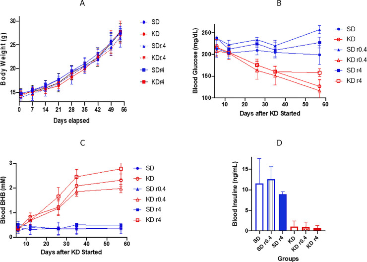
The mice in all SD groups and KD groups (with or without rapamycin) were given the same caloric energy from start point to the end (< 8 weeks). As shown in Fig 1A, the body weights of the mice in KD groups were not significantly different from those in SD groups at any time point during the study.

Fig 1.

Fig 1

The body weight (A), blood glucose (B), beta hydroxybutyrate (C), and insulin (D) levels are displayed (A) Body weight increased equally without differences in all groups. (B) Each glucose data point is a daily average of 3–9 mice, and each mouse was measured 3 times in that particular day at 3 different time points (9 am, 1 pm, and 5 pm). (C) Each beta hydroxybutyrate data point is a daily average of 3–9 mice with single measurement per mouse. (D) The insulin levels were measured when the mice were moribund. The serum was used to measure insulin level with an ELISA method. The blue lines or bars represent the data from SD groups. The red lines or bars are the data from KD groups. r0.4 and r4 means rapamycin at the dose 0.4 mg/kg and 4 mg/kg for 2 weeks, respectively.

KD reduced the blood glucose level in mice

Blood serum glucose concentrations were measured in all mice after one week of dietary intervention and at 4 additional intervals (Fig 1B). Glucose was lower in all KD groups (KD, KD r0.4, and KD r4)) than corresponding SD groups, and decreased further during 3 weeks of KD feeding. Blood glucose was significantly lower than that of mice in all SD groups at all time points. Rapamycin at a low dose did not have a clear effect on the glucose level. A higher dose of rapamycin (4 mg/kg) enhanced the glucose levels slightly in both SD r4 and KD r4. At day 57 the mildly elevated glucose ratios of KD r4/KD and SD r4/SD were 158/127 (p = 0.0042) and 227/199 (p = 0.0223), respectively (Fig 1B).

KD increased the blood beta-hydroxybutyrate level in mice

After 3 weeks, mice in all KD groups showed at least a four-fold elevation of serum BHB concentrations compared with SD mice. These elevations all reached statistical significance (p <0.005). Rapamycin did not affect the BHB level (Fig 1C).

KD reduced the blood insulin concentration in mice

At study termination (see Methods), we collected the blood from each mouse and measured insulin levels. As Fig 1D demonstrates, the insulin serum concentrations in all SD mice groups were 8 to 20-fold higher than the levels in the respective KD mice groups (p<0.0005). The paired comparison is shown in Table 1.

Table 1. Blood insulin level and comparison.

Compared Groups Mean Insulin Levels (ng/ml) P Value
SD vs KD 11.55: 1.08 0.0001
SD r0.4 vs KD r0.4 12.55: 0.96 0.0039
SD r4 vs KD r4 8.90: 0.62 <0.0001

KD inhibited tumor growth and prolonged mouse longevity

Mice were fed SD or KD from approximately 6 weeks of age until they reached a moribund condition. All tumor sizes were measured in each mouse weekly from day 1 to day 56, the time period during which no mice had yet reached moribundity. The combined tumor volumes (sum of all measurable tumor volumes in each mouse) of KD mice were smaller than that of SD mice (mean KD 506 mm3 vs, mean SD 1262 mm3, p <0.0001, 2- way ANOVA; Fig 2A). Rapamycin further enhanced the tumor growth inhibition: the mean combined tumor volume of KD r0.4 and KD r4 versus KD was 359 mm3 vs. 506 mm3 (p = 0.0049) and 195 vs. 506 (p < 0.0001), respectively.

Fig 2. Tumor size and survival.

Fig 2

Animal numbers at start of study diet: SD = 9, SD+r0.4 = 3; SD +r4 = 5; KD = 9; KD+r0.4 = 4; KD+r4 = 4; Tumor size (A) was measured once a week. Volume (mm3) was calculated as 0.5 L x S2 (L and S is the longest and shortest dimensions). The sum of all visible tumor volumes in each mouse was used as its tumor volume, and each point represents tumor volumes from 3–9 mice. Longevity (B) was determined to the time a mouse became moribund. The blue and red lines are the data from SD and KD groups, respectively. r0.4 and r4 represents rapamycin at the dose 0.4 mg/kg and 4 mg/kg for 2 weeks, respectively.

As shown in Fig 2B, the median survival of KD mice increased to 78 days as compared to 65 days for SD mice, a 20% increase (p = 0.0002, log rank test). KD, when combined with rapamycin at dose 4 mg/kg further increased the median survival to 95 days when compared to KD diet alone (78 days, as above, p = 0.002); and vs. SD r4 (81 days, p = 0.0049). See Fig 2.

KD reduced metastases in the lungs of mice

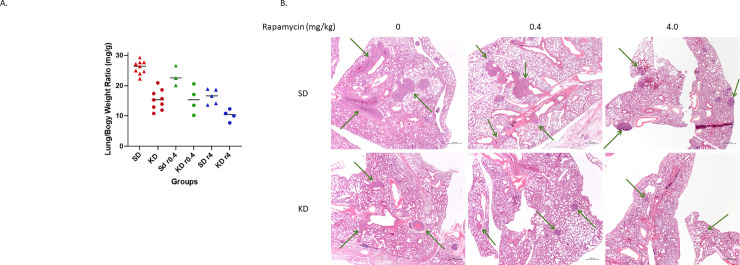
Moribund mice were euthanized and their major organs were resected as described in Methods. Only lungs were found to have metastatic tumors among all organ systems. Lungs were weighed before further pathological evaluation. The lung/body weight ratio of each mouse was also calculated as it is positively related to the lung tumor number (n) and/or size [22]. The data is shown in Fig 3A and Table 2.

Fig 3. The lung metastases.

Fig 3

Moribund mice were weighed, and their lungs were resected and weighed after taking ≥ 1 ml blood out from cardiac puncture. (A) The lung-to-body weight ratio from each group is presented. Round dots represent KD groups. Triangle dots represent SD groups. r0.4 and r4 represent rapamycin at doses of 0.4 mg/kg and 4 mg/kg separately. (B) The lung tissue pathology images are shown. The top row demonstrates lung tissue sections from SD, SDr0.4, and SDr4 groups. The bottom row shows lung tissue sections from KD, KDr0.4, and KDr4. The arrows point out tumor nodules. The magnification is 2.5 x for all pictures. The overall mass of tumors is reduced in the KD images.

Table 2. Lung/body weight ratios (mg/g).

Comparison Average Ratio P Value
SD/KD 26.0/15.3 < 0.0001
SD r0.4/KD r0.4 23.1/15.4 0.0542
SD r4/KD r4 16.4/10.2 0.0045
KD/KD r4 15.3/10.2 0.0163

There is a significant difference of average lung/body weight ratios between SD and KD groups: 26.0 ± 2.2 mg/g vs. 15.3 ± 3.3 mg/g, p < 0.0001. SD/KD with rapamycin at the lower dose of 0.4 mg/kg trended toward further inhibition of lung metastasis (p = 0.0542), with lung/body weight ratio 23.1 ± 3.3 vs. 15.4 ± 4.5 mg/g. Rapamycin at a higher dose (4 mg/kg), when combined with KD showed more significant reduction of lung metastases: SD r4 vs. KD r4 was 16.4 ± 2.5: 10.2± 1.9 mg/g, p = 0.0045, and KD vs. KD r4 was 15.3 ± 3.3: 10.2 ± 1.9, p = 0.0163, respectively. See Fig 3. (above).

The size and number of lung metastatic nodules were also measured in microscopic tumor slices from all mice. See S1 Table. These data provide general, but not statistically significant support for smaller overall metastases in all groups of the KD vs. SD animals. There is clearly limited statistical power due to small sample size; there are also large standard deviations. Both therapy groups have similar numbers of metastases. deviation.

Discussion

We have tested the effect of two diets, one very low in carbohydrate (≤ 0.1% of Calories); the other a standard diet (i.e. 80% calories from carbohydrate), on cancer growth and animal survival in a spontaneous breast cancer model in mice. Strict limitation of carbohydrate was effective at inhibiting serum insulin and glucose and inducing ketosis. While there were no ‘cures’, average overall tumor mass was reduced. The ketogenic group demonstrated prolonged survival as well. We also compared the effects of two different doses of rapamycin when added to the diets. Rapamycin delayed tumor growth in a dose-dependent manner and improved survival in both dietary groups. Greatest effects were seen when KD and rapamycin were combined.

In all groups, with or without rapamycin, the KD mice demonstrated longer survival, lower serum glucose and lower insulin concentrations than the corresponding SD mice. The total lung tumor mass in the KD animals was significantly and substantially smaller than in the corresponding SD mice. In view of significantly longer survival of the KD animals, both of these results are consistent with slower tumor growth in KD vs. SD.

Strict insulin inhibition can result in two principle effects, both of which have the potential to induce cancer cell programmed cell death and to reduce proliferation of cancer cells. First, reduced blood insulin concentrations at the cancer cell membrane results in less binding to the insulin receptor with resulting downstream inhibition of the PI3K-Akt-mTOR (PAM) signaling cascade [23], as well as the RAS-RAF-MEK-ERK pathway [24]. We therefore propose that reduced insulin concentration due to a ketogenic diet provides the potential to enhance programmed cell death by inhibiting the PAM cascade, and to reduce proliferation via both pathways [25,26]. A general caveat, of course, is that well-known, common mutations causing constitutive activation of PAM protein signals (e.g. PI3KCa) will resist programmed cell death and allow proliferation to continue [27]. Nonetheless those malignancies without these mutations can be therapeutically susceptible to the insulin inhibiting effects.

Second, hepatic ketogenesis due to insulin inhibition increases blood levels of the ketone bodies beta-hydroxybutyrate and acetoacetate, both of which have demonstrated histone deacetylase inhibitor effects at the cellular level. HDAC inhibitors are known to be capable of reducing cancer cell proliferation as well as enhancing programmed cell death [2830].

Reduced proliferation was indeed observed as seen in the reduced extent of lung metastatic mass to total body mass ratios with KD in all groups. However, while necrosis in the KD groups was also detected in tumor specimens, meaningful differences with SD could not be identified definitively with our small sample size.

The potential of ketogenic diets to inhibit cancers has been suggested, mainly in animal models, for at least four decades [31,32]. Human data has been limited mostly to a few case reports or small clinical trials [5,3336]. Additional studies have been reported more recently [37]. Meanwhile, in the past decade, interest has grown in insulin inhibition as a potential cancer therapeutic adjunct. Metformin, for example, has been applied toward this end with promising results in the neoadjuvant setting for breast cancer [38], but when applied therapeutically has demonstrated limited benefit [39]. Dietary carbohydrate restriction and metformin both reduce insulin secretion and glucose concentration, but the overall effect of carbohydrate restriction has additional effects beyond those of metformin. This is observed in its ability to induce formation of ketone bodies, known HDAC inhibitors. Ketone body formation speaks to the potential for translation to humans, as the 2 mM maximum extent of ketosis achieved in our mouse model is exceeded in people for whom 4–5 mM are quite achievable.

Nonetheless, one cannot assume that our mouse model results will translate to humans. Many mouse models, including our spontaneous breast tumor mouse with a B6 phenotype background, have been shown to have a degree of insulin resistance [40]. This resistance is reflected by a) persistent, high glucose concentrations in the SD group as well as b) delayed return of glucose concentrations to normal in the KD group (see persistent high glucose at 11 days in Fig 1, suggesting ongoing insulin resistance effect). Therefore, some of the relative survival disadvantage of the SD group may have resulted from a heightened insulin growth effect on the tumors. But it is worth noting, too, that the KD group might also have lived longer without prolonged high glucose levels. In any event, obesity with insulin resistance is commonplace among many patients with breast cancer, particularly in post-menopausal women [41]. The ultimate response to a KD in humans with breast cancer therefore demands controlled trials for those with normal as well as abnormal insulin sensitivity.

The results support the potential value of ketogenic diets in cancer therapy when coupled with existing agents, permitting additive or synergistic effects with toxic drugs. Successful translation, then, could result in reduction of drug doses while improving overall therapeutic effectiveness, thus extending patient survival while improving the quality of life during that period of greater longevity.

A ketogenic diet combined with existing drugs may provide a promising approach to increase the therapeutic effects of existing cancer therapies at lower levels of overall toxicity.

Supporting information

S1 Table. Size and number of metastases in lungs from microscopic slide.

(DOCX)

S1 File

(DOCX)

S2 File

(DOCX)

S3 File

(DOCX)

Acknowledgments

The authors are grateful to Linda Jelicks and Wade Koba for helpful suggestions and consistent support and to Dr. Igor Koman for his contribution and support.

Data Availability

All relevant data are within the manuscript and its Supporting Information files.

Funding Statement

Generous funding for this project was provided by ST Balchug, a commercial company which operates in the real estate sector. In addition, the study was supported in part by the CTSA Grant UL1TR002556 from the National Center for Advancing Translational Sciences (NCATS), a component of the National Institutes of Health (NIH). The funders had no role in study design, data collection and analysis, decision to publish, or preparation of the manuscript.

References

  • 1.Venkateswaran V. Haddad A. Fleshner NF, et al. Association of Diet-Induced Hyperinsulinemia With Accelerated Growth of Prostate Cancer (LNCaP) Xenografts, JNCI: Journal of the National Cancer Institute, 99, (23) 1793–1800, 10.1093/jnci/djm231 [DOI] [PubMed] [Google Scholar]
  • 2.Klement and Kämmerer Is there a role for carbohydrate restriction in the treatment and prevention of cancer? Nutrition & Metabolism 2011, 8:75 http://www.nutritionandmetabolism.com/content/8/1/75 [DOI] [PMC free article] [PubMed] [Google Scholar]
  • 3.Goodwin PJ. Obesity, insulin resistance and breast cancer outcomes. Breast. 2015;24 Suppl 2:S56–9. Epub 2015/08/19. 10.1016/j.breast.2015.07.014 [DOI] [PubMed] [Google Scholar]
  • 4.Klement RJ. Beneficial effects of ketogenic diets for cancer patients: a realist review with focus on evidence and confirmation. Med Oncol. 2017;34(8):132 Epub 2017/06/28. 10.1007/s12032-017-0991-5 . [DOI] [PubMed] [Google Scholar]
  • 5.Fine EJ, Segal-Isaacson CJ, Feinman RD, Herszkopf S, Romano MC, Tomuta N, et al. Targeting insulin inhibition as a metabolic therapy in advanced cancer: a pilot safety and feasibility dietary trial in 10 patients. Nutrition. 2012;28(10):1028–35. 10.1016/j.nut.2012.05.001 . [DOI] [PubMed] [Google Scholar]
  • 6.Fine EJ, Feinman RD: Insulin, Carbohydrate Restriction, Metabolic Syndrome and Cancer. Expert Rev Endocrinol Metab 2014, 9(6). 10.1586/17446651.2014.960392 [DOI] [PubMed] [Google Scholar]
  • 7.Hsieh MH, Choe JH, Gadhvi J, Kim YJ, Arguez MA, Palmer M, et al. p63 and SOX2 Dictate Glucose Reliance and Metabolic Vulnerabilities in Squamous Cell Carcinomas. Cell Rep. 2019;28(7):1860–78 e9. 10.1016/j.celrep.2019.07.027 . [DOI] [PMC free article] [PubMed] [Google Scholar]
  • 8.Hopkins BD, Pauli C, Du X, Wang DG, Li X, Wu D, et al. Suppression of insulin feedback enhances the efficacy of PI3K inhibitors. Nature. 2018;560(7719):499–503. Epub 2018/07/28. 10.1038/s41586-018-0343-4 [DOI] [PMC free article] [PubMed] [Google Scholar]
  • 9.Basho RK, Yam C, Gilcrease M, Murthy RK, Helgason T, Karp DD, et al. Comparative Effectiveness of an mTOR-Based Systemic Therapy Regimen in Advanced, Metaplastic and Nonmetaplastic Triple-Negative Breast Cancer. Oncologist. 2018;23(11):1300–9. Epub 2018/08/25. 10.1634/theoncologist.2017-0498 [DOI] [PMC free article] [PubMed] [Google Scholar]
  • 10.Jain V, Patel RK, Kapadia Z, Galiveeti S, Banerji M, Hope L. Drugs and hyperglycemia: A practical guide. Maturitas. 2017;104:80–3. Epub 2017/09/20. 10.1016/j.maturitas.2017.08.006 . [DOI] [PubMed] [Google Scholar]
  • 11.Schindler CE, Partap U, Patchen BK, Swoap SJ. Chronic rapamycin treatment causes diabetes in male mice. Am J Physiol Regul Integr Comp Physiol. 2014;307(4):R434–43. 10.1152/ajpregu.00123.2014 [DOI] [PMC free article] [PubMed] [Google Scholar]
  • 12.https://www.accessdata.fda.gov/scripts/cder/daf/index.cfm?event=overview.process&ApplNo=021083
  • 13.Klement(2018), Fasting, Fats and Physics: Combining ketogenic diets and radiation therapy against cancer. Complementary Medicine Research 25(2):102–113; 10.1159/000484045 Epub 2017. November 9 [DOI] [PubMed] [Google Scholar]
  • 14.Kim HS, Masko EM, Poulton SL, et al. Carbohydrate restriction and lactate transporter inhibition in a mouse xenograft model of human prostate cancer. BJU Int. 2012;110(7):1062‐1069. 10.1111/j.1464-410X.2012.10971.x [DOI] [PMC free article] [PubMed] [Google Scholar]
  • 15.Iyikesici MS. Feasibility study of metabolically supported chemotherapy with weekly carboplatin/paclitaxel combined with ketogenic diet, hyperthermia and hyperbaric oxygen therapy in metastatic non-small cell lung cancer. Int J Hyperthermia. 2019;36(1):446–55. 10.1080/02656736.2019.1589584 . [DOI] [PubMed] [Google Scholar]
  • 16.van der Louw E, Olieman JF, van den Bemt P, Bromberg JEC, Oomen-de Hoop E, Neuteboom RF, et al. Ketogenic diet treatment as adjuvant to standard treatment of glioblastoma multiforme: a feasibility and safety study. Ther Adv Med Oncol. 2019;11:1758835919853958 Epub 2019/07/02. 10.1177/1758835919853958 [DOI] [PMC free article] [PubMed] [Google Scholar]
  • 17.Khodabakhshi A, Akbari ME, Mirzaei HR, Mehrad-Majd H, Kalamian M, Davoodi SH. Feasibility, Safety, and Beneficial Effects of MCT-Based Ketogenic Diet for Breast Cancer Treatment: A Randomized Controlled Trial Study. Nutr Cancer. 2019:1–8. Epub 2019/09/10. 10.1080/01635581.2019.1650942 . [DOI] [PubMed] [Google Scholar]
  • 18.Woodhouse C, Ward T, Gaskill-Shipley M, Chaudhary R. Feasibility of a modified Atkins diet in glioma patients during radiation and its effect on radiation sensitization. Curr Oncol. 2019;26(4):e433–e8. Epub 2019/09/25. 10.3747/co.26.4889 [DOI] [PMC free article] [PubMed] [Google Scholar]
  • 19.Nutrient Requirements of Laboratory Animals: Fourth Revised Edition, 1995. Washington (DC)1995. [PubMed]
  • 20.Guy CT, Cardiff RD, Muller WJ. Induction of mammary tumors by expression of polyomavirus middle T oncogene: a transgenic mouse model for metastatic disease. Mol Cell Biol. 1992;12(3):954–61. 10.1128/mcb.12.3.954 [DOI] [PMC free article] [PubMed] [Google Scholar]
  • 21.Shishido SN, Delahaye A, Beck A, Nguyen TA. The anticancer effect of PQ1 in the MMTV-PyVT mouse model. Int J Cancer. 2014;134(6):1474–83. 10.1002/ijc.28461 [DOI] [PMC free article] [PubMed] [Google Scholar]
  • 22.Zou Y, Zong G, Ling YH, Hao MM, Lozano G, Hong WK, et al. Effective treatment of early endobronchial cancer with regional administration of liposome-p53 complexes. J Natl Cancer Inst. 1998;90(15):1130–7. 10.1093/jnci/90.15.1130 . [DOI] [PubMed] [Google Scholar]
  • 23.Boone E, Ansley J, Schulte M, Maleah Holland A: Validity and Reliability of the Keto-Mojo Blood Ketone and Glucose Meter; https://cdn.shopify.com/s/files/1/2010/7777/files/Augusta_University_Validity_and_Reliability_of_Keto_Mojo_Meter.pdf?2169
  • 24.Senapati P, Kato H, Lee M, Leung A, Thai C, Sanchez A, et al. Hyperemia promotes aberrant histone acetylation in triple-negative breast cancer. Epigenetics Chromatin. 2019;12(1):44 Epub 2019/07/19. 10.1186/s13072-019-0290-9 [DOI] [PMC free article] [PubMed] [Google Scholar]
  • 25.Nagini S, Sophia J, Mishra R. Glycogen synthase kinases: Moonlighting proteins with theranostic potential in cancer. Semin Cancer Biol. 2019;56:25–36. Epub 2018/01/09. 10.1016/j.semcancer.2017.12.010 . [DOI] [PubMed] [Google Scholar]
  • 26.Radogna F, Dicato M, Diederich M. Cancer-type-specific crosstalk between autophagy, necroptosis and apoptosis as a pharmacological target. Biochem Pharmacol. 2015;94(1):1–11. Epub 2015/01/07. 10.1016/j.bcp.2014.12.018 . [DOI] [PubMed] [Google Scholar]
  • 27.Kalaany NY, Sabatini DM. Tumours with PI3K activation are resistant to dietary restriction [published correction appears in Nature. 2020 May;581(7807):E2]. Nature. 2009;458(7239):725–731. 10.1038/nature07782 [DOI] [PMC free article] [PubMed] [Google Scholar]
  • 28.Thakur B, Kumar Y, Bhatia A. Programmed necrosis and its role in management of breast cancer. Pathol Res Pract. 2019;215(11):152652 Epub 2019/10/02. 10.1016/j.prp.2019.152652 . [DOI] [PubMed] [Google Scholar]
  • 29.Shimazu T, Hirschey MD, Newman J, He W, Shirakawa K, Le Moan N, et al. Suppression of oxidative stress by beta-hydroxybutyrate, an endogenous histone deacetylase inhibitor. Science. 2013;339(6116):211–4. Epub 2012/12/12. 10.1126/science.1227166 [DOI] [PMC free article] [PubMed] [Google Scholar]
  • 30.Wilson AJ, Chueh AC, Togel L, Corner GA, Ahmed N, Goel S, et al. Apoptotic sensitivity of colon cancer cells to histone deacetylase inhibitors is mediated by an Sp1/Sp3-activated transcriptional program involving immediate-early gene induction. Cancer Res. 2010;70(2):609–20. Epub 2010/01/14. 10.1158/0008-5472.CAN-09-2327 [DOI] [PMC free article] [PubMed] [Google Scholar]
  • 31.Tisdale MJ, Brennan RA, Fearon KC. Reduction of weight loss and tumour size in a cachexia model by a high fat diet. Br J Cancer. 1987;56(1):39–43. Epub 1987/07/01. 10.1038/bjc.1987.149 [DOI] [PMC free article] [PubMed] [Google Scholar]
  • 32.Beck SA, Tisdale MJ. Nitrogen excretion in cancer cachexia and its modification by a high fat diet in mice. Cancer Res. 1989;49(14):3800–4. Epub 1989/07/15. Klement, R.J., Brehm, N. & Sweeney, R.A. Ketogenic diets in medical oncology: a systematic review with focus on clinical outcomes. Med Oncol 37, 14 (2020). 10.1007/212032-020-1337-2. [DOI] [PubMed] [Google Scholar]
  • 33.Nebeling LC, Miraldi F, Shurin SB, Lerner E. Effects of a ketogenic diet on tumor metabolism and nutritional status in pediatric oncology patients: two case reports. J Am Coll Nutr. 1995;14(2):202–8. Epub 1995/04/01. 10.1080/07315724.1995.10718495 . [DOI] [PubMed] [Google Scholar]
  • 34.Champ CE, Palmer JD, Volek JS, Werner-Wasik M, Andrews DW, Evans JJ, et al. Targeting metabolism with a ketogenic diet during the treatment of glioblastoma multiforme. J Neurooncol. 2014;117(1):125–31. Epub 2014/01/21. 10.1007/s11060-014-1362-0 . [DOI] [PubMed] [Google Scholar]
  • 35.Seyfried TN, Flores R, Poff AM, D'Agostino DP, Mukherjee P. Metabolic therapy: a new paradigm for managing malignant brain cancer. Cancer Lett. 2015;356(2 Pt A):289–300. Epub 2014/07/30. 10.1016/j.canlet.2014.07.015 . [DOI] [PubMed] [Google Scholar]
  • 36.Zahra A, Fath MA, Opat E, Mapuskar KA, Bhatia SK, Ma DC, et al. Consuming a Ketogenic Diet while Receiving Radiation and Chemotherapy for Locally Advanced Lung Cancer and Pancreatic Cancer: The University of Iowa Experience of Two Phase 1 Clinical Trials. Radiat Res. 2017;187(6):743–54. Epub 2017/04/25. 10.1667/RR14668.1 [DOI] [PMC free article] [PubMed] [Google Scholar]
  • 37.Klement RJ, Brehm N, Sweeney RA. Ketogenic diets in medical oncology: A systematic review with focus on clinical outcomes. Med Oncol. 2020. January 11;37(2):14 10.1007/s12032-020-1337-2 [DOI] [PubMed] [Google Scholar]
  • 38.Niraula S, Dowling RJ, Ennis M, et al. Metformin in early breast cancer: a prospective window of opportunity neoadjuvant study. Breast Cancer Res Treat. 2012;135(3):821–830. 10.1007/s10549-012-2223-1 [DOI] [PubMed] [Google Scholar]
  • 39.Pimentel I, Lohmann AE, Ennis M, et al. A phase II randomized clinical trial of the effect of metformin versus placebo on progression-free survival in women with metastatic breast cancer receiving standard chemotherapy. Breast. 2019;48:17–23. 10.1016/j.breast.2019.08.003 [DOI] [PubMed] [Google Scholar]
  • 40.Goren HJ, Kulkarni RN, Kahn CR. Glucose homeostasis and tissue transcript content of insulin signaling intermediates in four inbred strains of mice: C57BL/6, C57BLKS/6, DBA/2, and 129X1. Endocrinology. 2004. July;145(7):3307–23. 10.1210/en.2003-1400 Epub 2004 Mar 24. . [DOI] [PubMed] [Google Scholar]
  • 41.Kabat GC, Kim MY, Lee JS, Ho GY, Going SB, Beebe-Dimmer J, et al. Metabolic Obesity Phenotypes and Risk of Breast Cancer in Postmenopausal Women. Cancer Epidemiol Biomarkers Prev. 2017. Dec;26(12):1730–1735. 10.1158/1055-9965.EPI-17-0495 Epub 2017 Sep 22. ; PMCID: PMC6986334. [DOI] [PMC free article] [PubMed] [Google Scholar]

Decision Letter 0

Salvatore V Pizzo

2 Jun 2020

PONE-D-20-11420

The Effect of a Ketogenic Diet and Synergy with Rapamycin in a Mouse Model of Breast Cancer.

PLOS ONE

Dear Dr. Fine,

Thank you for submitting your manuscript to PLOS ONE. After careful consideration, we feel that it has merit but does not fully meet PLOS ONE’s publication criteria as it currently stands. Therefore, we invite you to submit a revised version of the manuscript that addresses the points raised during the review process.

Please submit your revised manuscript by Jul 17 2020 11:59PM. If you will need more time than this to complete your revisions, please reply to this message or contact the journal office at plosone@plos.org. When you're ready to submit your revision, log on to https://www.editorialmanager.com/pone/ and select the 'Submissions Needing Revision' folder to locate your manuscript file.

Please include the following items when submitting your revised manuscript: 

Both reviewers have identified a number of issues which need to be addressed if the authors plan to submit a revised manuscript. The final comment of reviewer 2 in particular needs to be considered. As you can see the reviewer seeks clarification as to how the measurements could have been blind if only one individual othdid all of the measurements.

  • A rebuttal letter that responds to each point raised by the academic editor and reviewer(s). You should upload this letter as a separate file labeled 'Response to Reviewers'.

  • A marked-up copy of your manuscript that highlights changes made to the original version. You should upload this as a separate file labeled 'Revised Manuscript with Track Changes'.

  • An unmarked version of your revised paper without tracked changes. You should upload this as a separate file labeled 'Manuscript'.

If you would like to make changes to your financial disclosure, please include your updated statement in your cover letter. Guidelines for resubmitting your figure files are available below the reviewer comments at the end of this letter.

If applicable, we recommend that you deposit your laboratory protocols in protocols.io to enhance the reproducibility of your results. Protocols.io assigns your protocol its own identifier (DOI) so that it can be cited independently in the future. For instructions see: http://journals.plos.org/plosone/s/submission-guidelines#loc-laboratory-protocols

We look forward to receiving your revised manuscript.

Kind regards,

Salvatore V Pizzo

Academic Editor

PLOS ONE

Journal Requirements:

When submitting your revision, we need you to address these additional requirements.

1. Please ensure that your manuscript meets PLOS ONE's style requirements, including those for file naming. The PLOS ONE style templates can be found at

https://journals.plos.org/plosone/s/file?id=wjVg/PLOSOne_formatting_sample_main_body.pdf and

https://journals.plos.org/plosone/s/file?id=ba62/PLOSOne_formatting_sample_title_authors_affiliations.pdf

2. At this time, we request that you  please report additional details in your Methods section regarding animal care, as per our editorial guidelines:

(a) Please state the specific source of the mice used in the study (e.g. Jackson laboratories). In addition, please state the total number of mice used in the study

(b) Please provide details of animal welfare (e.g., shelter, food, water, environmental enrichment)

Thank you for your attention to these requests.

3. Please note that PLOS does not permit references to “data not shown.” Authors should provide the relevant data within the manuscript, the Supporting Information files, or in a public repository. If the data are not a core part of the research study being presented, we ask that authors remove any references to these data.

4. To comply with PLOS ONE submission guidelines, in your Methods section, please provide additional information regarding your statistical analyses. For more information on PLOS ONE's expectations for statistical reporting, please see https://journals.plos.org/plosone/s/submission-guidelines.#loc-statistical-reporting

5. Thank you for stating the following in the Acknowledgments Section of your manuscript:

'The authors are very grateful to ST Balchug who provided private philanthropic funding for these investigations. In addition, the study was supported in part by the CTSA Grant UL1TR002556 from the National Center for Advancing Translational Sciences (NCATS), a component of the National Institutes of Health (NIH)'

We note that you have provided funding information that is not currently declared in your Funding Statement. However, funding information should not appear in the Acknowledgments section or other areas of your manuscript. We will only publish funding information present in the Funding Statement section of the online submission form.

a. Please remove any funding-related text from the manuscript and let us know how you would like to update your Funding Statement. Currently, your Funding Statement reads as follows:

'The funding for this study and for all the authors was provided by a private philanthropic donor (mentioned in the Acknowledgments). Accordingly there was no national grant award number. The sponsor does not have a website for philanthropic giving. The sponsor made no effort to influence any aspect of the study design, data collection and analysis, decision to publish, or preparation of the manuscript.'

b. Additionally, because some of your funding information pertains to commercial funding, we ask you to provide an updated Competing Interests statement, declaring all sources of commercial funding.

In your Competing Interests statement, please confirm that your commercial funding does not alter your adherence to PLOS ONE Editorial policies and criteria by including the following statement: "This does not alter our adherence to PLOS ONE policies on sharing data and materials.” as detailed online in our guide for authors  http://journals.plos.org/plosone/s/competing-interests.  If this statement is not true and your adherence to PLOS policies on sharing data and materials is altered, please explain how.

c. Please include the updated Competing Interests Statement and Funding Statement in your cover letter. We will change the online submission form on your behalf.

Please know it is PLOS ONE policy for corresponding authors to declare, on behalf of all authors, all potential competing interests for the purposes of transparency. PLOS defines a competing interest as anything that interferes with, or could reasonably be perceived as interfering with, the full and objective presentation, peer review, editorial decision-making, or publication of research or non-research articles submitted to one of the journals. Competing interests can be financial or non-financial, professional, or personal. Competing interests can arise in relationship to an organization or another person. Please follow this link to our website for more details on competing interests: http://journals.plos.org/plosone/s/competing-interests

6. We note that you have indicated that data from this study are available upon request. PLOS only allows data to be available upon request if there are legal or ethical restrictions on sharing data publicly. For information on unacceptable data access restrictions, please see http://journals.plos.org/plosone/s/data-availability#loc-unacceptable-data-access-restrictions.

In your revised cover letter, please address the following prompts:

a) If there are ethical or legal restrictions on sharing a de-identified data set, please explain them in detail (e.g., data contain potentially identifying or sensitive patient information) and who has imposed them (e.g., an ethics committee). Please also provide contact information for a data access committee, ethics committee, or other institutional body to which data requests may be sent.

b) If there are no restrictions, please upload the minimal anonymized data set necessary to replicate your study findings as either Supporting Information files or to a stable, public repository and provide us with the relevant URLs, DOIs, or accession numbers. Please see http://www.bmj.com/content/340/bmj.c181.long for guidelines on how to de-identify and prepare clinical data for publication. For a list of acceptable repositories, please see http://journals.plos.org/plosone/s/data-availability#loc-recommended-repositories.

We will update your Data Availability statement on your behalf to reflect the information you provide.

7. Please include captions for your Supporting Information files at the end of your manuscript, and update any in-text citations to match accordingly. Please see our Supporting Information guidelines for more information: http://journals.plos.org/plosone/s/supporting-information

[Note: HTML markup is below. Please do not edit.]

Reviewers' comments:

Reviewer's Responses to Questions

Comments to the Author

1. Is the manuscript technically sound, and do the data support the conclusions?

The manuscript must describe a technically sound piece of scientific research with data that supports the conclusions. Experiments must have been conducted rigorously, with appropriate controls, replication, and sample sizes. The conclusions must be drawn appropriately based on the data presented.

Reviewer #1: Yes

Reviewer #2: No

**********

2. Has the statistical analysis been performed appropriately and rigorously?

Reviewer #1: Yes

Reviewer #2: I Don't Know

**********

3. Have the authors made all data underlying the findings in their manuscript fully available?

The PLOS Data policy requires authors to make all data underlying the findings described in their manuscript fully available without restriction, with rare exception (please refer to the Data Availability Statement in the manuscript PDF file). The data should be provided as part of the manuscript or its supporting information, or deposited to a public repository. For example, in addition to summary statistics, the data points behind means, medians and variance measures should be available. If there are restrictions on publicly sharing data—e.g. participant privacy or use of data from a third party—those must be specified.

Reviewer #1: No

Reviewer #2: Yes

**********

4. Is the manuscript presented in an intelligible fashion and written in standard English?

PLOS ONE does not copyedit accepted manuscripts, so the language in submitted articles must be clear, correct, and unambiguous. Any typographical or grammatical errors should be corrected at revision, so please note any specific errors here.

Reviewer #1: Yes

Reviewer #2: Yes

**********

5. Review Comments to the Author

Please use the space provided to explain your answers to the questions above. You may also include additional comments for the author, including concerns about dual publication, research ethics, or publication ethics. (Please upload your review as an attachment if it exceeds 20,000 characters)

Reviewer #1: The paper "The effect of a ketogenic diet and synergy with rapamycin in a mouse model of breast

cancer" is an important contribution to the growing literature on ketogenic therapy in cancer.

The research question is well described and relevant given the fact that rapamycin has received a lot of attention as a

calorie restriction mimetic and anti-ageing drug.

The study has several strengths, among them the naturally occurence of the murine breast tumors instead of xenografted ones,

measurement of relevant outcome variables and good description of the methods and results.

The major limitation is the small sample size in some of the treatment groups (n=4), but the

treatments were chosen as to still provide sufficiantly clear outcome differences.

The most obvious limitation when reading the paper was that many references did not support the claims made.

I therefore recommend publication of this paper after some points are corrected by the authors. I think this will

further improve the scientific rigor and clarity of argumentation.

- Lines 37-39: The authors refer to insulin inhibition, suggesting therefby from the outset

that this is a major mechanism how ketogenic diets work. However, none of the four references cited

have measured insulin levels or discussed this mechanism in great detail. Ref. 1 has no authors listed , I suppose

it is a review by Allen et al. 2014, Redox Biol 2:963-970 ? Ref.4 did not show any benefit of the ketogenic diet

and therefore does not support the claim of the authors made in that sentence. Please cite more relevant studies showing

tumor growth inhibition potentially achieved via insulin decrease (e.g. Venkateswaran et al. 2007, JNCI 99:1793-1800)

or drop the statement about insulin here.

- Line 73: The authors refer to additive effects of a ketogenic diet with chemotherapy in mice, but cite Iyikesici et al (ref.12) which is

a human study, Gluschnaider et al. (ref.13) which did not combine a ketogenic diet with chemotherapy, and Schartz et al. (ref. 14) which is

a review not specifically on the ketogenic diet. Thus, none of these references support the claims made. Instead, I would recommend citing

the following studies:

Klement (2018), Complementary Medicine Research 25(2):102-113; Morscher et al. (2016), Oncotarget 7:17060–17073;

Kim et a. (2012), BJU International 110:1062-1069

PLease also delete "for widely metastatic disease" (line 72), this is not true for all studies

- Same line (73): Ref.4 and ref. 15 have shown no benefit of the ketogenic diet, hence do not support the claim made

- Line 103: Please define IACUC at first usage.

- Line 123: Instead of "etc." please name what has been tested

- Line 157: The t-test assumes normal distribution of the data. Have you tested if this is valid? I doubt that with such small sample size this

could be done. Instaed, I would suggest using the Mann Whitney U test here, too.

- Lines 172-173: Please read this sentence again, it appears there is a word missing and "mouse" should be "mice"

- Line 193 (caption to Fig. 1) The description of panel A (body weight) is missing, and (A) should be (B), (B) should be (C) and (C) should be (D).

- Line 210: 2-way ANOVA should also be mentioned in the Methods section

- Line 285: The abbreviation HDACi is never used after definition and can be dropped; in line 300 use "HDAC" instead of "histone deacetylase"

- Lines 293-294: "Human data has been sparse" --> First RCTs have been conducted! Please cite Klement et al. 2020, Med Oncol 37:14 who

have summarized clincal studies with relevant outcomes

- Figure 1: axis label "Days elapsed" refers to which time point?

- Data availability statement was missing

Reviewer #2: The manuscript entitled “The Effect of a Ketogenic Diet and Synergy with Rapamycin in a Mouse Model of Breast Cancer.” PONE-D-20-11420 The authors attempt to demonstrate that ketogenic diet combined with rapamycin inhibits breast cancer growth. There are both major and minor problems with this paper that make a thorough review difficult.

Some minor problems that need to be addressed: Line 73 of introduction states, “effects in humans” and then lists references that are feasibility studies, and do not show effectiveness. Researchers must be very careful with language when discussing human trials. Line 82 list the protein/carb/fat content but does not discuss the kind of fat used. We have found that the lipid/PUFA content of the KD is very important for tumor growth inhibition. Please list the complete content of the diet. Line 183 a decimal is missing. Figure 1B, the initial blood glucose is very high in all the animals. Even after 11 days the glucose level is very high in KD groups. This is very surprising and counterintuitive. Sometimes blood glucose in mice spikes if they are scared and the time of the blood draw and you don’t act very quickly. Therefore, hemoglobin A1C might be a better indicator of true glucose control. Figure 1D insulin spelling on the axis and no indication of statistics performed. Figure 2A how many mice were in each group. It should be in the legend. The way that you measure tumor growth hides important data. Did you count the number of tumors arising on each mouse (tells us if initiation is affected); vs tumor volume of a single tumor (growth inhibition).

Some major problems to be addressed: line 108 indicates that there was only one investigator measure the mice. How could you keep that person blinded? Tumor size measurements has been shown to be subject to UNintentional biasing. It is essential that the investigator be blinded. Also, the general health of the mice needs to be investigated. Were they lethargic? Was there a change in their coat? Is there any other indication that the mice we made ill by the treatment? Lastly, the lung needs to be examined by a blinded pathologist to determine the number and stage of the nodules.

In general, there might be interesting information within the text but because the data is not adequately described it is impossible for me to adequately review it.

**********

6. PLOS authors have the option to publish the peer review history of their article (what does this mean?). If published, this will include your full peer review and any attached files.

If you choose “no”, your identity will remain anonymous but your review may still be made public.

Do you want your identity to be public for this peer review? For information about this choice, including consent withdrawal, please see our Privacy Policy.

Reviewer #1: Yes: Rainer J. Klement

Reviewer #2: Yes: Melissa Fath

[NOTE: If reviewer comments were submitted as an attachment file, they will be attached to this email and accessible via the submission site. Please log into your account, locate the manuscript record, and check for the action link "View Attachments". If this link does not appear, there are no attachment files.]

While revising your submission, please upload your figure files to the Preflight Analysis and Conversion Engine (PACE) digital diagnostic tool, https://pacev2.apexcovantage.com/. PACE helps ensure that figures meet PLOS requirements. To use PACE, you must first register as a user. Registration is free. Then, login and navigate to the UPLOAD tab, where you will find detailed instructions on how to use the tool. If you encounter any issues or have any questions when using PACE, please email PLOS at figures@plos.org. Please note that Supporting Information files do not need this step.

Attachment

Submitted filename: Plos one review KD 6.2020.docx

PLoS One. 2020 Dec 3;15(12):e0233662. doi: 10.1371/journal.pone.0233662.r002

Author response to Decision Letter 0


19 Aug 2020

1. Please ensure that your manuscript meets PLOS ONE's style requirements, including those for file naming. The PLOS ONE style templates can be found at

https://journals.plos.org/plosone/s/file?id=wjVg/PLOSOne_formatting_sample_main_body.pdf and

https://journals.plos.org/plosone/s/file?id=ba62/PLOSOne_formatting_sample_title_authors_affiliations.pdf

We believe we have observed such requirements.

At this time, we request that you please report additional details in your Methods section regarding animal care, as per our editorial guidelines:

(a) Please state the specific source of the mice used in the study (e.g. Jackson laboratories). In addition, please state the total number of mice used in the study

(b) Please provide details of animal welfare (e.g. shelter, food, water, environmental enrichment).

Thank you for your attention to these requests.

Existing text was noted as Jax Lab (a subdivision of Jackson Laboratories which provide the animals), but changed to Jackson Laboratories.

Total numbers of mice used now indicated, and animal welfare description expanded.

3. Please note that PLOS does not permit references to “data not shown.” Authors should provide the relevant data within the manuscript, the Supporting Information files, or in a public repository. If the data are not a core part of the research study being presented, we ask that authors remove any references to these data.

Done.

4. To comply with PLOS ONE submission guidelines, in your Methods section, please provide additional information regarding your statistical analyses. For more information on PLOS ONE's expectations for statistical reporting, please see https://journals.plos.org/plosone/s/submission-guidelines.#loc-statistical-reporting

5. Thank you for stating the following in the Acknowledgments Section of your manuscript:

'The authors are very grateful to ST Balchug who provided private philanthropic funding for these investigations. In addition, the study was supported in part by the CTSA Grant UL1TR002556 from the National Center for Advancing Translational Sciences (NCATS), a component of the National Institutes of Health (NIH)'

We note that you have provided funding information that is not currently declared in your Funding Statement. However, funding information should not appear in the Acknowledgments section or other areas of your manuscript. We will only publish funding information present in the Funding Statement section of the online submission form.

Removed from Acknowledgments

a. Please remove any funding-related text from the manuscript and let us know how you would like to update your Funding Statement. Currently, your Funding Statement reads as follows:

'The funding for this study and for all the authors was provided by a private philanthropic donor (mentioned in the Acknowledgments). Accordingly there was no national grant award number. The sponsor does not have a website for philanthropic giving. The sponsor made no effort to influence any aspect of the study design, data collection and analysis, decision to publish, or preparation of the manuscript.'

We’ve removed funding information from the Acknowledgments.

The Funding Statement will now reflect the philanthropic sponsor as well as the CTSA Grant UL1TR002556 from the National Center for Advancing Translational Sciences (NCATS), a component of the National Institutes of Health (NIH).

b. Additionally, because some of your funding information pertains to commercial funding, we ask you to provide an updated Competing Interests statement, declaring all sources of commercial funding.

In your Competing Interests statement, please confirm that your commercial funding does not alter your adherence to PLOS ONE Editorial policies and criteria by including the following statement: "This does not alter our adherence to PLOS ONE policies on sharing data and materials.” as detailed online in our guide for authors http://journals.plos.org/plosone/s/competing-interests. If this statement is not true and your adherence to PLOS policies on sharing data and materials is altered, please explain how.

Above comments are incorrect. The philanthropic funding is not from a commercial source. The point above is therefore moot.

c. Please include the updated Competing Interests Statement and Funding Statement in your cover letter. We will change the online submission form on your behalf.

Please know it is PLOS ONE policy for corresponding authors to declare, on behalf of all authors, all potential competing interests for the purposes of transparency. PLOS defines a competing interest as anything that interferes with, or could reasonably be perceived as interfering with, the full and objective presentation, peer review, editorial decision-making, or publication of research or non-research articles submitted to one of the journals. Competing interests can be financial or non-financial, professional, or personal. Competing interests can arise in relationship to an organization or another person. Please follow this link to our website for more details on competing interests: http://journals.plos.org/plosone/s/competing-interests

Not applicable.

6. We note that you have indicated that data from this study are available upon request. PLOS only allows data to be available upon request if there are legal or ethical restrictions on sharing data publicly. For information on unacceptable data access restrictions, please see http://journals.plos.org/plosone/s/data-availability#loc-unacceptable-data-access-restrictions.

We remove the statement that data are available on request. There are no relevant data required for the submitted paper, which is now complete as is.

In your revised cover letter, please address the following prompts:

a) If there are ethical or legal restrictions on sharing a de-identified data set, please explain them in detail (e.g., data contain potentially identifying or sensitive patient information) and who has imposed them (e.g., an ethics committee). Please also provide contact information for a data access committee, ethics committee, or other institutional body to which data requests may be sent.

b) If there are no restrictions, please upload the minimal anonymized data set necessary to replicate your study findings as either Supporting Information files or to a stable, public repository and provide us with the relevant URLs, DOIs, or accession numbers. Please see http://www.bmj.com/content/340/bmj.c181.long for guidelines on how to de-identify and prepare clinical data for publication. For a list of acceptable repositories, please see http://journals.plos.org/plosone/s/data-availability#loc-recommended-repositories.

We will update your Data Availability statement on your behalf to reflect the information you provide.

The above statements are relevant to human studies, but not to an animal study.

7. Please include captions for your Supporting Information files at the end of your manuscript, and update any in-text citations to match accordingly. Please see our Supporting Information guidelines for more information: http://journals.plos.org/plosone/s/supporting-information

[Note: HTML markup is below. Please do not edit.]

Reviewers' comments:

Reviewer's Responses to Questions

Comments to the Author

1. Is the manuscript technically sound, and do the data support the conclusions?

Reviewer #1: Yes

Reviewer #2: No

2. Has the statistical analysis been performed appropriately and rigorously?

Reviewer #1: Yes

Reviewer #2: I Don't Know

3. Have the authors made all data underlying the findings in their manuscript fully available?

Reviewer #1: No

Reviewer #2: Yes

4. Is the manuscript presented in an intelligible fashion and written in standard English?

Reviewer #1: Yes

Reviewer #2: Yes

5. Review Comments to the Author

Reviewer #1:

The major limitation is the small sample size in some of the treatment groups (n=4), but the treatments were chosen as to still provide sufficiEntly clear outcome differences.

As the reviewer says, the outcome is clear. The main point is that in studies like these we have control of the independent variables. There is clear separation of the different experimental groups

The most obvious limitation when reading the paper was that many references did not support the claims made. I therefore recommend publication of this paper after some points are corrected by the authors. I think this will further improve the scientific rigor and clarity of argumentation.

- Lines 37-39: The authors refer to insulin inhibition, suggesting thereby from the outset that this is a major mechanism how ketogenic diets work. However, none of the four references cited have measured insulin levels or discussed this mechanism in great detail. Ref. 1 has no authors listed , I suppose it is a review by Allen et al. 2014, Redox Biol 2:963-970 ?

- Ref.4 did not show any benefit of the ketogenic diet and therefore does not support the claim of the authors made in that sentence.

-

Please cite more relevant studies showing tumor growth inhibition potentially achieved via insulin decrease (e.g. Venkateswaran et al. 2007, JNCI 99:1793-1800)

or drop the statement about insulin here.

We have changed the initial references. We are grateful to the reviewer for pointing us to Venkateswaran. References 1-4 are now: (Note: the text now refers to 1-7 since references 5-7 also refer to insulin involvement):

NEW REFERENCES

1. Venkateswaran, V. Haddad, A. Fleshner, NF, et al. Association of Diet-Induced Hyperinsulinemia With Accelerated Growth of Prostate Cancer (LNCaP) Xenografts, JNCI: Journal of the National Cancer Institute, 99, (23) 1793–1800, https://doi.org/10.1093/jnci/djm231

2. Klement and Kämmerer Is there a role for carbohydrate restriction in the treatment and prevention of cancer? Nutrition & Metabolism 2011, 8:75 http://www.nutritionandmetabolism.com/content/8/1/75

3. Goodwin PJ. Obesity, insulin resistance and breast cancer outcomes. Breast. 2015;24 Suppl 2:S56-9. Epub 2015/08/19. doi: 10.1016/j.breast.2015.07.014. PubMed PMID: 26283600. **FORMER REF 35**

6. Fine EJ, Feinman RD: Insulin, Carbohydrate Restriction, Metabolic Syndrome and Cancer. Expert Rev Endocrinol Metab 2014, 9(6).

- Line 73: The authors refer to additive effects of a ketogenic diet with chemotherapy in mice, but cite Iyikesici et al (ref.12) which is

a human study, Gluschnaider et al. (ref.13) which did not combine a ketogenic diet with chemotherapy, and Schartz et al. (ref. 14) which is

a review not specifically on the ketogenic diet. Thus, none of these references support the claims made. Instead, I would recommend citing

the following studies:

Klement (2018), Complementary Medicine Research 25(2):102-113; Morscher et al. (2016), Oncotarget 7:17060–17073;

Kim et a. (2012), BJU International 110:1062-1069

These are now references 12 and 13 and we refer only to these 2 and have moved reference 12 to reference 15 and we now include the references as (15-17)

PLease also delete "for widely metastatic disease" (line 72), this is not true for all studies

We are pointing out references which report on use of a ketogenic diet in widely metastatic disease, even if only some of their referenced papers report on these. The relative sparsity of data for KD’s in metastatic disease is the point.

- Same line (73): Ref.4 and ref. 15 have shown no benefit of the ketogenic diet, hence do not support the claim made

Reference 4 has been replaced with

1. Klement RJ. Beneficial effects of ketogenic diets for cancer patients: a realist review with focus on evidence and confirmation. Med Oncol. 2017;34(8):132. Epub 2017/06/28. doi: 10.1007/s12032-017-0991-5. PubMed PMID: 28653283.

Fine EJ, Feinman RD: Insulin, Carbohydrate Restriction, Metabolic Syndrome and Cancer. Expert Rev Endocrinol Metab 2014, 9(6)

- Line 103: Please define IACUC at first usage.

-

“IACUC (Institutional Animal Care and Use Committee)” has been inserted.

- Line 123: Instead of "etc." please name what has been tested

We have changed the text to read:

“was used to measure insulin level with an ELISA based on chemiluminescence.”

We’ve eliminated text describing lab data not relevant to the study (BUN, bicarbonate, creatinine)

- Line 157: The t-test assumes normal distribution of the data. Have you tested if this is valid? I doubt that with such small sample size this could be done. Instaed, I would suggest using the Mann Whitney U test here, too.

We have added the Mann Whitney U test which confirms the results.

- Lines 172-173: Please read this sentence again, it appears there is a word missing and "mouse" should be “mice"

Changed to

“As shown in Figure 1b., serum glucose concentrations decreased slightly in all mice after one week. A further decrease was observed in all KD groups (KD, KD r0.4, and KD r4), decreasing further for the remainder of period of KD feeding. “

- Line 193 (caption to Fig. 1) The description of panel A (body weight) is missing, and (A) should be (B), (B) should be (C) and (C) should be (D).

- Corrected, as below:

Fig 1. Comparison of KD and SD groups on (A) body weight,(B), blood glucose and (C) beta hydroxybutyrate and (D) insulin. Each glucose data point is a daily average of 3-9 mice, and each mouse was measured at 3 different time points (9 am, 1 pm, and 5 pm) each day. Each beta hydroxybutyrate data point is a daily average of 3-9 mice with single measurement per mouse. The insulin levels were measured with an ELISA method when the mice were moribund. The blue lines or bars represent the data from SD groups. The red lines or bars are the data from KD groups. r0.4 and r4 means rapamycin at the dose 0.4 mg/kg and 4 mg/kg for 2 weeks, respectively.

- Line 210: 2-way ANOVA should also be mentioned in the Methods section

Done.

Line 145.

The combined tumor volume represented by the sum of all visible tumor volumes was used as a surrogate measure of the overall growth rate of the primary tumors and was analyzed with 2-way ANOVA (fig. 2A) . (This measure does not include additional growth due to metastases)

Also added to Legend which now reads:

Figure 2. Tumor size and survival. Tumor size (A) was measured once a week. Volume (mm3) was calculated as 0.5 L x S2 (L and S is the longest and shortest dimensions). The sum of all visible tumor volumes in each mouse was used as its tumor volume, and each point represents tumor volumes from 3-9 mice. mean KD 506 mm3 vs, mean SD 1262 mm3, p <0.0001, 2 way ANOVA

- Line 285: The abbreviation HDACi is never used after definition and can be dropped; in line 300 use "HDAC" instead of "histone deacetylase”

Changed.

- Lines 293-294: "Human data has been sparse" --> First RCTs have been conducted! Please cite Klement et al. 2020, Med Oncol 37:14 who

have summarized clinlcal studies with relevant outcomes.

New Reference 31. Old reference 31 now reference 35. We agree that we were understating the case, considering the accumulating evidence [31] and references therein; although the field must be considered in its early stages.

- Figure 1: axis label "Days elapsed" refers to which time point?

Label changed to DAYS AFTER KD STARTED

- Data availability statement was missing

“Data available from the authors upon request.” Has been added to end of MS.

Reviewer #2:

The manuscript entitled “The Effect of a Ketogenic Diet and Synergy with Rapamycin in a Mouse Model of Breast Cancer.” PONE-D-20-11420 The authors attempt to demonstrate that ketogenic diet combined with rapamycin inhibits breast cancer growth. There are both major and minor problems with this paper that make a thorough review difficult.

Some minor problems that need to be addressed: Line 73 of introduction states, “effects in humans” and then lists references that are feasibility studies, and do not show effectiveness. Researchers must be very careful with language when discussing human trials.

As described in answer to Reviewer #1 we have changed the references.

Line 82 lists the protein/carb/fat content but does not discuss the kind of fat used. We have found that the lipid/PUFA content of the KD is very important for tumor growth inhibition. Please list the complete content of the diet.

The diet composition is now listed. Both diets contained fat derived from cocoa butter, obviously with the same ratio of saturated, monounsaturated and polyunsaturated fats; of course, the total amount of fat was very different. It is certainly plausible that other ratios of PUFA to saturated fats would have effects beyond those we’ve shown. It would be interesting and important to understand those effects, but that is a different study than we sought to accomplish.

Line 183 a decimal is missing.

Fixed.

Figure 1B, the initial blood glucose is very high in all the animals. Even after 11 days the glucose level is very high in KD groups. This is very surprising and counterintuitive. Sometimes blood glucose in mice spikes if they are scared and the time of the blood draw and you don’t act very quickly. Therefore, hemoglobin A1C might be a better indicator of true glucose control.

This is a good point and interesting observation. This is not a common breed of mouse and the high glucose may be related to the physiology but, at this point, it is a reproducible measurement. It has also been our observation in other mouse and rat experiments that high and irregular glucose concentrations can be observed. Despite the high glucose value, it was interesting, and we believe important, that insulin remained low in the ketogenic diet group, and not in the standard diet group. It appears that in at least some rodent breeds glucose levels are not regulated nearly as tightly as in humans. We did not measure glucagon, and perhaps that would have some bearing.

Figure 1D insulin spelling on the axis

Fixed.

and no indication of statistics performed. Figure 2A how many mice were in each group. It should be in the legend.

Done.

The way that you measure tumor growth hides important data. Did you count the number of tumors arising on each mouse (tells us if initiation is affected); vs tumor volume of a single tumor (growth inhibition).

**: “Because there are many tumors of different sizes, an ideal presentation is not obvious. It would surely be interesting to understand initiation better, but this was not the point of the study.

Some major problems to be addressed: line 108 indicates that there was only one investigator measure the mice. How could you keep that person blinded? Tumor size measurements has been shown to be subject to UNintentional biasing. It is essential that the investigator be blinded.

Tumor size was measured with calipers. We do not believe bias was involved but we must acknowledge that it’s possible to harbor an inadvertent bias, ie to ‘wish’ for a particular result. So we thank the reviewer for pointing out this possibility.

However, even if bias of any kind was involved at the time of caliper measurements, lung weight (vastly tumor) and body were weighed at post-mortem. There is no personal judgement involved in weight measurements. If bias was involved at the point of animal euthanasia (i.e. if tumors were measured as too large in order to inadvertently delay the euthanasia) then the weight measurement of metastases to the lungs should not have turned out lower than in the standard diet group.

The above said, we appreciate the insight. Whereas we did not even consider the idea of blinding the caliper measurements in the present study, we will certainly do so in future studies. I have double checked with the IACUC who have informed me that this is indeed the standard.

Also, the general health of the mice needs to be investigated. Were they lethargic? Was there a change in their coat? Is there any other indication that the mice we made ill by the treatment? Lastly, the lung needs to be examined by a blinded pathologist to determine the number and stage of the nodules.

The general health of the mice was indeed evaluated daily, as in the text. In particular, the ability of the mice to reach their food or water (or lethargy, i.e. the inability to reach the food or water) was assessed regularly, as indicated. The coat was evaluated weekly, but no changes were observed. Since inability to reach food was the justification for euthanasia, as determined by our IACUC, we did not indicate animal coat quality in the text; a clear coat did not change our decision. The animals were dragging due to a huge burden of primary and especially metastatic disease, as their lung to total body weight ratios in our table indicate. The coat quality, which may otherwise be important, in this instance had no role in determining the endpoint of this study.

The lungs were indeed evaluated by a blinded pathologist (co-author Dr. Fineberg), as indicated in the text. But the most fundamental data, from the perspective of tumor metastatic mass, was the measurement of lung weight. A pathologic figure was provided, but only as representative of the lung data, as explicitly stated in the legend. Actual counting of mets and sizes of mets by the pathologist were statistically far less meaningful than overall metastatic lung mass and weight. The overall tumor mass provides the integral of tumor mass from what would have to be innumerable slides. The measure of total metastatic mass to determine overall metastatic information was approved by our pathology co-author who read and reviewed the manuscript, as indicated in our submission.

In general, there might be interesting information within the text but because the data is not adequately described it is impossible for me to adequately review it.

We respectfully disagree. We’ve added the additional information requested, but the data are qualitatively as well as quantitatively different between groups and highly consistent across experimental measurements. The experimental design determines how long the mice survived and the difference in metabolic parameters is clear. This is the point of the experiment. Representative tissue from pathologic sections are shown in Figure 4, but, as in the comment above, these kinds of data were not considered adequate for statistical evaluation of a process in a mouse model of breast cancer which by design metastasizes specifically to the lungs.

Decision Letter 1

Salvatore V Pizzo

4 Sep 2020

PONE-D-20-11420R1

The Effect of a Ketogenic Diet and Synergy with Rapamycin in a Mouse Model of Breast Cancer.

PLOS ONE

Dear Dr. Fine,

Thank you for submitting your manuscript to PLOS ONE. After careful consideration, we feel that it has merit but does not fully meet PLOS ONE’s publication criteria as it currently stands. Therefore, we invite you to submit a revised version of the manuscript that addresses the points raised during the review process.

Both reviewers found a number of minor issues which should be considered before the manuscript can be accepted for publication.

Please submit your revised manuscript by Oct 19 2020 11:59PM. If you will need more time than this to complete your revisions, please reply to this message or contact the journal office at plosone@plos.org. When you're ready to submit your revision, log on to https://www.editorialmanager.com/pone/ and select the 'Submissions Needing Revision' folder to locate your manuscript file.

Please include the following items when submitting your revised manuscript:

  • A rebuttal letter that responds to each point raised by the academic editor and reviewer(s). You should upload this letter as a separate file labeled 'Response to Reviewers'.

  • A marked-up copy of your manuscript that highlights changes made to the original version. You should upload this as a separate file labeled 'Revised Manuscript with Track Changes'.

  • An unmarked version of your revised paper without tracked changes. You should upload this as a separate file labeled 'Manuscript'.

If you would like to make changes to your financial disclosure, please include your updated statement in your cover letter. Guidelines for resubmitting your figure files are available below the reviewer comments at the end of this letter.

If applicable, we recommend that you deposit your laboratory protocols in protocols.io to enhance the reproducibility of your results. Protocols.io assigns your protocol its own identifier (DOI) so that it can be cited independently in the future. For instructions see: http://journals.plos.org/plosone/s/submission-guidelines#loc-laboratory-protocols

We look forward to receiving your revised manuscript.

Kind regards,

Salvatore V Pizzo

Academic Editor

PLOS ONE

[Note: HTML markup is below. Please do not edit.]

Reviewers' comments:

Reviewer's Responses to Questions

Comments to the Author

1. If the authors have adequately addressed your comments raised in a previous round of review and you feel that this manuscript is now acceptable for publication, you may indicate that here to bypass the “Comments to the Author” section, enter your conflict of interest statement in the “Confidential to Editor” section, and submit your "Accept" recommendation.

Reviewer #1: (No Response)

Reviewer #2: (No Response)

**********

2. Is the manuscript technically sound, and do the data support the conclusions?

The manuscript must describe a technically sound piece of scientific research with data that supports the conclusions. Experiments must have been conducted rigorously, with appropriate controls, replication, and sample sizes. The conclusions must be drawn appropriately based on the data presented.

Reviewer #1: Yes

Reviewer #2: Yes

**********

3. Has the statistical analysis been performed appropriately and rigorously?

Reviewer #1: Yes

Reviewer #2: Yes

**********

4. Have the authors made all data underlying the findings in their manuscript fully available?

The PLOS Data policy requires authors to make all data underlying the findings described in their manuscript fully available without restriction, with rare exception (please refer to the Data Availability Statement in the manuscript PDF file). The data should be provided as part of the manuscript or its supporting information, or deposited to a public repository. For example, in addition to summary statistics, the data points behind means, medians and variance measures should be available. If there are restrictions on publicly sharing data—e.g. participant privacy or use of data from a third party—those must be specified.

Reviewer #1: No

Reviewer #2: Yes

**********

5. Is the manuscript presented in an intelligible fashion and written in standard English?

PLOS ONE does not copyedit accepted manuscripts, so the language in submitted articles must be clear, correct, and unambiguous. Any typographical or grammatical errors should be corrected at revision, so please note any specific errors here.

Reviewer #1: Yes

Reviewer #2: No

**********

6. Review Comments to the Author

Please use the space provided to explain your answers to the questions above. You may also include additional comments for the author, including concerns about dual publication, research ethics, or publication ethics. (Please upload your review as an attachment if it exceeds 20,000 characters)

Reviewer #1: The authors have addressed all my previous comments satisfactorily except for one. In my previous comments I have stated:

Please cite Klement et al. 2020, Med Oncol 37:14 who

have summarized clinlcal studies with relevant outcomes.

However, the new reference 37 is not the correct one and has no relevance for the sentence in which it is used (line 323: "Additional trials have been reported more recently [37]"). Please change this reference.

Reviewer #2: (No Response)

**********

7. PLOS authors have the option to publish the peer review history of their article (what does this mean?). If published, this will include your full peer review and any attached files.

If you choose “no”, your identity will remain anonymous but your review may still be made public.

Do you want your identity to be public for this peer review? For information about this choice, including consent withdrawal, please see our Privacy Policy.

Reviewer #1: Yes: Rainer J. Klement

Reviewer #2: No

[NOTE: If reviewer comments were submitted as an attachment file, they will be attached to this email and accessible via the submission site. Please log into your account, locate the manuscript record, and check for the action link "View Attachments". If this link does not appear, there are no attachment files.]

While revising your submission, please upload your figure files to the Preflight Analysis and Conversion Engine (PACE) digital diagnostic tool, https://pacev2.apexcovantage.com/. PACE helps ensure that figures meet PLOS requirements. To use PACE, you must first register as a user. Registration is free. Then, login and navigate to the UPLOAD tab, where you will find detailed instructions on how to use the tool. If you encounter any issues or have any questions when using PACE, please email PLOS at figures@plos.org. Please note that Supporting Information files do not need this step.

Attachment

Submitted filename: Plos one re-review KD 8.2020.docx

PLoS One. 2020 Dec 3;15(12):e0233662. doi: 10.1371/journal.pone.0233662.r004

Author response to Decision Letter 1


1 Oct 2020

The manuscript entitled “The Effect of a Ketogenic Diet and Synergy with Rapamycin in a Mouse Model of Breast Cancer.” PONE-D-20-11420 The authors attempt to demonstrate that ketogenic diet combined with rapamycin inhibits breast cancer growth. There are both major and minor problems with this paper that make a thorough review difficult.

Some minor problems that need to be addressed: Line 73 of introduction states, “effects in humans” and then lists references that are feasibility studies, and do not show effectiveness. Researchers must be very careful with language when discussing human trials.

As described in answer to Reviewer #1 we have changed the references.

I do not think you have adequately addressed my concerns. The first sentence and line 74 still imply effectiveness of KD in humans. None of the references 1-4 or 15-17 support effectiveness in human studies. There are numerous small studies that show feasibility of KD only. Reference 6 (probably changed for reference 5?) is a small pilot study that concludes nothing about prolonged survival. In addition, all references need to be double checked for accuracy.

Reference 5 and reference 6 do indeed need to be swapped. We have done this.

We owe Dr. Klement an apology for improperly describing data that he illustrated very well in his review (ref. 37), now properly included. This review as well as some of the other articles now cited were not available on-line in our institution’s library at the time we wrote our response (the library as well as the institution were having IT difficulties, partly due to COVID19 and staffing) but these references have since become available. We were in the unfortunate position of quoting articles that Dr. Klement recommended without the opportunity to read them properly.

In this comment I agree with the reviewer in many respects. We didn’t actually use the term effectiveness. The terms “effects” and “efficacy” are employed in several sentences, but not in reference to the articles Dr. Klement describes. The first sentence and line 74 both use the word “effects”, but in the context of animal studies, not humans. There is also a distinction between efficacy from effectiveness when used in a medical context. Effectiveness is an overall measure used when patients take drugs clinically, including an overall reduction of benefit when patients stop taking drugs due to side effects, for example. The term efficacy is limited to controlled trials, where behavior of the subjects is controlled, by definition, and where efficacy is measured among those who successfully take the medication.

In any event our study’s goal was to reduce side effects so that an increase in the overall effectiveness of combined therapies might be realized, perhaps eventually in humans. So, perhaps ironically, we should actually have used the term effectiveness, not efficacy, with reference to our hoped for future objective in patients. We have swapped in that term in the relevant sentences of the Introduction as well as in the Discussion.

Given these distinctions, while we agree with the reviewer that phase 1 trials are designed to measure feasibility and safety and, are not designed to measure efficacy or effectiveness, it is not at all unusual for such measures to be reported anyway. For illustrative purposes, in refs 15-17, itemized by Dr. Klement, the trials were designed to measure safety and feasibility, but various outcome effects (overall survival, for example) were indeed measured and reported.

I hope the above clarifies what we meant to say.

Line 82 list the protein/carb/fat content but does not discuss the kind of fat used. We have found that the lipid/PUFA content of the KD is very important for tumor growth inhibition. Please list the complete content of the diet.

The diet composition is now listed. Both diets contained fat derived from cocoa butter, obviously with the same ratio of saturated, monounsaturated and polyunsaturated fats; of course, the total amount of fat was very different. It is certainly plausible that other ratios of PUFA to saturated fats would have effects beyond those we’ve shown. It would be interesting and important to understand those effects, but that is a different study than we sought to accomplish.

Thank you for the though listing of fat/PUFA content. This is important data for other researchers wishing to build upon when answering the specific types of nutrients to include in a diet.

Line 183 a decimal is missing.

I did find a missing decimal on line 219 in the revised version and corrected this. Thank you. (I have line 183 in front of me and see no missing decimals. The number on line 183 is a statistical significance value of 0.05. This could be an issue of changing line numbers during editing.)

Figure 1B, the initial blood glucose is very high in all the animals. Even after 11 days the glucose level is very high in KD groups. This is very surprising and counterintuitive. Sometimes blood glucose in mice spikes if they are scared and the time of the blood draw and you don’t act very quickly. Therefore, hemoglobin A1C might be a better indicator of true glucose control.

This is a good point and interesting observation. This is not a common breed of mouse and the high glucose may be related to the physiology but, at this point, it is a reproducible measurement. It has also been our observation in other mouse and rat experiments that high and irregular glucose concentrations can be observed. Despite the high glucose value, it was interesting, and we believe important, that insulin remained low in the ketogenic diet group, and not in the standard diet group. It appears that in at least some rodent breeds glucose levels are not regulated nearly as tightly as in humans. We did not measure glucagon, and perhaps that would have some bearing.

Glucose and insulin level data if very important especially because this study might be translated into humans. It is important to discuss the limitations of model system. The original strain of mice in which this model system was derived, B6 have very high fasting and non-fasting glucose. They also have abnormal insulin responses. These mice might resemble diabetic or glucose intolerant humans more so than healthy humans. A good reference for this discussion is: Glucose Homeostasis and Tissue Transcript Content of Insulin Signaling Intermediates in Four Inbred Strains of Mice: C57BL/6, C57BLKS/6, DBA/2, and 129X1 Endocrinology, Volume 145, Issue 7, 1 July 2004. In Figure 1 the KD returns the glucose level down to what would be in wildtype strains or healthy humans not on a KD. For clarity please list the strain of mouse used in the abstract; add comparisons of glucose and insulin levels of wild type mice in the results; and the limitations to translation of the model system to humans in the discussion. Another important point for the discussion is that the diet in this study is very low in carbohydrates and does not resemble most human KDs.

We thank the reviewer for pointing out our omission. We have surely added that effects seen in mouse models cannot be assumed to apply to humans. However, if anything, a higher glucose level in a mouse vs. a human (on a KD) would argue for a better effect in the human than the mouse. Humans also can reach higher levels of ketosis than mice, even though they consume less restrictive ketogenic diets, so the effects in mice again may argue for an even better response in humans. Nonetheless, we agree that we cannot assume translation to the human without further study and we have indicated this.

We have observed wide variations in blood glucose responses in mice in previous studies of our own. We have chosen to not discuss these aspects of mouse physiologic behavior in detail. This is a complex subject and has no direct bearing on the primary results showing prolonged survival. We’ve pointed out that rapamycin itself causes hyperglycemia in humans, although not in mice in doses such as those administered. The observation that hyperglycemia without hyperinsulinemia was seen initially on the ketogenic diet in this mouse model and then reverted to normal glucose concentrations during the ketogenic diet is surely an unusual finding. The overall pattern however shows distinct differences between a continued KD vs. a continued SD, where in the latter neither the high glucose levels (nor the insulin levels) correct over time. The suggestion that an initial anxiety reaction (provoking epinephrine secretion with attendant hyperglycemia, for example) is quite plausible, but we did not obtain epinephrine levels.

Further discussion on the potential hormonal interactions would indeed be interesting but would nonetheless be largely speculative without additional data. At this point the speculation involved would be fodder for further criticism but would contribute little to our primary observations or the reasons for our study. This kind of discussion would be more appropriate to a comprehensive review article of hyperglycemia in mice and the variations seen in different mouse breeds.

In summary, the desired and expected physiologic effect of ketosis measured in our mouse model was, despite the extreme level of carbohydrate restriction, if anything, less pronounced than an expected effect in humans. We elicited 2 mM ketone body concentrations in mouse serum, whereas a lesser extent of CHO restriction normally achievable by humans can produce 4-5 mM or more. These differences between mouse and human models have no obvious bearing on our hypothesis or on our primary observations. Similar to the complex issue of glycemic responses in diverse mouse species above, this discussion would be more appropriate to a review article.

Figure 1D insulin spelling on the axis and no indication of statistics performed. Figure 2A how many mice were in each group.

It should be in the legend.

The number of mice in each group is not in the figure legend. The abstract should read 3-9 mice per group on line 20. Line 194 still incorrectly says 6 groups of 34 mice each. It is upsetting to this reviewer that the problem was not adequately addressed the first time. It looks like only 7 mice are in the survival graph in the SD.

We apologize for not recognizing these errors, presumably resulting from the mental blur of reviewing the same text too many times. We’ve corrected the numbers in the paper as well as added the recommended numbers to the abstract. The numbers of mice in the SD at start are 9, as indicated.

The way that you measure tumor growth hides important data. Did you count the number of tumors arising on each mouse (tells us if initiation is affected); vs tumor volume of a single tumor (growth inhibition).

**: “Because there are many tumors of different sizes, an ideal presentation is not obvious. It would surely be interesting to understand initiation better, but this was not the point of the study.

Fine, but if you have the lungs you could report the number of nodules.

Thank you for your comment. We have included data from a pathologic slide that was evaluated. It is in our supplemental data.

Tumor initiation is indeed interesting, and it is represented in our model by the spontaneous tumor initiation in breast tissue. Metastases do not reflect initiation as they arise due to additional mutations which permit breaking off from the primary tumor, lodging and growing in distant tissues, etc. The lung metastases were innumerable and these numbers were not reported by the pathologist. Larger nodule sizes were reported in the supplemental data, but these data were limited and differences between groups were not statistically significant, as indicated in the text. As you can see from the representative slide, many were microscopic. The bulk of the paraffin block was not used for staining. It was saved for RNA sequencing analysis and proteomics, both sent to other laboratories. These analyses were delayed by COVID and could not be included in this paper. At this point we don’t know if these samples will ever be analyzed or even recovered.

Some major problems to be addressed: line 108 indicates that there was only one investigator measure the mice. How could you keep that person blinded? Tumor size measurements has been shown to be subject to UNintentional biasing. It is essential that the investigator be blinded. Also, the general health of the mice needs to be investigated.

Tumor size was measured with calipers. We do not believe bias was involved but we must acknowledge that it’s possible to harbor an inadvertent bias, ie to ‘wish’ for a particular result. So we thank the reviewer for pointing out this possibility. However, even if bias of any kind was involved at the time of caliper measurements, lung weight (vastly tumor) and body were weighed at post-mortem. There is no personal judgement involved in weight measurements. If bias was involved at the point of animal euthanasia (i.e. if tumors were measured as too large in order to inadvertently delay the euthanasia) then the weight measurement of metastases to the lungs should not have turned out lower than in the standard diet group. The above said, we appreciate the insight. Whereas we did not even consider the idea of blinding the caliper measurements in the present study, we will certainly do so in future studies. I have double checked with the IACUC who have informed me that this is indeed the standard.

Thank you for your considered response. I agree that the large difference in the tumor volume was unlikely due to unintentional bias. I also think that lung weight is valid measurement to report. The line about an individual measurer should be left in the manuscript so the limitations of the study are clearly understood.

Thank you for your remark as well. We have indicated the individual measurer.

Were they lethargic? Was there a change in their coat? Is there any other indication that the mice we made ill by the treatment?

The general health of the mice was indeed evaluated daily, as in the text. In particular, the ability of the mice to reach their food or water (or lethargy, i.e. the inability to reach the food or water) was assessed regularly, as indicated. The coat was evaluated weekly, but no changes were observed. Since inability to reach food was the justification for euthanasia, as determined by our IACUC, we did not indicate animal coat quality in the text; a clear coat did not change our decision. The animals were dragging due to a huge burden of primary and especially metastatic disease, as their lung to total body weight ratios in our table indicate. The coat quality, which may otherwise be important, in this instance had no role in determining the endpoint of this study.

I understand that the condition of the mice regarding tumor burden was evaluated but what about diet and rapamycin. Did you see a change in the mice before the tumor endpoint that might indicate that they did not tolerate the diet or drug?

We’ve indicated all the changes that were observed. None of the already described changes could be attributed to rapamycin, and none suggested intolerance to either diet or to the drug. As mentioned, there were changes neither in the coat quality nor were there behavioral changes other than those already mentioned. We saw no intolerance to rapamycin or diet and saw no reason to raise these issues, particularly while mice have long been studied with ketogenic diets as well as with rapamycin at moderate doses without such intolerance reported. (Ketogenic diets administered by Tisdale et al, as in our reference go back to the 1980’s. Rapamycin has been used in mice studies for years, and just recently reported in Nature, (our ref. 8) without such effects noted.)

Lastly, the lung needs to be examined by a blinded pathologist to determine the number and stage of the nodules.

The lungs were indeed evaluated by a blinded pathologist (co-author Dr. Fineberg), as indicated in the text. But the most fundamental data, from the perspective of tumor metastatic mass, was the measurement of lung weight. A pathologic figure was provided, but only as representative of the lung data, as explicitly stated in the legend. Actual counting of mets and sizes of mets by the pathologist were statistically far less meaningful than overall metastatic lung mass and weight. The overall tumor mass provides the integral of tumor mass from what would have to be innumerable slides. The measure of total metastatic mass to determine overall metastatic information was approved by our pathology co-author who read and reviewed the manuscript, as indicated in our submission.

Line 172 should say blinded pathologist. For figure 4, the number of mets in the lung is interesting and important and should be included especially considering the experimental limitations on the other data. If the data is good enough to be included as a qualitative figure than it should be quantified.

The number of overall mets was innumerable but was quantified per high power field by the pathologist based on the slide she evaluated. It is not reasonable to extrapolate macroscopic numbers from one or several microscopic slides. We have discussed this with our pathologist. The reason we reported the weight of the lung metastases was precisely because an attempt at enumeration of metastases from slides had serious statistical limitations. We indicated this in the text. We have included the pathologic enumeration of metastases from slides as provided to us by our blinded pathologist in the Supplemental Data.

There are still multiple minor issues including but not comprehensively; line 194 says 4 week old mice started the diet while line 237 says 6 week old. It is never explained if the mice go off both the treatment and the diet after 5 weeks or do they continue on the diet until death? Too many period on line 201. Line 88 has grams 3 different ways. More than one font is used.

Thank you for noticing these inconsistencies. The mice were received at approximately 4 weeks of age from Jackson Labs and were started on their diets at approximately 6 weeks, on which they remained until death by euthanasia. This information has been clarified in the text. Multiple “gram” spellings have been condensed to ‘gm’ in all cases. I can’t find extra periods on line 201 or anywhere nearby. My apologies, as I’ve now been over that vicinity many times. I have also double checked the font and it remains Arial 12 throughout the paragraphs, with the exception of the Volume equation (in the Tumor and Survival Measurement section) which was most clearly portrayed using Cambria Math font.

Attachment

Submitted filename: Response to reviewers 9-30-2020.docx

Decision Letter 2

Salvatore V Pizzo

19 Oct 2020

PONE-D-20-11420R2

The Effect of a Ketogenic Diet and Synergy with Rapamycin in a Mouse Model of Breast Cancer.

PLOS ONE

Dear Dr. Fine,

Thank you for submitting your manuscript to PLOS ONE. After careful consideration, we feel that it has merit but does not fully meet PLOS ONE’s publication criteria as it currently stands. Therefore, we invite you to submit a revised version of the manuscript that addresses the points raised during the review process.

Serious objections have been raised in review, and one reviewer has recommended rejection.  We are willing to consider a revised manuscript but these issues need to be addressed. 

Please submit your revised manuscript by Dec 03 2020 11:59PM. If you will need more time than this to complete your revisions, please reply to this message or contact the journal office at plosone@plos.org. When you're ready to submit your revision, log on to https://www.editorialmanager.com/pone/ and select the 'Submissions Needing Revision' folder to locate your manuscript file.

Please include the following items when submitting your revised manuscript:

  • A rebuttal letter that responds to each point raised by the academic editor and reviewer(s). You should upload this letter as a separate file labeled 'Response to Reviewers'.

  • A marked-up copy of your manuscript that highlights changes made to the original version. You should upload this as a separate file labeled 'Revised Manuscript with Track Changes'.

  • An unmarked version of your revised paper without tracked changes. You should upload this as a separate file labeled 'Manuscript'.

If you would like to make changes to your financial disclosure, please include your updated statement in your cover letter. Guidelines for resubmitting your figure files are available below the reviewer comments at the end of this letter.

If applicable, we recommend that you deposit your laboratory protocols in protocols.io to enhance the reproducibility of your results. Protocols.io assigns your protocol its own identifier (DOI) so that it can be cited independently in the future. For instructions see: http://journals.plos.org/plosone/s/submission-guidelines#loc-laboratory-protocols

We look forward to receiving your revised manuscript.

Kind regards,

Salvatore V Pizzo

Academic Editor

PLOS ONE

[Note: HTML markup is below. Please do not edit.]

Reviewers' comments:

Reviewer's Responses to Questions

Comments to the Author

1. If the authors have adequately addressed your comments raised in a previous round of review and you feel that this manuscript is now acceptable for publication, you may indicate that here to bypass the “Comments to the Author” section, enter your conflict of interest statement in the “Confidential to Editor” section, and submit your "Accept" recommendation.

Reviewer #1: (No Response)

Reviewer #2: (No Response)

**********

2. Is the manuscript technically sound, and do the data support the conclusions?

The manuscript must describe a technically sound piece of scientific research with data that supports the conclusions. Experiments must have been conducted rigorously, with appropriate controls, replication, and sample sizes. The conclusions must be drawn appropriately based on the data presented.

Reviewer #1: Yes

Reviewer #2: No

**********

3. Has the statistical analysis been performed appropriately and rigorously?

Reviewer #1: Yes

Reviewer #2: I Don't Know

**********

4. Have the authors made all data underlying the findings in their manuscript fully available?

The PLOS Data policy requires authors to make all data underlying the findings described in their manuscript fully available without restriction, with rare exception (please refer to the Data Availability Statement in the manuscript PDF file). The data should be provided as part of the manuscript or its supporting information, or deposited to a public repository. For example, in addition to summary statistics, the data points behind means, medians and variance measures should be available. If there are restrictions on publicly sharing data—e.g. participant privacy or use of data from a third party—those must be specified.

Reviewer #1: Yes

Reviewer #2: Yes

**********

5. Is the manuscript presented in an intelligible fashion and written in standard English?

PLOS ONE does not copyedit accepted manuscripts, so the language in submitted articles must be clear, correct, and unambiguous. Any typographical or grammatical errors should be corrected at revision, so please note any specific errors here.

Reviewer #1: Yes

Reviewer #2: Yes

**********

6. Review Comments to the Author

Please use the space provided to explain your answers to the questions above. You may also include additional comments for the author, including concerns about dual publication, research ethics, or publication ethics. (Please upload your review as an attachment if it exceeds 20,000 characters)

Reviewer #1: My previous comment appears to NOT have been taken into account; I accept the paper, but would like the authors to change reference 37 into Klement et al. 2020, Med Oncol 37:14 (https://pubmed.ncbi.nlm.nih.gov/31927631/).

Reviewer #2: I do not feel that the authors adequately addressed my concerns from prior reviews. Importantly, my concerns with discussing the limitations of the murine model regarding translation to humans. The murine mouse model they have used is based on a diabetic mouse and this is important for the translation and deserved mention in the discussion. They have added reference 37, which is a review paper that only cites murine model data and they have written that it is a human trial. In both previous reviews I asked them to be cautious about overstating human trial results. Instead of heeding my advice they lectured me on the difference of effectiveness and efficacy. The added pathology quantification data is important but underrepresented by putting it in supplemental.

**********

7. PLOS authors have the option to publish the peer review history of their article (what does this mean?). If published, this will include your full peer review and any attached files.

If you choose “no”, your identity will remain anonymous but your review may still be made public.

Do you want your identity to be public for this peer review? For information about this choice, including consent withdrawal, please see our Privacy Policy.

Reviewer #1: Yes: Rainer J. Klement

Reviewer #2: No

[NOTE: If reviewer comments were submitted as an attachment file, they will be attached to this email and accessible via the submission site. Please log into your account, locate the manuscript record, and check for the action link "View Attachments". If this link does not appear, there are no attachment files.]

While revising your submission, please upload your figure files to the Preflight Analysis and Conversion Engine (PACE) digital diagnostic tool, https://pacev2.apexcovantage.com/. PACE helps ensure that figures meet PLOS requirements. To use PACE, you must first register as a user. Registration is free. Then, login and navigate to the UPLOAD tab, where you will find detailed instructions on how to use the tool. If you encounter any issues or have any questions when using PACE, please email PLOS at figures@plos.org. Please note that Supporting Information files do not need this step.

PLoS One. 2020 Dec 3;15(12):e0233662. doi: 10.1371/journal.pone.0233662.r006

Author response to Decision Letter 2


21 Oct 2020

Reviewer #1: My previous comment appears to NOT have been taken into account; I accept the paper but would like the authors to change reference 37 into Klement et al. 2020, Med Oncol 37:14 (https://pubmed.ncbi.nlm.nih.gov/31927631/).

Response: Our apologies. We have changed the reference as requested.

Reviewer #2: I do not feel that the authors adequately addressed my concerns from prior reviews. Importantly, my concerns with discussing the limitations of the murine model regarding translation to humans. The murine mouse model they have used is based on a diabetic mouse and this is important for the translation and deserved mention in the discussion. They have added reference 37, which is a review paper that only cites murine model data and they have written that it is a human trial. In both previous reviews I asked them to be cautious about overstating human trial results. Instead of heeding my advice they lectured me on the difference of effectiveness and efficacy. The added pathology quantification data is important but underrepresented by putting it in supplemental.

Response: We have changed reference 37 as also requested by Reviewer #1. The new review indeed addresses human studies.

We also have added the reference recommended by Reviewer #2 regarding transgenic mice, particularly the insulin resistance of our mouse strain’s genetic background. Despite our initial skepticism about the added value of this information, it is indeed relevant, and we have added to the discussion accordingly.

We disagree that the pathology quantification data adds to the fundamental information required in the paper. The numbers of tumors and the size information is too sparse to be statistically significant. The statistically significant data is already within the fundamental figures included. These data are based on macroscopic weight measurements, not a single microscopic slide. But if the editors wish to move this slide into an additional fundamental figure for the paper, we will accept their judgment.

Attachment

Submitted filename: Response to Reviewers v. 4 10-21-2020.docx

Decision Letter 3

Salvatore V Pizzo

9 Nov 2020

The Effect of a Ketogenic Diet and Synergy with Rapamycin in a Mouse Model of Breast Cancer.

PONE-D-20-11420R3

Dear Dr. Fine,

We’re pleased to inform you that your manuscript has been judged scientifically suitable for publication and will be formally accepted for publication once it meets all outstanding technical requirements.

Within one week, you’ll receive an e-mail detailing the required amendments. When these have been addressed, you’ll receive a formal acceptance letter and your manuscript will be scheduled for publication.

An invoice for payment will follow shortly after the formal acceptance. To ensure an efficient process, please log into Editorial Manager at http://www.editorialmanager.com/pone/, click the 'Update My Information' link at the top of the page, and double check that your user information is up-to-date. If you have any billing related questions, please contact our Author Billing department directly at authorbilling@plos.org.

If your institution or institutions have a press office, please notify them about your upcoming paper to help maximize its impact. If they’ll be preparing press materials, please inform our press team as soon as possible -- no later than 48 hours after receiving the formal acceptance. Your manuscript will remain under strict press embargo until 2 pm Eastern Time on the date of publication. For more information, please contact onepress@plos.org.

Kind regards,

Salvatore V Pizzo

Academic Editor

PLOS ONE

Additional Editor Comments (optional):

Reviewers' comments:

Acceptance letter

Salvatore V Pizzo

16 Nov 2020

PONE-D-20-11420R3

The Effect of a Ketogenic Diet and Synergy with Rapamycin in a Mouse Model of Breast Cancer.

Dear Dr. Fine:

I'm pleased to inform you that your manuscript has been deemed suitable for publication in PLOS ONE. Congratulations! Your manuscript is now with our production department.

If your institution or institutions have a press office, please let them know about your upcoming paper now to help maximize its impact. If they'll be preparing press materials, please inform our press team within the next 48 hours. Your manuscript will remain under strict press embargo until 2 pm Eastern Time on the date of publication. For more information please contact onepress@plos.org.

If we can help with anything else, please email us at plosone@plos.org.

Thank you for submitting your work to PLOS ONE and supporting open access.

Kind regards,

PLOS ONE Editorial Office Staff

on behalf of

Dr. Salvatore V Pizzo

Academic Editor

PLOS ONE

Associated Data

    This section collects any data citations, data availability statements, or supplementary materials included in this article.

    Supplementary Materials

    S1 Table. Size and number of metastases in lungs from microscopic slide.

    (DOCX)

    S1 File

    (DOCX)

    S2 File

    (DOCX)

    S3 File

    (DOCX)

    Attachment

    Submitted filename: Plos one review KD 6.2020.docx

    Attachment

    Submitted filename: Plos one re-review KD 8.2020.docx

    Attachment

    Submitted filename: Response to reviewers 9-30-2020.docx

    Attachment

    Submitted filename: Response to Reviewers v. 4 10-21-2020.docx

    Data Availability Statement

    All relevant data are within the manuscript and its Supporting Information files.


    Articles from PLoS ONE are provided here courtesy of PLOS

    RESOURCES